
Children’s National gives first commercial dose of new FDA-approved gene therapy for Duchenne muscular dystrophy
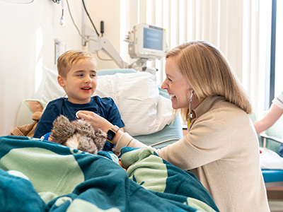
On the day before his 6th birthday, Hiram, 5, was the first patient ever with DMD to receive the drug after the U.S. Food and Drug Administration (FDA) approved its use last month.
Children’s National Hospital is the first pediatric hospital to administer a commercial dose of Elevidys (delandistrogene moxeparvovec-rokl), the first gene therapy for the treatment of pediatric patients with Duchenne muscular dystrophy (DMD).
On the day before his 6th birthday, Hiram, 5, was the first patient ever with DMD to receive the drug after the U.S. Food and Drug Administration (FDA) approved its use last month.
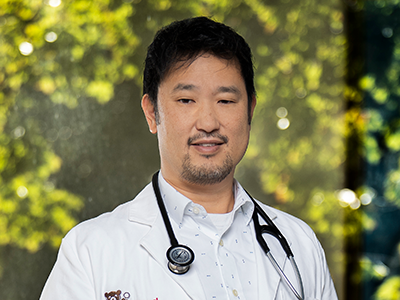
“The approval of Elevidys opens a new door for young patients with DMD and their families,” says Sarah Wright, D.O., neuromuscular neurologist at Children’s National. “This disease has had limited targeted treatments to date which can help alter the trajectory of disease.”
The background
On June 22, the FDA approved the use of Elevidys for patients 4 through 5 years of age with DMD with a confirmed mutation in the DMD gene who do not have a pre-existing medical reason preventing treatment with this therapy.
DMD is a rare and progressive genetic neuromuscular disease that predominantly affects males. It is caused by genetic changes in the DMD gene that affects the muscles, leading to muscle wasting that gets worse over time. Symptoms include progressive weakness and loss (atrophy) of both skeletal and heart muscle. Muscle weakness is usually noticeable in early childhood when signs like delayed ability to sit, stand or walk, and difficulties learning to speak manifest in a patient.
How it works
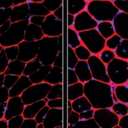
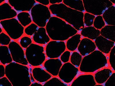
Elevidys is a one-time intravenous gene therapy that aims to delay or halt the progression of DMD by delivering a modified, functional version of dystrophin to muscle cells. The dystrophin gene is the largest known human gene.
“Elevidys is a viral vector (the ‘envelope to deliver the gene of interest’) mediated gene therapy that allows for the introduction of a gene that codes for a shortened form of dystrophin protein, or microdystrophin,” Dr. Wright explains. While not a cure for DMD, trials of Elevidys have demonstrated increases in dystrophin expression and improved functional results in young children with the disease.
“We have years of dedicated work on the part of researchers, clinician leaders and advocacy organizations in the field of muscular dystrophy to thank for this ground-breaking moment,” says Dr. Wright. “The approval of Elevidys offers families of patients ages 4-5 with DMD the option to receive this gene therapy that is designed to target the underlying cause of the disease.”
“The time-sensitivity of this medication illustrates the importance of going to a top academic pediatric hospital early on in neurologic care,” adds Elizabeth Wells, M.D., senior vice president of the Center for Neuroscience and Behavioral Medicine at Children’s National.
What’s next
The neuromuscular team at Children’s National is looking forward to offering this therapy to young patients with DMD and to the completion of additional trials/results for therapies in the DMD drug development pipeline.
“The research and approval of novel therapies provides more options for our DMD patients and their families, which is a critical step toward improving the lives of patients with DMD,” Dr. Wright says.


 Children’s National Hospital announced a $96 million investment from an anonymous donor family to transform rare childhood brain tumor research and care. The donation, which strengthens our globally recognized leadership in the field, is one of the largest in the hospital’s history.
 Children’s National Hospital in Washington, D.C., was ranked #5 in the nation on the U.S. News & World Report 2023-24 Best Children’s Hospitals annual rankings. This marks the seventh straight year Children’s National has made the Honor Roll list. The Honor Roll is a distinction awarded to only 10 children’s hospitals nationwide.
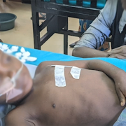
 Children’s National Hospital named


 A new validated self-report tool called the Gender Self-Report provides researchers a way to characterize the gender of research participants beyond their binary designated sex at birth.