
Harnessing children’s immune systems to fight their own brain tumors
Dalia Haydar, Pharm.D., Ph.D., principal investigator for the Program for Cell Enhancement and Technologies for Immunotherapies, recently joined Children’s National Hospital to help develop breakthrough treatments that hopefully will be a key in the fight against pediatric brain tumors. She brings her deep experience at St. Jude Children’s Research Hospital to the Center for Cancer and Immunology Research (CCIR) to help support the NexTGen team’s 10-year, $25-million Cancer Grand Challenges award.
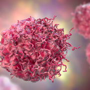
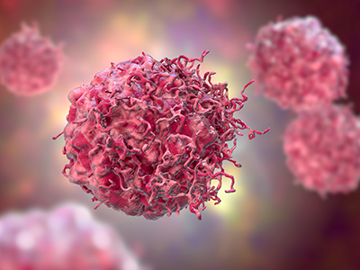
Dalia – literally – has drive: She commutes 180 miles round trip from her home in Hershey, Pa., to her lab. She says she is grateful to be at one of the few research institutions in the world that is researching how to harness the power of CAR T-cell therapies to attack solid tumors in kids. While these therapies have been approved to treat leukemia and other blood cancers, solid tumors have proven far more stubborn. Haydar has tremendous hope that she and the team led by CCIR Director Catherine Bollard, M.D., M.B.Ch.B., will change that.
Q: Could you explain the importance of this research?
A: Unfortunately, once a patient is diagnosed with a brain tumor, especially a kid, there’s very little we can do. Using chemotherapy or radiation therapy has big disadvantages because of developmental delays and other side effects. We are hoping this kind of immunotherapy – where we take the patient’s own immune cells and engineer them in the lab to attack their cancer – will eradicate their very harsh and aggressive tumors, without causing significant adverse effects.
Q: How are researchers at Children’s National going to attack solid tumors with a treatment originally designed for blood cancers?
A: We have a lot of resources and expertise at Children’s National that we are trying to put together to develop a therapy that would cure brain tumors. Unfortunately, solid tumors are hard to treat and there are several challenges for any kind of immunotherapy. But right now, being at a place where all the necessary resources, support and expertise are available, we are hoping to address each of these challenges, and we are determined to do something in a meaningful timeframe to push that survival curve toward the advantage of those kids.
Q: How soon can this work be done?
A: Within two or three years, we are hopeful we’ll be able to identify the best working regimen of this CAR T-cell immunotherapy and investigate if it will work in a patient. I foresee, in the next 5 to 10 years, that we’re hopefully going to have such therapy for kids with brain tumors.
Q: What has surprised most you in your work?
A: There are so many challenges in developing immunotherapies for kids with brain tumors. First, if it works for adults, it doesn’t necessarily work for kids. Some of the tumors in kids are more aggressive. We need to understand the tumor itself, besides understanding the immunotherapy we’re developing.
The other challenge is CAR-T immunotherapy is not like a pill or taking radiotherapy that is standardized for several patients. It’s a very expensive therapy. It’s taking the patient’s own immune cell, like a bone marrow transplant. We put it in the lab, re-engineer the cells without transforming them into a cancer cell, enable those immune cells to attack the cancer, and then put them back into the patient. There are a lot of steps you need to take to make sure you don’t artificially harm those cells or introduce contamination.
One of the most intriguing challenges for me is how we make immunotherapy work for kids who have different kinds of brain tumors – a medulloblastoma versus a glioma versus an embryonal tumor. This is one of the challenges that keeps me on my toes, and I’m hoping to answer.
Q: What is the power of being in a multi-center environment like the Children’s National Research Institute?
A: We have to do enough science on the bench to support any proposal for the therapy to move to the clinic. The last thing we want to do is to investigate a drug or therapy in patients without really knowing how it works and the potential adverse effects. Being able to work with researchers at different stages of the bench-to-bedside spectrum, as well as being able to have access to patient samples and innovative preclinical models, helps push the science forward in a shorter time frame.
 A clinical trial testing a new drug to increase growth in children with short stature. The first ever high-intensity focused ultrasound procedure on a pediatric patient with neurofibromatosis. A low dose gene therapy vector that restores the ability of injured muscle fibers to repair. These were among the most popular articles we published on Innovation District in 2022. Read on for our full top 10 list.


 Children’s National Hospital named