A simple smartphone photo may hold clues to sickle cell disease, a new study finds.
Artificial intelligence (AI) continues to move pediatric medicine forward, and now it’s showing that a simple smartphone photo may hold clues to sickle cell disease (SCD). Using machine learning, a team at Children’s National identified subtle facial differences linked to SCD in 308 children and young adults in Kinshasa, Democratic Republic of the Congo (DRC). Half of them had confirmed SCD, and each was matched with a control participant of the same age and sex.
What this means
In the study, published in PLOS Digital Health, the team’s model found 14 facial features that differed between the two groups. Six measurements around the eyes, nose and mouth did the most to tell participants with SCD apart from controls. Using those six, the model reached an accuracy of about 80% for the identification of children with SCD. Differences were most noticeable in older age groups.
Why it matters
SCD affects about 8 million people worldwide and is a major cause of childhood illness and death in sub-Saharan Africa. In places like the DRC, access to screening, confirmatory testing and specialist care is often limited. Non-invasive tools that use widely available smartphones could one day support standard clinical and community care.
Children’s National leads the way
To the team’s knowledge, this is the first machine learning study of facial features associated with SCD. The model is designed to be interpretable so clinicians and researchers can see exactly which facial measurements drive the results.
“Sickle cell disease is most common in the places where screening and specialist care are hardest to reach, so we asked whether a tool as widely available as a smartphone camera could help the care of children with the condition,” said Marius George Linguraru, DPhil, MA, MS, director of the Division of AI Research at Children’s National and senior author of the study. “Because our machine learning model is interpretable, we can see exactly which facial features are associated with sickle cell disease. That transparency is essential as we build digital tools meant to support, not replace, lab testing and clinical care.”
The work builds on the team’s earlier facial analysis research in populations with rare diseases that are underrepresented in digital health, including children in the DRC.
What’s next for the team
The team will need studies across multiple sites, over time and with detailed clinical data for further analysis. Those studies would test whether these facial features could serve as digital biomarkers for the non-invasive monitoring of disease progression and early detection of complications.
Children’s National led this research in collaboration with partners in the U.S., the Democratic Republic of Congo, Malawi and the United Kingdom, including the University of California, Irvine; Texas A&M University; Nationwide Children’s Hospital; the University of Glasgow; Kamuzu University of Health Sciences; George Washington University; the University of Colorado Anschutz Medical Campus; and Oregon Health & Science University.
https://innovationdistrict.childrensnational.org/wp-content/uploads/2026/11/400x300_HanleyMeany.jpg300400Innovation Districthttps://innovationdistrict.childrensnational.org/wp-content/uploads/2025/09/InnovationDistrict_CN_WebHeader-1396px-1030x151.pngInnovation District2026-11-10 09:40:442026-11-10 09:47:08Podcast: New Hope for Challenging Childhood Cancer: The Potential of Cell and Gene Therapy
By pinpointing a tumor’s specific methylation class, the test can give clinicians greater confidence in the diagnosis and better support a precision medicine approach.
Children’s National now offers an in-house DNA methylation profiling test that could give children with central nervous system (CNS) tumors a clearer diagnosis. Patrick Cimino, MD, PhD, the hospital’s inaugural Lucy Balian Rorke-Adams, MD, Professor of Molecular Neuropathology, led the effort to bring this advanced testing capability to patients, pathologists and care teams at Children’s National.
What this means
Targeted epigenetic profiling looks for a defined set of DNA signatures within a tumor. Methylation profiling takes a different approach, instead, analyzing chemical patterns that regulate gene activity across the genome. Together, those patterns create a molecular fingerprint to help distinguish tumors that look alike under a microscope.
That distinction can be especially helpful when tumors share overlapping histologic or genetic features and are difficult to diagnose. By pinpointing a tumor’s specific methylation class, the test can give clinicians greater confidence in the diagnosis and better support a precision medicine approach.
For Dr. Cimino, bringing the test to Children’s National marks an important step toward more precise, individualized care for children with brain tumors. “This test really helps you hone in on the actual diagnostic class the tumor belongs to, helping to move the needle for precision medicine,” he said.
The patient benefit
Brain tumors in children and adolescents can be difficult to diagnose because they are rare and biologically complex. A more precise classification can give neuro-oncologists, radiation oncologists and surgeons a clearer picture of the disease as they shape a treatment plan for each child.
“It transforms pediatric brain tumor diagnostics into something that is more accurate, allowing clinicians to have a better idea of what they’re treating,” Dr. Cimino said.
Why we’re excited
Because the assay is performed at Children’s National, samples no longer need to be sent to an outside institution – a step that can add time to the diagnostic process. “Having it in-house helps accelerate the time from tissue collection to diagnosis,” Dr. Cimino said.
What’s next
The test will also open new doors for research. Each sample generates about one million data points across the genome, but only a portion are needed to classify the tumor. The remaining data could help researchers explore biomarkers, improve risk stratification and identify tumor subtypes that have not yet been recognized. Children’s National also plans to accept referrals and samples from outside institutions, broadening access to advanced molecular diagnostics and creating more opportunities for collaboration.
A strategic philanthropic investment from an anonymous family to advance pediatric brain tumor research and care through the Brain Tumor Institute helped make the test’s validation and implementation possible. The effort strengthens the institute’s diagnostic and research capabilities and brings promising precision medicine tools closer to the children and families who may benefit.
For children with brain tumors, the goal is simple: a clearer diagnosis sooner and better information to guide the care that follows.
https://innovationdistrict.childrensnational.org/wp-content/uploads/2026/10/DNA-Methylation-feature.jpg300400Innovation Districthttps://innovationdistrict.childrensnational.org/wp-content/uploads/2025/09/InnovationDistrict_CN_WebHeader-1396px-1030x151.pngInnovation District2026-10-12 11:50:502026-10-12 12:13:33Dr. Patrick Cimino leads launch of new DNA methylation test for pediatric brain tumors
Children’s National faculty are helping shape the future of biomedical research through service on National Institutes of Health peer review panels.
Twelve Children’s National faculty members are helping shape the future of biomedical research through service on the National Institutes of Health (NIH) peer review panels. Recognized by NIH leadership for their work in 2025, these experts brought their knowledge and experience to the rigorous evaluation of grant proposals competing for federal funding. Their contributions help identify promising science, strengthen the research process and move discoveries with the potential to improve health one step closer to reality.
The big picture
NIH invests about $38 billion each year in medical research through competitive grants. Peer reviewers assess the research question, study plan, team and potential impact of each proposal before funding decisions are made.
NIH selects reviewers based on their research accomplishments, publications and other scientific contributions. Serving on a panel requires substantial time and preparation, from reading detailed applications and writing assessments to taking part in panel discussions.
Reviewers also gain first-hand knowledge of the NIH review process that they can share with colleagues preparing future applications. Their service supports the national research community while strengthening research at Children’s National.
Four Children’s National experts are taking on an even greater role in the NIH peer review process. As standing members of a study section, these leading scientists are formally appointed (often via the NIH Director or designees) to a chartered review group for a term lasting 4 to 6 years.
Fabbri serves on the Molecular Cancer Diagnosis and Classification study section, reviewing applications on the discovery, development and validation of molecular signatures of cancer.
Koutroulis serves on the Surgery, Trauma and Anesthesiology study section, reviewing grants on sepsis, wound healing, trauma, anesthetics and surgical complications.
Limperopoulos serves on the Developmental Brain Disorders study section, reviewing applications to investigate genetic, metabolic, immune, microbiome, infectious, environmental and behavioral factors in the fetal, neonatal, pediatric and adolescent brain that lead to abnormal brain development and function.
Patel serves on the National Institute for Child Health and Human Development study section.
Standing members make an ongoing commitment to review grant applications, take part in panel discussions and bring specialized knowledge to important funding decisions. Through this service, Drs. Fabbri, Koutroulis, Limperopoulos and Patel are helping the NIH identify promising research and support discoveries that could shape the future of health and medicine.
Why it matters
“Serving as an NIH peer reviewer is a meaningful commitment that strengthens the entire biomedical research community,” said Catherine Bollard, MBChB, MD, senior vice president and chief research officer at Children’s National. “Our faculty are helping uphold a rigorous, trusted process while bringing valuable insight back to Children’s National and supporting the next generation of discoveries that improve health.”
This service reflects the strength of the research community at Children’s National which includes experts across disciplines who not only pursue bold ideas but also help determine which ideas can move science forward. By bringing their knowledge, leadership and commitment to the NIH peer review, these faculty members are strengthening research nationwide and helping build a healthier future for children and families everywhere.
https://innovationdistrict.childrensnational.org/wp-content/uploads/2026/10/NIH-feature.jpg300400Innovation Districthttps://innovationdistrict.childrensnational.org/wp-content/uploads/2025/09/InnovationDistrict_CN_WebHeader-1396px-1030x151.pngInnovation District2026-10-01 12:24:512026-10-08 10:12:57Children’s National faculty help guide the future of federally funded research
Children’s National experts connect with pediatric oncology leaders from around the world at SIOP 2026 in San Antonio.
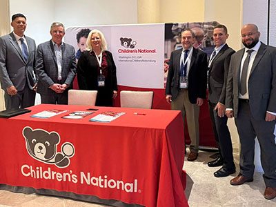
Children’s National demonstrated its leadership in pediatric oncology during the 2026 International Society of Paediatric Oncology (SIOP) gathering in San Antonio, with a prominent presence at the joint SLAOP–SIOP North America Pre-Congress and representation at the SIOP Congress.
Through its nationally recognized Cancer and Blood Disorders Center, Children’s National is helping shape the future of pediatric cancer care, research and innovation. Its specialists combine expertise across oncology, hematology, blood and marrow transplant, cellular therapy and survivorship, while collaborating with colleagues around the world to improve outcomes for children with cancer.
That leadership was especially visible at the first joint conference between SLAOP (Sociedad Latinoamericana de Oncología Pediátrica) and SIOP North America, which was held prior to the main congress. Jeffrey Dome, MD, PhD, Eugene Hwang, MD, Amy Hont, MD, and Adriana Fonseca-Sheridan, MD, shared Children’s National expertise with pediatric cancer leaders from North America and Latin America. The program fostered collaboration around pediatric cancer care, survivorship, access to treatment and clinical research.
At the SIOP Congress, Dr. Dome presented on behalf of Children’s National, while the organization’s booth provided an additional opportunity to connect with pediatric oncology leaders from around the world and showcase its expertise. As senior vice president of the Cancer and Blood Disorders Center and North American Continental President of SIOP, he brings both deep clinical expertise and an international leadership perspective to the effort to advance childhood cancer care.
In the following Q&A, Dr. Dome discusses how global collaboration is changing pediatric oncology, the strengths Children’s National brings to that work and the opportunities ahead to improve outcomes for children with cancer.
Q: You’ve had the opportunity to work with pediatric cancer leaders from around the world through your work with SIOP. How has that shaped the way you think about the future of childhood cancer care? A: As a junior faculty member, one of my mentors advised me that it is important to gain an international perspective to fully understand the treatment of pediatric cancer. I took that to heart and attended my first SIOP conference in 2006. The meeting was eye-opening to learn that the largely North American Children’s Oncology Group (COG) approach isn’t the only way to treat childhood cancer. Valuable lessons can be learned across the spectrum of high, middle and low-income settings.
Dr. Dome, North American Continental President of SIOP, represents Children’s National at SIOP 2026.
Q: Why are global partnerships and collaboration so important to advancing pediatric cancer research and treatment today? A: Global partnerships are important for several reasons. First, 80% of the global childhood cancer burden occurs outside the United States and other high-income countries; and survival rates are considerably lower outside high-income settings. We must work together to address this disparity. Second, many childhood cancers are rare, and molecular analysis has created ever-smaller subgroups. It takes international collaboration to effectively study such rare subgroups. Third, different areas of the world have different treatment approaches from us and it may be beneficial to adopt certain aspects of treatment in our treatment regimens. We can only fully understand the different approaches through open exchange of information.
Q: Children’s National is recognized as a leader in pediatric cancer care and research. What strengths does the organization bring to international collaborations aimed at improving outcomes for children with cancer? A: Children’s National is a recognized leader in several areas, including treatment of brain tumors, neuroblastoma, kidney cancers, sarcomas and high-risk leukemias. We are leaders in blood and marrow transplantation and gene therapy and pioneers in T-cell therapy for leukemias, brain tumors and solid tumors. We are proud to disseminate knowledge to assist others across the world.This year, you helped bring together experts from across North America and Latin America through the joint SLAOP-SIOP North America Pre-Congress. Why was that collaboration important?
The SLAOP-SIOP North America conference was the first-of –its-kind to build partnerships in our hemisphere. The conference program was generated in a fully collaborative manner, with input from leaders in both Latin America and North America. Each symposium had speakers from across the Americas, focusing on the current status, opportunities, and challenges specific to the different settings.
Q: What role do you see Children’s National playing in shaping the future of pediatric cancer care, both nationally and globally? A: I am proud that Children’s National sponsored both the main SIOP Congress and the SLAOP-SIOP-North America pre-conference and hope to continue that tradition in the future. As an international leader in pediatric oncology research and care, it is our responsibility to promote global efforts to disseminate information and foster partnerships to create new knowledge.
https://innovationdistrict.childrensnational.org/wp-content/uploads/2026/09/SIOP-2026-Feature.jpg300400Innovation Districthttps://innovationdistrict.childrensnational.org/wp-content/uploads/2025/09/InnovationDistrict_CN_WebHeader-1396px-1030x151.pngInnovation District2026-09-23 12:26:422026-09-23 12:30:02Children’s National Leaders Help Shape the Future of Pediatric Oncology at SIOP 2026
For children and teens with cancer, fertility preservation can offer something powerful: choices for the future. At Children’s National, Jennifer Levine, MD, MSW, helps families understand how treatment affects fertility and which options are available.
An internationally recognized expert in survivorship medicine, Dr. Levine directs the Comprehensive Cancer Wellness Program, working with specialists across Children’s National and beyond. She led the development of pediatric fertility risk assessment guidelines and is helping update them.
Here, she explains why access is challenging, what her research is revealing and how she hopes to make fertility preservation a routine part of pediatric cancer care.
Q:What drew you to fertility preservation research? A: I wanted to help families navigate a complicated system during an already overwhelming time. Fertility preservation can seem straightforward, but access requires care teams, researchers, education, policy and hospital systems to work together.
Q:Why is fertility preservation important for young patients with cancer? A: Survival comes first, of course, but pediatric cancer care also has to look ahead to life after treatment, including whether therapy could affect a patient’s ability to have biological children in the future. When there is a risk, families should have an opportunity to understand their options and protect future possibilities.
Q:What are the biggest barriers to fertility preservation care? A: Awareness has improved dramatically over the past 25 years, but it remains inconsistent. Cost is a major hurdle. Insurance coverage varies widely, and families may face preauthorization delays, confusing billing or out-of-pocket expenses. Timing is another significant challenge.
Q:What is your research helping us better understand about fertility preservation? A: Much of my research examines what keeps young people from receiving care and how we can make it easier to obtain. Right now, I’m looking at costs from both a big-picture view and from what families experience directly. I work with other researchers to analyze insurance data to understand the financial burden on families. I also lead and contribute to studies that follow in real time what happens when families seek fertility preservation. One Children’s National study, funded by the Rally Foundation, is helping us understand the real-world insurance process and costs of sperm banking. Together, this work is affirming that access is shaped not only by whether fertility preservation is medically possible, but also by timing, coverage, out-of-pocket costs and the systems families must navigate before treatment begins. I am also researching ways to protect fertility. I co-lead a national Children’s Oncology Group study of whether a hormone-suppressing drug called Triptorelin can protect ovarian function in teens and young adults receiving chemotherapy.
Q:Why does the insurance research focus on sperm banking? A: Sperm banking is generally the simplest and least expensive fertility preservation option, making it a practical starting point. My colleagues and I have advocated for all post-pubertal males to have the opportunity to bank sperm, regardless of their estimated fertility risk, because that option might not be available later.
Q:What makes risk assessment so difficult in children? A: It is not always possible to predict who will experience infertility. Risk varies by diagnosis, treatment, age and sex. Some patients are clearly at low or high risk, but many fall into a gray area. Cost makes that uncertainty even harder for families to navigate.
Q: How does Children’s National support fertility preservation care? A: We have a strong cancer wellness team, powered with help from philanthropy. We offer or coordinate the major fertility preservation options, and a navigator helps families understand their choices and move through the process. Many committed individuals at Children’s National make this work possible, including specialists in oncology, urology, adolescent gynecology, reproductive endocrinology, stem cell transplant and anesthesiology.
Q: What does the future of fertility preservation care look like? A: My goal is for fertility preservation to become an accessible, routine part of cancer care. Every patient family should be offered an informed conversation about risk and options. Stronger research, policy and clinical systems can create a smoother path to care.
https://innovationdistrict.childrensnational.org/wp-content/uploads/2026/09/Jennifer-Levine-feature.jpg300400Innovation Districthttps://innovationdistrict.childrensnational.org/wp-content/uploads/2025/09/InnovationDistrict_CN_WebHeader-1396px-1030x151.pngInnovation District2026-09-23 11:50:202026-09-23 15:07:17Q&A with Jennifer Levine, MD, MSW: Preserving choices for cancer survivors
Children’s National in Washington, D.C., was once again ranked as a top hospital in the nation by the U.S. News & World Report 2026-27 Best Children’s Hospitals annual rankings. This marks the 10th straight year Children’s National has made the Honor Roll, a distinction awarded to only 10 children’s hospitals nationwide.
Children’s National also ranked in all 11 specialty services and is the highest U.S. News ranked children’s hospital in Washington, D.C, Maryland and Virginia. Recently, U.S. News introduced pediatric & adolescent behavioral health as a service line in its rankings. While there are no ordinal rankings for behavioral health, the Children’s National program was named one of the top 50 programs in the country for the third year in a row.
“We’re so proud to reach this milestone of being named among the nation’s top children’s hospitals for the 10th year in a row,” said Michelle Riley-Brown, MHA, FACHE, president and chief executive officer of Children’s National. “Our leadership in specialties like neurology, cancer, neonatology, and diabetes and endocrinology underscores our belief that kids are our everything. It also shows what’s possible when our teams bring their expertise, commitment and heart to make sure every child receives the best care.”
The annual rankings are the most comprehensive source of quality-related information on U.S. pediatric hospitals and recognize the nation’s top 50 pediatric hospitals based on a scoring system developed by U.S. News.
“Earning a place on U.S. News’ Best Children’s Hospitals list is a testament to Children’s National’s dedication to clinical excellence and patient safety,” said Ben Harder, chief of health analysis and managing editor at U.S. News. “These rankings highlight the standout hospitals in each specialty and each region.”
The bulk of the score for each specialty service is based on quality and outcomes data. The process includes a survey of relevant specialists across the country who are asked to list hospitals they believe provide the best care for patients with the most complex conditions.
The Children’s National specialties ranked in the top 10 nationally by U.S. News are:
https://innovationdistrict.childrensnational.org/wp-content/uploads/2026/09/USNWR-Badges-2026-27-CNRI-1.jpg385685Innovation Districthttps://innovationdistrict.childrensnational.org/wp-content/uploads/2025/09/InnovationDistrict_CN_WebHeader-1396px-1030x151.pngInnovation District2026-09-15 01:00:552026-09-11 14:02:41Children’s National ranked among top children’s hospitals nationwide
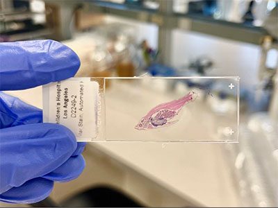
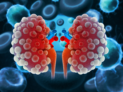
The study found that hospitals generally diagnose and stage pediatric kidney tumors accurately, but identified an important gap in recognizing a rare, aggressive form of Wilms tumor.
A large Children’s Oncology Group (COG) study found that hospitals generally diagnose and stage pediatric kidney tumors accurately, but identified an important gap in recognizing a rare, aggressive form of Wilms tumor.
Published in Pediatric Blood & Cancer, the study found that diffuse anaplastic Wilms tumor was missed by local institutions in about one-third of cases, highlighting the value of expert pathology review for difficult-to-diagnose tumors.
The big picture
Researchers analyzed 6,311 patients enrolled in the COG AREN03B2 Renal Tumor Biology and Classification Study from 2006 to 2019. They compared assessments by local pathologists, radiologists and surgeons with real-time reviews by central experts.
These evaluations determine a tumor’s diagnosis, stage and risk classification, which helped assign patients to appropriate clinical trials for treatment, while also helping care teams select the most appropriate treatment outside research studies.
What the study found
Overall, local institutions and central experts agreed on the pathological diagnosis in 91.1% of cases, the assessment of lung nodules on CT scans in 96.1% and surgical staging in 90%. When researchers considered all risk factors together, local institutions assigned the same treatment-risk category as central reviewers 87% of the time.
The largest discrepancy involved diffuse anaplastic Wilms tumor, an aggressive and treatment-resistant subtype that comprises about 5 to 10% of Wilms tumors. Local institutions correctly identified 181 of 269 cases, or 67%. Because more intensive treatment is needed to achieve successful outcomes for anaplastic Wilms tumor, an accurate identification of this subtype is especially important.
“Accurately identifying anaplastic Wilms tumor is critical because it influences treatment, and ultimately, patient outcome,” said Jeffrey Dome, MD, PhD, co-senior author of the study and senior vice president of the Cancer and Blood Disorders Center at Children’s National. “While our findings show that pediatric kidney tumors are diagnosed and staged accurately in most cases, they also demonstrate the value of expert pathology review when evaluating rare, aggressive tumor features that can be difficult to recognize.”
Why it matters
The findings show that COG institutions assess most pediatric kidney tumors with a high degree of accuracy. They also demonstrate where specialized expertise can make a difference.
For children with diffuse anaplastic Wilms tumor, an accurate diagnosis is particularly important because tumor classification helps determine treatment intensity. Missing anaplastic features could result in a child receiving less intensive treatment than their disease requires.
What’s next
The researchers say expert pathology consultation remains important for institutions with limited experience evaluating pediatric kidney tumors. Current research seeks to identify anaplastic histology by detecting molecular changes in circulating tumor DNA. There are also initiatives to harness artificial intelligence to identify anaplastic Wilms tumor based on the appearance of histology slides.
https://innovationdistrict.childrensnational.org/wp-content/uploads/2026/09/Kidney-tumor-feature.png300400Innovation Districthttps://innovationdistrict.childrensnational.org/wp-content/uploads/2025/09/InnovationDistrict_CN_WebHeader-1396px-1030x151.pngInnovation District2026-09-10 13:30:332026-09-10 13:31:38Study identifies diagnostic gap in an aggressive type of Wilms tumor
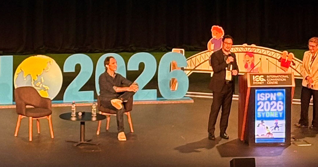
Roger J. Packer, MD, director of the Brain Tumor Institute and director of the Gilbert Family Neurofibromatosis Institute at Children’s National, founded ISPNO in 1986 and continues to help shape the future of pediatric neuro-oncology.
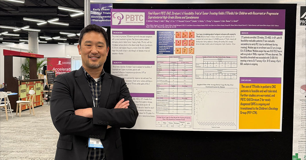
Eugene Hwang, MD, participates in a featured debate on the role of artificial intelligence in cancer care and research at ISPNO 2026.
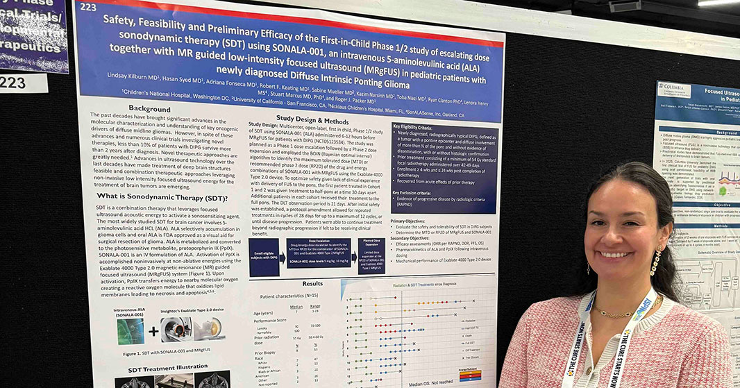
Adriana Fonseca, MD, presents research at ISPNO 2026, contributing to international efforts to advance care for children with brain tumors.
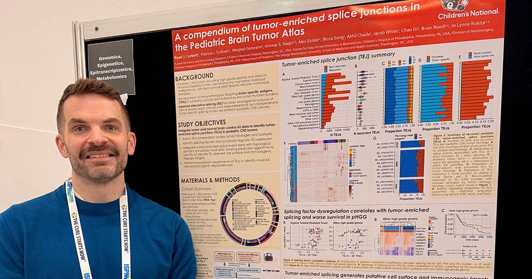
Ryan Corbett, PhD, shares research findings during a poster session at the 22nd International Symposium on Pediatric Neuro-Oncology.
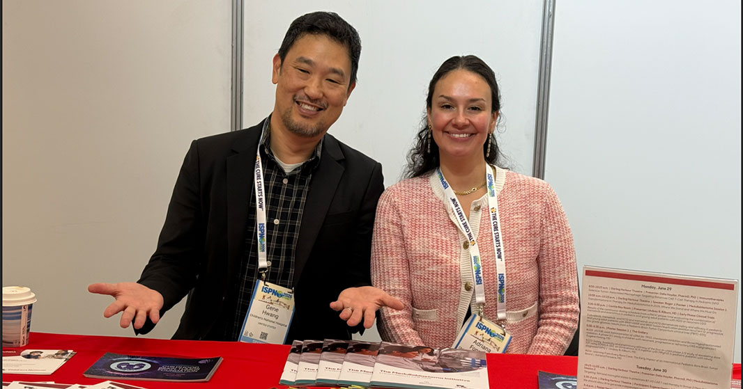
Eugene Hwang, MD, and Adriana Fonseca, MD, represent Children’s National at the institution’s booth during ISPNO 2026.
Eugene Hwang, MD, presenting emerging research at a poster session at ISPNO 2026.
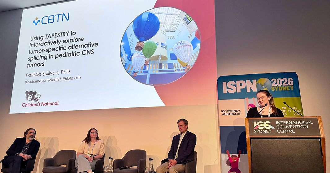
Patricia Sullivan, PhD, presents research on TAPESTRY, an interactive platform designed to explore tumor-specific alternative splicing in pediatric CNS tumors.
When Roger J. Packer, MD, organized the first International Symposium on Pediatric Neuro-Oncology (ISPNO) in 1986, he saw an opportunity to bring together experts from around the world who shared a common goal: advancing care and outcomes for children with brain tumors.
Four decades later, that vision has evolved into the world’s premier pediatric neuro-oncology meeting, fostering collaboration across disciplines, accelerating the exchange of scientific discoveries and helping shape the future of the field.
That vision was on display once again at the 22nd International Symposium on Pediatric Neuro-Oncology, held June 28 through July 1, 2026, in Sydney, Australia. More than 1,000 attendees from 56 countries gathered for four days of scientific presentations, collaboration and discussion focused on improving care for children with central nervous system tumors.
“When we first conceptualized and had the first meeting, we convened a very small group, about 100 to 150 people interested in pediatric brain tumors,” Dr. Packer says. “It was a mix of neurosurgeons, neurologists, oncologists and radiation oncologists.”
The first major international symposium followed in Philadelphia, drawing more than 350 attendees, far exceeding expectations and helping establish the foundation for what would become ISPNO.
“We expected 200 people, and we got 350,” Dr. Packer recalls. “From there, we decided to make this a truly international meeting that would reach all disciplines involved in the care of children with brain tumors.”
Unlike many professional organizations, ISPNO was intentionally designed as a symposium rather than a society. Its purpose was simple: bring together basic scientists, translational researchers and clinicians to openly share ideas and discoveries.
“The goal has not been anything other than the rapid interchange of ideas and growing the field,” Dr. Packer says.
That approach helped transform ISPNO into a global forum that today attracts well over 1,000 participants while expanding opportunities for clinicians and investigators from developing countries to participate and share their work.
A front-row seat to the molecular era
Over the last 40 years, pediatric neuro-oncology has undergone dramatic transformation. Dr. Packer recalls a turning point roughly a decade ago when researchers began sharing groundbreaking discoveries about the biology of pediatric brain tumors.
“There had been this explosion in our understanding of the biology of childhood medulloblastomas and an inkling that a similar explosion would occur in childhood gliomas,” he says. “That was entering the molecular era of childhood brain tumor care.”
Those insights ushered in a new generation of biologically driven therapies that have changed how tumors are classified and treated. What was once a field largely defined by surgery, radiation and chemotherapy is increasingly shaped by molecular targeted therapies and immunotherapy.
“Over the past two meetings, immunotherapy, especially cellular immunotherapy, has increasingly revolutionized the field and given us great hope,” Dr. Packer says.
The symposium itself has reflected that evolution, drawing growing numbers of molecular biologists, immunologists and translational researchers alongside neurosurgeons, oncologists and neurologists.
Children’s National driving the next generation of discovery
Many of the advances highlighted at ISPNO mirror work underway at Children’s National.
“I think it’s amazing to me that in the last two years, because of new resources, philanthropy and new ideas, we’ve been able to open eight to 10 new biologically based either cellular therapy or molecular targeted therapy protocols,” Dr. Packer says. “About half of those will be emanating directly from work being done here and led by our investigators.”
Among those efforts are Children’s National-led CAR T-cell studies and novel approaches combining focused ultrasound technology with cellular therapies.
“We’re the first to take the technology of focused ultrasound and combine it with cellular therapy,” Dr. Packer says. “It’s very exciting.”
That spirit of innovation was evident throughout the 2026 symposium, where Children’s National experts presented research spanning targeted therapies, cellular immunotherapy, artificial intelligence, neurofibromatosis and rare brain tumors.
Children’s National at ISPNO 2026
Children’s National physicians and researchers contributed across the scientific program as presenters, moderators, panel participants and poster authors. Highlights included:
Presentations and Leadership Roles
Roger J. Packer, MD, Transformations in Therapy for Medulloblastoma in 2026: Where We Stand and Where We Must Go
Roger J. Packer, MD, Presenter during the symposium Awards Ceremony
Dalia Haydar, PharmD, PhD, Selective Tumor-Associated Macrophage Targeting Enhances CAR T-Cell Therapy in Pediatric DIPG
Dalia Haydar, PharmD, PhD, Targeting TAM and Microglial States to Potentiate B7-H3 CAR T-Cell Therapy in Group 3 Medulloblastoma
Dalia Haydar, PharmD, PhD, Engineering Next-Generation CAR T Cells for Pediatric Brain Tumors: Dual Targeting Strategies
Lindsay B. Kilburn, MD, Results from a PNOC phase I trial evaluating trametinib and everolimus in pediatric and young adult patients with high-grade glioma or recurrent low-grade glioma
Lindsay B. Kilburn, MD, Results from Pediatric Brain Tumor Consortium trial PBTC-055 evaluating targeted therapies for recurrent or progressive low-grade glioma
Eugene Hwang, MD, debate participant in We should allow AI to generate independent recommendations in cancer care and research
Poster Presentations
Marius George Linguraru, DPhil, MA, MSc, Local Magnetic Resonance Imaging Radiomic Features of the Anterior Visual Pathway Differentiate Observation Versus Treatment in Children with NF1-OPG
Marius George Linguraru, DPhil, MA, MSc, CAVS-NF1: Web-Based AI Tool for Longitudinal Volumetric Analysis of Anterior Visual Pathway in NF1
Eugene Hwang, MD, JACOB: Joint Administration of cDNA for TP53, Checkpoint Inhibition and Boost/Hypofractionated Radiation
Lindsay B. Kilburn, MD, Safety, Feasibility and Preliminary Efficacy of the First-in-Child Phase 1/2 Study of Escalating Dose Sonodynamic Therapy Using SONALA-001 Together With MR-Guided Focused Ultrasound in Pediatric Patients With Newly Diagnosed DIPG
Adriana Fonseca, MD, Learning From Every Rare Case: The Early Impact of an International Pediatric Rare Brain Tumor Board
Adriana Fonseca, MD, CNS Tumor Type Prevalence According to Age Group: An Analysis of 21,000 Cases Confirmed by Methylation Profiling
Dipak Poria, PhD, Adriana Fonseca, MD, Building Preclinical Models for Ultra-Rare Pediatric Brain Tumors: Early Outputs of the International Rare Brain Tumor Program
Dipak Poria, PhD, Adriana Fonseca, MD, Establishing Patient-Derived and iPSC-Organoid Models of High-Risk Childhood Astroblastoma
Dalia Haydar, PharmD, PhD, From Design to DEFEAT: Optimizing Dual-Target B7H3/GPC2 CAR T Cells for Pediatric Brain Tumors
Dalia Haydar, PharmD, PhD, TCR-CAR T Cells as a Novel Approach for Pediatric High-Grade Glioma Therapy
Elton VanNoy, Modulating DNA Methylation During CAR T Manufacturing to Enhance Immunotherapy for Pediatric Glioma
Kaleem Coleman, MicroRNA-Mediated Reprogramming of the Tumor Microenvironment: Enhancing CAR T-Cell Therapy for Diffuse Intrinsic Pontine Glioma
Conrad Russell Cruz, Harnessing the Noncanonical Peptidome of Diffuse Midline Glioma for Adoptive T-Cell Therapies
Ryan Corbett, PhD, A Compendium of Tumor-Enriched Splice Junctions in the Pediatric Brain Tumor Atlas
Patricia Sullivan, PhD, Using TAPESTRY to Interactively Explore Tumor-Specific Alternative Splicing in Pediatric CNS Tumors
Looking toward the next 40 years
While much has changed since the first symposium, Dr. Packer believes the field is only beginning to realize the potential of recent scientific advances.
Survival rates for some pediatric brain tumors have improved dramatically, and treatments are becoming increasingly precise and less harmful. Looking ahead, he expects molecular therapies and immunotherapies to reshape outcomes for children facing the most challenging diagnoses.
“I think the next decade will bring transformational steps,” he says. “We will take tumor types that are currently essentially incurable or have a very low likelihood of long-term survival to a point where we can treat them effectively and save the majority of children.”
For Dr. Packer, however, the symposium’s greatest accomplishment is not the size of the meeting or the number of attendees. It is the culture of collaboration that has defined ISPNO from the beginning.
“My advice is to think globally and not siloed,” he says. “Ensure the sharing of knowledge freely without barriers.”
Forty years after helping launch ISPNO, that philosophy continues to guide both the symposium and the work taking place at Children’s National. As new discoveries move from the laboratory to clinical trials and ultimately to patients, the institution remains at the forefront of efforts to improve outcomes and quality of life for children with brain tumors around the world.
https://innovationdistrict.childrensnational.org/wp-content/uploads/2026/09/Packer-feature.jpg300400Innovation Districthttps://innovationdistrict.childrensnational.org/wp-content/uploads/2025/09/InnovationDistrict_CN_WebHeader-1396px-1030x151.pngInnovation District2026-09-01 14:35:122026-09-01 14:37:09Forty years of collaboration, discovery and progress in pediatric neuro-oncology
A chemotherapy-free stem cell transplant may offer a safer path to curing children with sickle cell disease.
For children with sickle cell disease (SCD), a stem cell transplant from a matched sibling donor can offer a cure, but the intensive chemotherapy traditionally used before transplant can cause significant short- and long-term side effects.
A new multicenter clinical trial led by Children’s National and published in Blood Advances points to a potentially safer path forward. Researchers evaluated a regimen that avoids chemotherapy and found it was feasible, well tolerated and did not result in graft-versus-host disease (GVHD), one of transplantation’s most serious complications.
While additional work is needed to improve long-term transplant success, the findings mark an important step toward safer curative therapy for children with SCD.
Why it matters
For decades, chemotherapy has been considered an essential part of preparing patients for stem cell transplant. While effective, it can carry risks ranging from short-term illness to long-term health complications.
This study challenges this long-standing approach by showing that a matched sibling donor transplant may be possible without chemotherapy. Researchers also found that no patients developed GVHD, a potentially life-threatening complication in which donor immune cells attack the patient’s body.
The study also captured how patients were feeling in the weeks after transplant, offering an early look at the patient experience during recovery.
“One of the most exciting findings was that patients’ reported quality of life did not decline in the early period after transplant,” Robert Nickel, MD, study author and pediatric hematologist at Children’s National, said. “That is noteworthy because we typically expect patients to experience significant side effects and a temporary worsening in how they feel during the weeks immediately following transplant.””
Moving the field forward
Researchers found that approximately one in five patients experienced graft rejection, causing SCD to return.
The ongoing trial has been modified to improve donor cell engraftment while preserving the regimen’s favorable safety profile.
The next phase will focus on strengthening long-term transplant success.
“Our goal is to make curative therapy for sickle cell disease safer without compromising its effectiveness,” said Dr. Nickel. “These results show that a chemotherapy-free transplant is possible and help us understand how to improve engraftment so more children can benefit.”
Children’s National leads the way
Children’s National led the international multicenter trial and enrolled the largest number of patients. The study is among the first to evaluate this approach in children with SCD — an option not currently available at most pediatric transplant centers.
The research was supported by the Doris Duke Foundation.
https://innovationdistrict.childrensnational.org/wp-content/uploads/2023/03/sickle-cell-disease-feature.png300400Innovation Districthttps://innovationdistrict.childrensnational.org/wp-content/uploads/2025/09/InnovationDistrict_CN_WebHeader-1396px-1030x151.pngInnovation District2026-07-22 11:44:002026-07-22 11:45:16Chemotherapy-free transplant approach offers new hope for children with sickle cell disease
A first-in-human clinical trial led by Children’s National showed encouraging early results for a new T-cell immunotherapy in children and young adults with aggressive brain tumors.
Researchers report encouraging early results from a first-in-human clinical trial led by Children’s National using a new T-cell immunotherapy for children and young adults with some of the deadliest brain tumors, including diffuse intrinsic pontine glioma (DIPG) and relapsed central nervous system (CNS) tumors. These findings, published in Nature Medicine, are particularly significant given the challenges faced in treating pediatric brain tumors, which remain the leading cause of cancer-related deaths in children. Immunotherapies have been shown to work in blood cancers but rarely succeed in solid tumors, especially brain tumors.
“This study represents an important step toward developing safer and more effective T-cell therapies for children with devastating brain cancers,” said Catherine Bollard, MBChB, MD, senior vice president and chief research officer at Children’s National, and co-senior author of the study. “Even in this early-stage trial focused on safety, we were encouraged to see lasting clinical benefit in several patients who otherwise had very few options.”
The Phase 1 study evaluated a novel multi-targeted T-cell therapy designed to strike three proteins commonly found in pediatric brain tumors – WT1, PRAME and Survivin. Investigators say the results provide early evidence that the patient’s own immune cells delivered through the bloodstream can reach and fight tumors in the brain while producing fewer severe side effects than some existing engineered immune therapies. Many other brain tumor immunotherapies require direct injection into the brain or cerebrospinal fluid.
“We were excited to see that we could preserve safety and quality of life while generating anti-tumor responses by attacking three targets at once,” said Eugene Hwang, MD, chief of Oncology at Children’s National and co-senior author of the study.
Key takeaways:
The multi-target design may help address tumor heterogeneity, one of the major barriers to successful treatment of aggressive childhood cancers.
The trial successfully established a feasible manufacturing process, identified a maximum tolerated dose and defined an early safety profile – key milestones needed to advance the therapy into future Phase 2 studies.
Researchers analyzed patients with DIPG or relapsed brain cancers, showing both responses and prolonged disease control, with some patients remaining disease-free years after treatment.
“We are so proud of the team at Children’s National and inspired by what this breakthrough means for children with brain cancer,” said Callie and Brad Nierenberg, founders of Chance for Life, which helped to fund the study. “Supporting this trial has been one of the most meaningful investments Chance for Life has made.”
“Pediatric tumors are one of the greatest challenges in cancer research, with children still facing extremely limited treatment options, and existing treatments often causing severe side effects,” said David Scott, director of Cancer Grand Challenges, which supports the NexTGen team, a group of international collaborators co-led by Dr. Bollard that is taking on the ‘solid tumors in children’ Cancer Grand Challenge.
This research was also supported by generous funders reflecting the strength of the community in fighting these deadly cancers, including: the Chance for Life Foundation; anonymous private investors supporting the Children’s National Hospital Brain Tumor Institute; Cancer Research UK, the National Cancer Institute and The Mark Foundation for Cancer Research, through the Cancer Grand Challenges award (NCI/CRUK OT2CA278700); the GW T32 Cancer Biology Program (NIH/NCI T32 CA247756); the Rally Foundation; the Willie Strong Foundation; and the Warrior Jace Foundation.
https://innovationdistrict.childrensnational.org/wp-content/uploads/2026/07/brain-tumor-feature.jpg300400Innovation Districthttps://innovationdistrict.childrensnational.org/wp-content/uploads/2025/09/InnovationDistrict_CN_WebHeader-1396px-1030x151.pngInnovation District2026-07-01 11:20:282026-07-01 11:21:36Innovative cell therapy offers new hope for children with aggressive brain tumors
Using advanced zebrafish genetic models, Dr. Vasileva can track cancer cells as they grow and spread within living organisms, offering an unprecedented view into tumor development.
What if researchers could watch pediatric cancers develop in real time and uncover the earliest moments when healthy cells become tumors? That question drives Elena Vasileva, PhD, a principal investigator at Children’s National Research Institute, whose work explores how childhood cancers begin, evolve and interact with the cells around them.
Using advanced zebrafish genetic models, Dr. Vasileva can track cancer cells as they grow and spread within living organisms, offering an unprecedented view into tumor development. Her research combines molecular oncology, developmental biology and emerging technologies such as single-cell analysis to better understand the vulnerabilities of pediatric cancers and identify new opportunities for treatment.
As she builds her laboratory at Children’s National with the support from the Center of Cancer and Immunology Research and Brain Tumor Institute, Dr. Vasileva is focused on advancing precision pediatric oncology and developing new research models for childhood cancers that remain understudied. Here, she shares what drew her to Children’s National, the promise of zebrafish models and the future of pediatric cancer research.
Q: What brought you to Children’s National? A: I was looking for a highly collaborative environment where scientists and clinicians work closely together to solve important problems. Children’s National stood out because of its strong pediatric oncology program and translational focus. I was excited by the opportunity to work alongside leaders in the field like Drs. Catherine Bollard, Roger Packer and Muller Fabbri, and contribute to research that can ultimately improve care for children.
Q: What is the focus of your research program? A: My research sits at the intersection of molecular oncology and developmental biology. I’m interested in understanding how pediatric tumors initiate, develop and progress, as well as how they interact with the surrounding cellular environment. By uncovering the molecular mechanisms that drive these processes, we hope to identify new therapeutic opportunities.
Q: How do zebrafish models help advance pediatric cancer research? A: Zebrafish allow us to introduce mutations found in human pediatric cancers and observe tumor development in real time. Because the animals are transparent during development, we can visualize how cancer cells grow, spread and interact with other cells. Importantly, childhood cancers arise in growing organisms, so zebrafish provide a dynamic developmental context that closely aligns with the biology we are trying to understand.
Zebrafish provide a dynamic developmental context that closely aligns with the biology researchers are trying to understand.
Q: What advances in the field are you most excited about? A: Technologies such as single-cell transcriptomics, proteomics, spatial biology and computational analysis are transforming how we study tumors. We can now examine individual cells within a tumor and better understand why some respond to treatment while others do not. These tools are helping drive a shift toward precision pediatric oncology and more personalized therapies.
Q: What role could artificial intelligence play in cancer research? A: Artificial intelligence has tremendous potential to help researchers analyze tumor architecture and cellular morphology more efficiently. It can distinguish different cell types and detect subtle changes very quickly, which could help us better understand tumor heterogeneity and identify new biological insights.
Q: What challenges remain in pediatric oncology? A: Pediatric cancers remain significantly underfunded, and many rare childhood cancers still lack strong research models. Because pediatric cancers are biologically different from adult cancers, discoveries in adult oncology cannot always be applied to children. Continued collaboration, awareness and investment are essential to advancing research and developing better treatments.
Q: What are your goals for the future? A: I hope to build a robust research program that develops new zebrafish models for pediatric cancers, particularly those that currently lack effective experimental systems. My long-term goal is to bridge fundamental biological discovery with translational impact by identifying tumor vulnerabilities and testing new therapeutic strategies that can benefit patients.
https://innovationdistrict.childrensnational.org/wp-content/uploads/2026/06/zebrafish-CNRI.jpg385685Innovation Districthttps://innovationdistrict.childrensnational.org/wp-content/uploads/2025/09/InnovationDistrict_CN_WebHeader-1396px-1030x151.pngInnovation District2026-06-17 13:53:072026-06-17 13:54:14Rethinking pediatric cancer treatment: Q&A with Elena Vasileva, PhD
Main stage at the International Society for Cell & Gene Therapy 2026 conference in Dublin, Ireland. Photo by Roger Kenny.
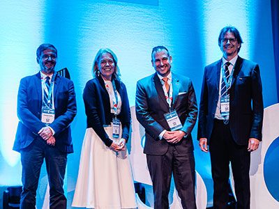
Patrick Hanley, PhD, chief and director of the Cellular Therapy Program at Children’s National Hospital, was recognized with the inaugural 2026 Mid-Career Award from the International Society for Cell & Gene Therapy (ISCT), one of the leading global organizations advancing cell and gene therapy research and clinical translation.
The award honors mid-career leaders who have made “significant and sustained contributions to the advancement of cell and gene therapy translation,” recognizing measurable impacts on science, innovation and patient outcomes. In its award letter, leaders praised Dr. Hanley’s “sustained scientific excellence, international standing and transformative leadership within the Society.”
Dr. Hanley was formally recognized during the organization’s 2026 annual meeting in Dublin, where leaders in cell and gene therapy from around the world gathered to discuss the latest advances shaping the future of medicine. Children’s National researchers made a strong showing throughout the meeting, with seven combined presentations, posters and panels, highlighting the institution’s growing leadership in advancing cell and gene therapy research.
Dr. Hanley’s work has helped advance the field through leadership in the discovery, optimization and manufacturing of novel cell and gene therapies for clinical application. The award citation highlighted his oversight of more than 650 cellular therapy products and his leadership of centralized manufacturing efforts supporting multicenter trials led by the Children’s Oncology Group and the Blood and Marrow Transplant Clinical Trials Network.
At Children’s National, Dr. Hanley leads research focused on developing advanced cellular immunotherapies for children with cancer and other serious diseases. His work centers on engineering immune cells that can better recognize and fight disease while improving the safety, scalability and accessibility of these therapies for pediatric patients.
His research program has helped drive progress in areas including CAR T-cell therapy, virus-specific T-cell therapies and other next-generation cellular immunotherapy approaches. Dr. Hanley and his collaborators are working to accelerate the translation of promising discoveries into clinical trials designed specifically for children.
The organization also recognized Dr. Hanley’s longstanding leadership and service within the field, including roles as Cytotherapy commissioning editor, North America regional vice president, co-chair of the Workforce Development Committee, co-chair of the Immuno-Gene Therapy Committee and co-founder of the Early-Stage Professionals Committee.
Among the additional Children’s National researchers recognized at the meeting was Kajal Chaudhry, PhD, who received a Technologist Award for research focused on next-generation cellular immunotherapy approaches for pediatric solid tumors. Her award-winning abstract, Multimodal CAR-TA T Cell Therapy Combining B7H3 and PRAME Targeting With TNG–TGFBTII Armoring Elicits Enhanced Anti-tumor Potency in Solid Tumor Models, explored strategies to enhance the effectiveness of engineered immune cells against difficult-to-treat cancers.
Children’s National also earned the conference’s Best Poster Award, presented to Conrad Russell Y. Cruz, MD, PhD, recognizing additional research excellence from the institution.
Together, the honors underscore Children’s National Hospital’s continued investment in translating cutting-edge discoveries into new therapies for children and reflect growing international recognition of its leadership across discovery science, translational research and clinical innovation in cell and gene therapy.
https://innovationdistrict.childrensnational.org/wp-content/uploads/2026/05/ISCT-2026-Dublin-0517-feature.jpg300400Innovation Districthttps://innovationdistrict.childrensnational.org/wp-content/uploads/2025/09/InnovationDistrict_CN_WebHeader-1396px-1030x151.pngInnovation District2026-05-26 10:27:372026-05-27 11:25:38Patrick Hanley, PhD, receives inaugural international award from ISCT
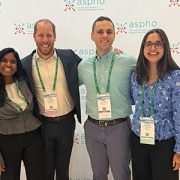
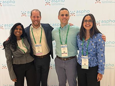
Children’s National fellows shared their research at ASPHO 2026, contributing to important conversations in pediatric hematology/oncology.
Children’s National Hospital had a strong presence at the 2026 ASPHO Conference, held April 29th – May 2nd in Minneapolis, Minnesota, where pediatric hematology/oncology specialists gathered to share the latest clinical, translational and survivorship research and to advance collaboration across the field. This year’s meeting offered an important platform for Children’s National oncology faculty and fellows to contribute to national conversations through invited presentations, abstract-driven sessions and society leadership roles.
Jennifer Levine, MD, medical director of the Cancer Survivorship Program, presented during the Presidential Symposium, “Addressing Financial Distress in Pediatric Hematology/Oncology: Implications for Cancer, Sickle Cell Disease, and Survivorship.” Her talk, “The Financial Burden of Fertility Preservation among CAYA with Cancer,” highlighted an important dimension of survivorship care and reflected Children’s National Hospital’s commitment to advancing patient-centered oncology research.
Lea Mayer, DNP, APRN, CPNP-PC, CPHON, pediatric nurse practitioner, was named chair of the ASPHO Advanced Practice Provider Special Interest Group and contributed as a panel expert and poster co-presenter.
Liny John, MD, MSE, MHS, represented Children’s National in ASPHO leadership through service on the Education Committee and as chair of the Accreditation Sub-Committee.
Children’s National fellows also contributed to the scientific program. Alan Siegel, MD, received an Early Career Travel Award and delivered a platform presentation on immune subset dynamics following selumetinib treatment for NF1 plexiform neurofibromas. Anisha Gopu-Gilboy, MD and Audrey Ward, MD presented posters that further showcased the breadth of fellow-led clinical scholarship at Children’s National.
By sharing expertise on a national stage, the oncology team is helping shape the future of the field and reinforcing leadership in research, education and innovation at Children’s National.
https://innovationdistrict.childrensnational.org/wp-content/uploads/2026/05/ASPHO-group-feature.jpg300400Innovation Districthttps://innovationdistrict.childrensnational.org/wp-content/uploads/2025/09/InnovationDistrict_CN_WebHeader-1396px-1030x151.pngInnovation District2026-05-19 09:50:302026-08-05 12:27:42Children’s National oncology team showcases research and leadership at ASPHO 2026
Researchers at Children’s National Hospital are helping advance pediatric AI with a first-of-its-kind open-access brain tumor imaging dataset designed to improve radiology and precision medicine for children.
Artificial intelligence (AI) is moving quickly in medicine, and radiology is one of the easiest places to see that progress. But in pediatrics, the path forward is more complicated. Children are not just smaller versions of adult patients. They have different diseases, different biology and different care processes, which means AI has to be built with that reality in mind from the beginning.
That challenge is clear in a new effort focused on pediatric brain tumors. While rare, these tumors remain the most common solid tumors in children and the leading cause of cancer-related mortality in the pediatric population. Compared to adult brain tumors, they behave differently across biology, anatomy and clinical care, which makes diagnosis, monitoring and treatment response especially complex. AI has the potential to improve all of this. But progress has been slow, largely because the field has been working without the kind of large, standardized and accessible datasets that make AI actually work at scale.
Dive deeper
A new international effort, co-led by Marius George Linguraru, DPhil, MA, MSc, of Children’s National Hospital and Anahita Fathi Kazerooni, PhD of Children’s Hospital of Philadelphia, is helping change that. The Brain Tumor Segmentation in Pediatrics dataset, known as BraTS-PEDs, is the first large-scale, open-access benchmark dataset designed specifically for pediatric brain tumor segmentation and analysis. It brings together MRI data from multiple imaging sequences from 457 pediatric patients with high-grade gliomas, collected across several institutions and international research consortia.
Each case includes a full set of clinically relevant imaging sequences, including pre- and postcontrast T1-weighted, T2-weighted and T2-FLAIR MRI. Experts labeled different parts of each tumor using established pediatric neuro-oncology guidelines. They combined automated tools designed for pediatric cases with careful review and refinement by neuroradiologists. The result is a dataset that is not only large but consistent in a way the field has not had before.
Just as important, the dataset is structured to support real-world use. It is divided into training, validation and hidden testing subsets, allowing researchers to benchmark models in a reproducible way and evaluate how well those models generalize across institutions.
Why this matters
This kind of resource addresses one of the biggest barriers in pediatric AI. Without enough high-quality, standardized data, even the most promising algorithms struggle to translate into clinical use.
BraTS-PEDs changes that by giving researchers a shared foundation. It allows for consistent method comparison, supports model development across institutions and opens the door for integrating imaging with molecular and clinical data, which is where things start to move toward precision medicine in a meaningful way.
For pediatric radiology, that shift is critical. AI is not just about improving image analysis. It is about building tools that can help clinicians better understand disease, track how it evolves and make more informed decisions over time. Dr. Linguraru’s team has created public tools to provide open access to the volumetric analysis of pediatric brain tumors to doctors and researchers everywhere.
What comes next
At Children’s National, this work fits into a broader effort to rethink how AI is developed and deployed in pediatric care. Through its Division of AI Research, the focus is on creating a clinically grounded ecosystem where data, technology and clinical expertise are tightly connected. The goal is to move beyond isolated projects and build infrastructure that allows ideas to move from concept to application in a way that is scalable and sustainable. As Dr. Linguraru notes, “AI tools can play a key role in radiology, but radiologists must be able to trust in the systems’ design and receive adequate training.”
Radiology is already leading the way when it comes to AI in medicine, but pediatrics is still catching up in terms of data and infrastructure. Efforts like BraTS-PEDs are a step toward closing that gap. By creating shared, standardized resources, they make it possible to build and evaluate models in a way that is consistent, collaborative and clinically relevant. For children, that matters. Getting AI right in pediatrics is not just about innovation. It is about making sure the tools being developed actually reflect the patients they are meant to serve.
This research, Brain Tumor Segmentation in Pediatrics (BraTS-PEDs): A Multi-Institutional Benchmark Dataset for Pediatric Neuro-Oncology, was published in Radiology: Artificial Intelligence. Authors from Children’s National Hospital include Marius George Linguraru, DPhil, MA, MSc; Zhifan Jiang, PhD; Xinyang Liu, PhD; Brian Rood, MD; and Roger J. Packer, MD; in collaboration with colleagues from multiple institutions and international consortia working to advance artificial intelligence in pediatric neuro-oncology.
https://innovationdistrict.childrensnational.org/wp-content/uploads/2026/05/brain-tumors-CNRI.png385685Innovation Districthttps://innovationdistrict.childrensnational.org/wp-content/uploads/2025/09/InnovationDistrict_CN_WebHeader-1396px-1030x151.pngInnovation District2026-05-12 16:26:182026-05-12 16:28:16Advancing clinical deployment of artificial intelligence in pediatric radiology
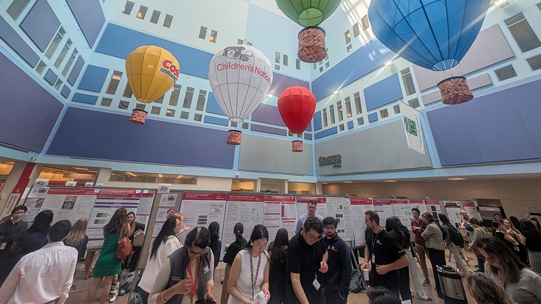
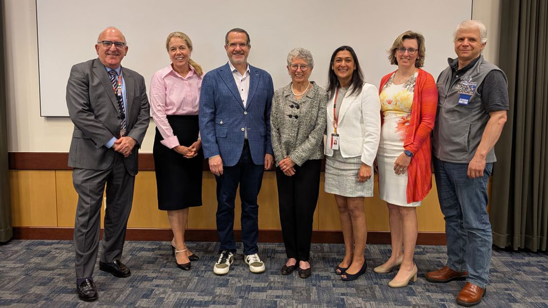
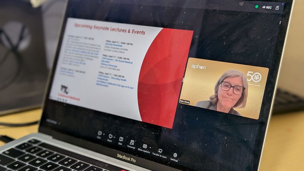
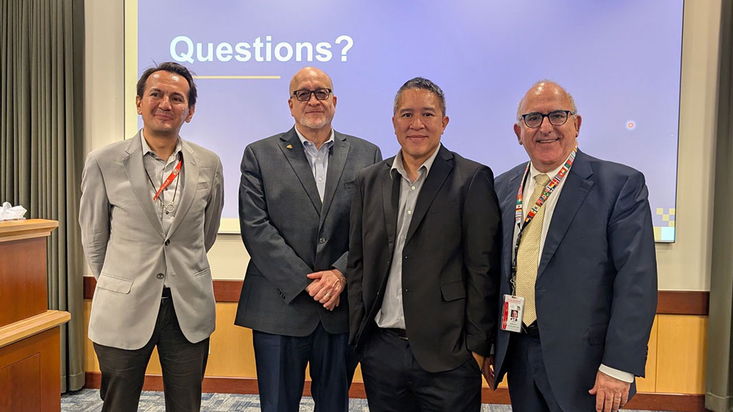
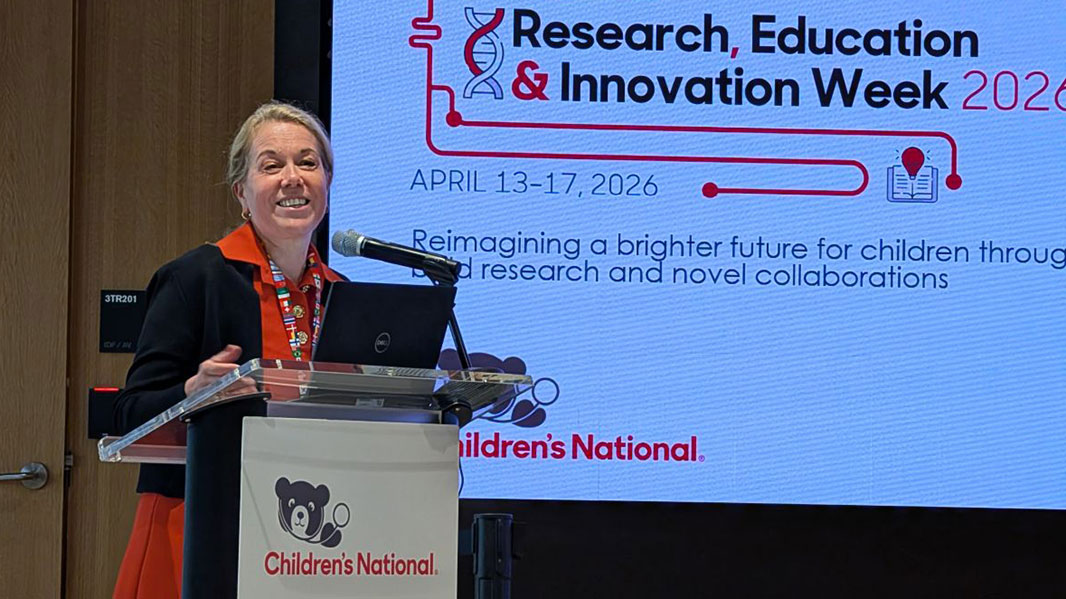
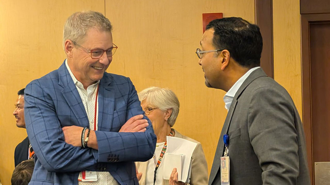
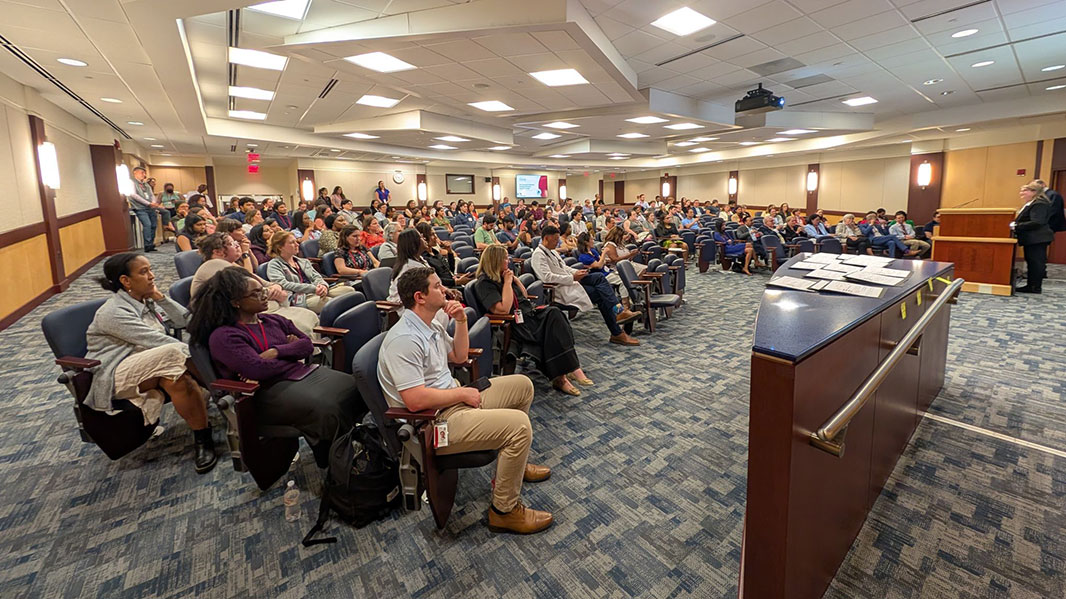
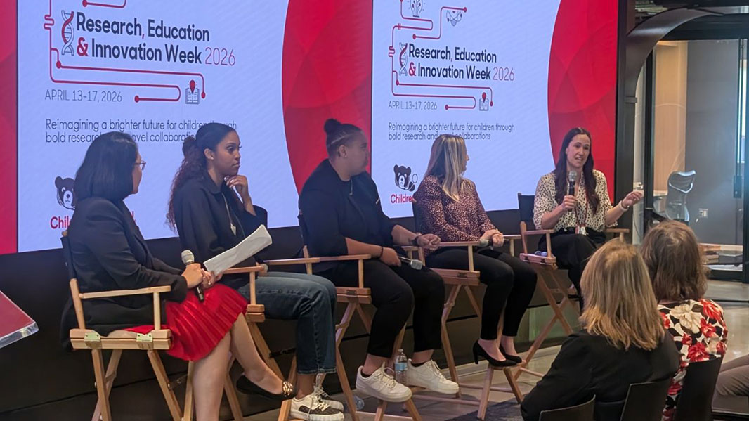
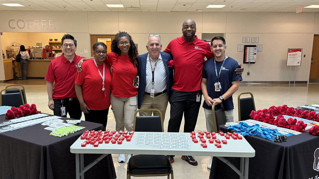
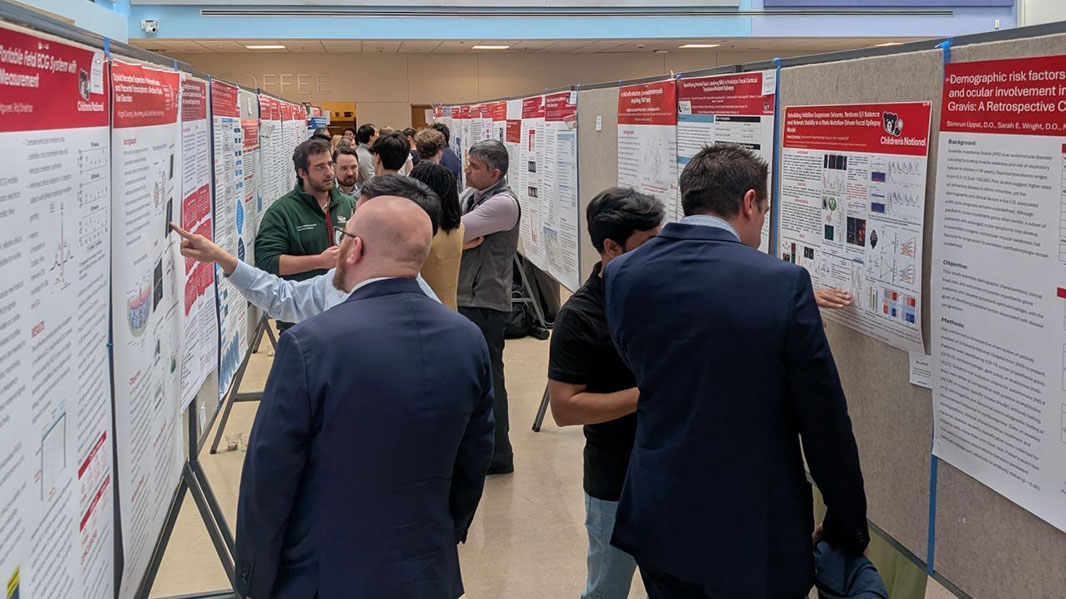
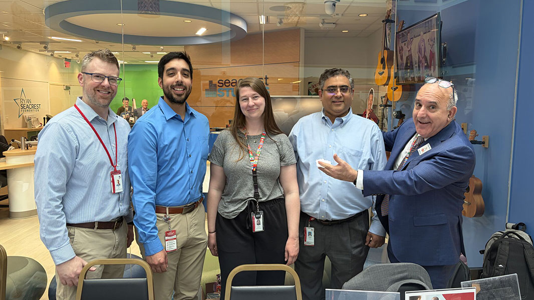
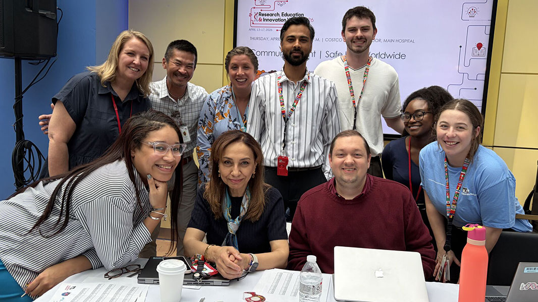
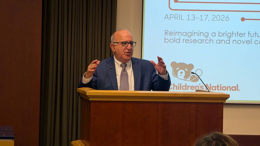
For five days in April, the hallways, auditoriums and virtual platforms of Children’s National Hospital became a space for the kind of honest and necessary conversations that move pediatric science forward. Research, Education & Innovation (REI) Week 2026, held April 13–17, brought together investigators, clinicians, trainees and partners across disciplines to grapple with the theme: reimagining a brighter future for children through bold research and novel collaborations. REI Week remains a cornerstone of the institution’s academic mission 16 years in and this year’s edition made clear that progress in pediatric health demands not just discovery, but clarity of purpose and the courage to push forward.
Making the case for pediatric-first development
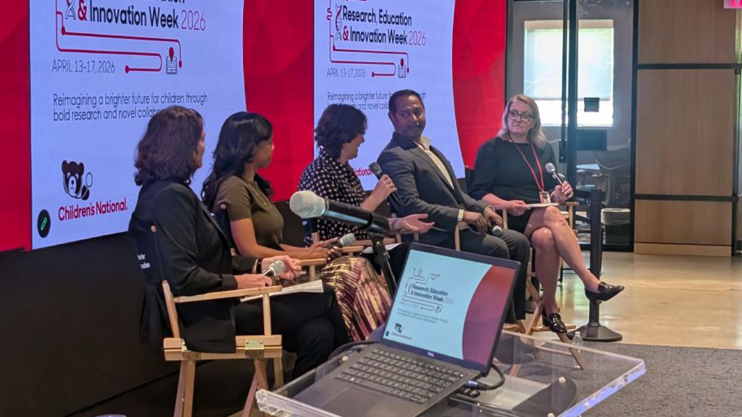
Innovation Day opened the week at the Research & Innovation Campus with a panel discussion on drug and device development. Panelists were clear that making pediatric drug and device development a genuine priority is essential to delivering safe, effective care.
A media and social media panel pushed researchers toward another uncomfortable question: why should anyone outside this building care about what we do? Speakers explored how to frame research so it resonates beyond academic circles, making the case that storytelling and audience awareness are not soft skills but a part of the job.
A Shark Tank-style session showcased diverse artificial intelligence (AI) project proposals aimed at advancing pediatric care and research, with teams presenting bold ideas in a competitive format.
Standing up for science and innovation
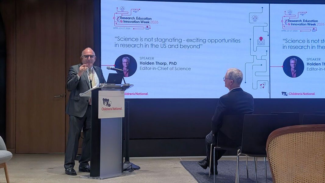
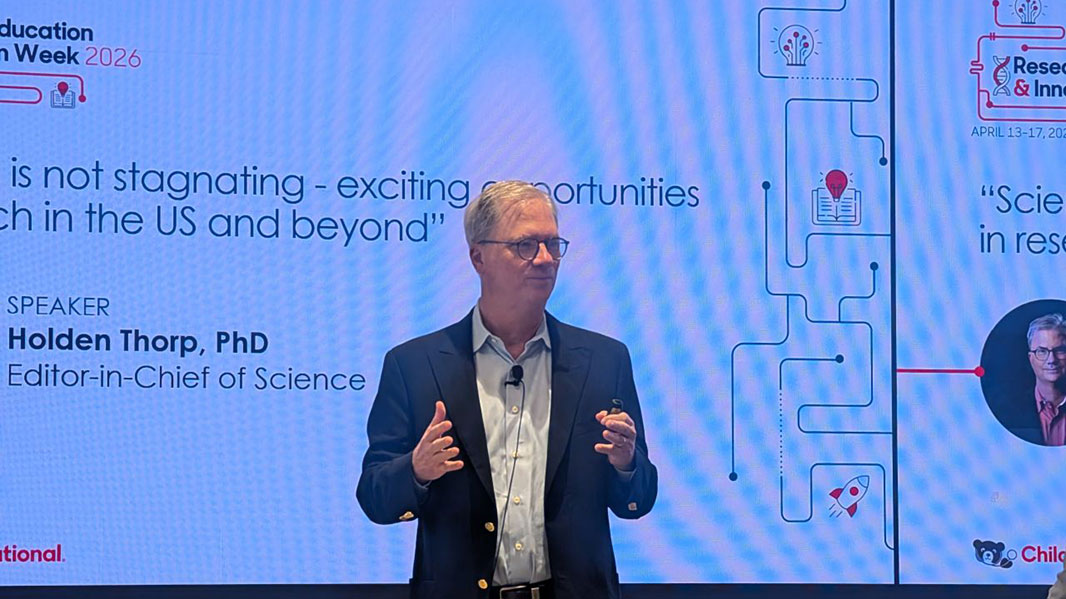
Holden Thorp, PhD, editor-in-chief of Science, set a defining tone for the week in his Monday keynote, speaking frankly about the importance of resisting external pressures and standing up for scientific integrity. His message — that science must hold its ground even when it’s difficult — echoed throughout the days that followed.
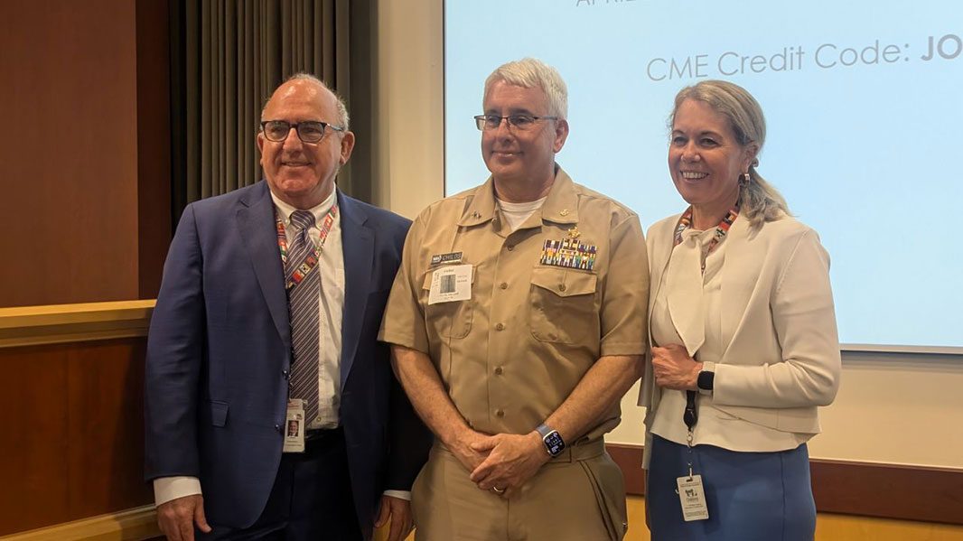
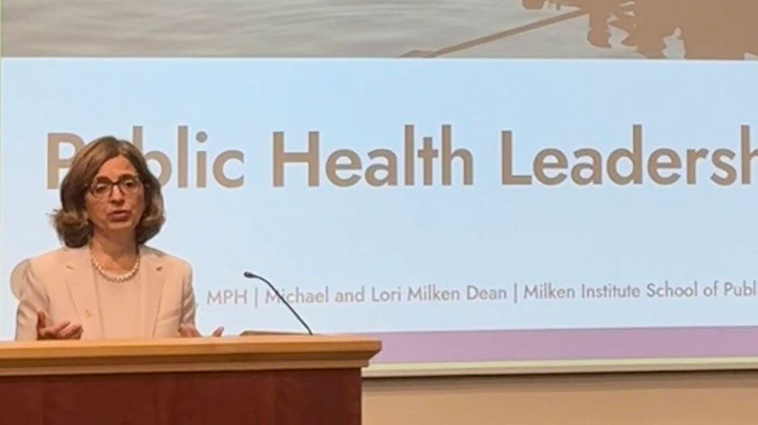
Richard Childs, MD, assistant U.S. surgeon general and scientific director of the National Heart, Lung and Blood Institute, reinforced why sustained investment matters, highlighting how resources and infrastructure at the National Institutes of Health (NIH) enable the kinds of accomplishments that transform care. Kelly Gebo, MD, MPH, dean of the Milken Institute School of Public Health at George Washington University, grounded leadership in something simpler: “Listen before acting. Stay grounded in values. Adapt to change. Explore new funding. Communicate clearly. Lead with empathy.”
A trust problem, not a knowledge problem
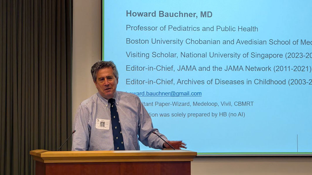
Midweek programming zeroed in on research communication and didn’t flinch. Howard Bauchner, MD, vice chairman of pediatrics at the Boston University School of Medicine, delivered the Larrie Greenberg Grand Rounds Lecture with a call for a cultural shift: quality, not quantity, of publications must become the norm for the research enterprise to regain credibility.
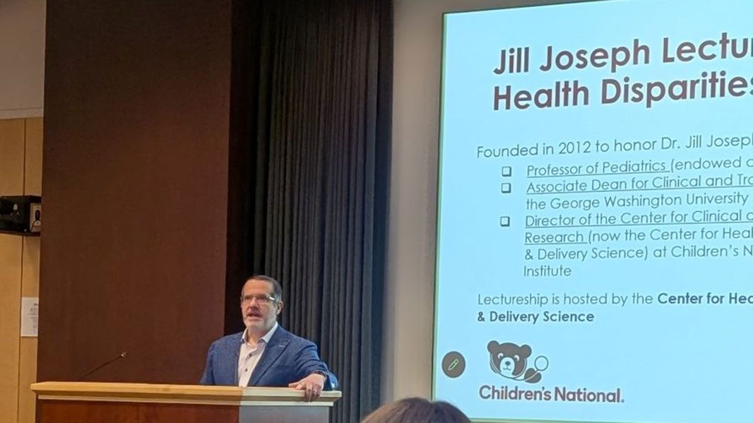
The Jill Joseph Grand Rounds Lecture with Aaron Carroll, MD, MS, president and CEO of AcademyHealth, reframed how the field thinks about science dissemination. The problem, Dr. Carroll argued, is not that the public lacks knowledge, it is that they lack trust. He urged a shift toward solutions-based research and challenged researchers to understand the difference between relative and absolute risk, noting that headlines almost always reach relative figures. If science wants to grab attention responsibly, it has to understand how attention actually works.
Peter Hotez, MD, PhD, professor of pediatrics and molecular virology and microbiology at Baylor College of Medicine, co-director of the Texas Children’s Hospital Center for Vaccine Development and dean of the National School of Tropical Medicine, brought the stakes into sharp relief in his lecture on vaccines and immunizations in an era of anti-science, describing his personal and professional journey, including writing “Vaccines Did Not Cause Rachel’s Autism”, as part of an ongoing effort to counter misinformation and advocate for the families most affected by it.
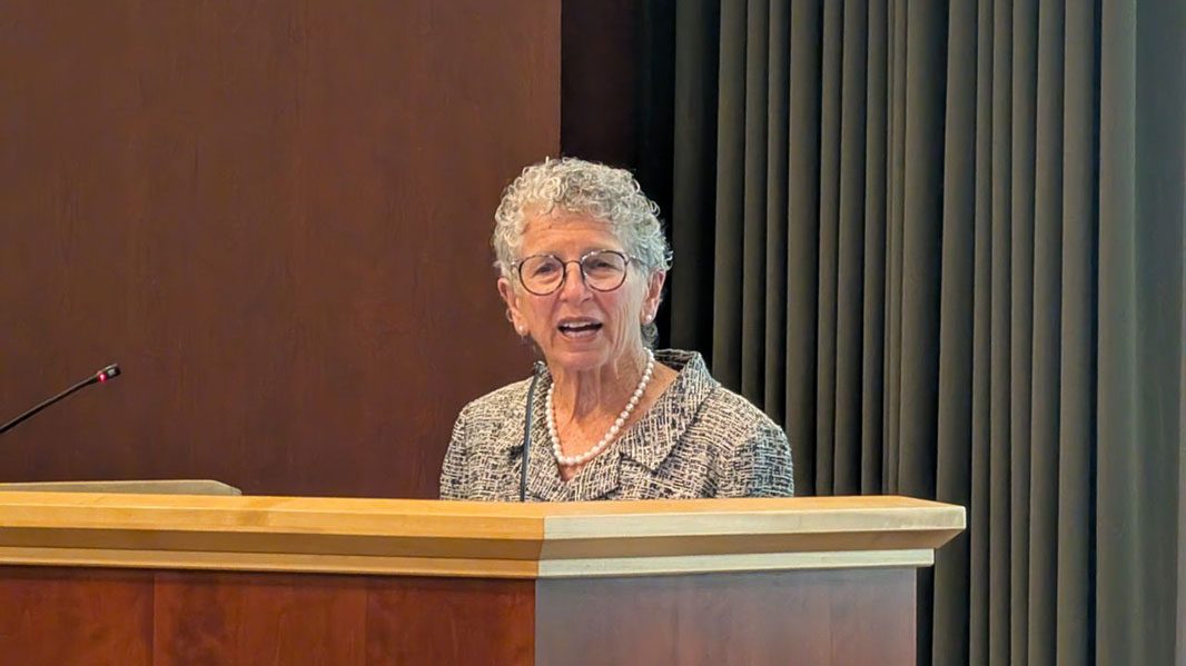
Lauren Clark, PhD, RN, FAAN, professor and Shapiro Family Endowed Chair in Developmental Disability Studies at UCLA’s Joe C. Wen School of Nursing, closed the communication thread with a pointed observation: if researchers never leave their own scientific community, they are missing the boat. She emphasized that plain language is a strategy, not a compromise.
From global health to the clinic floor
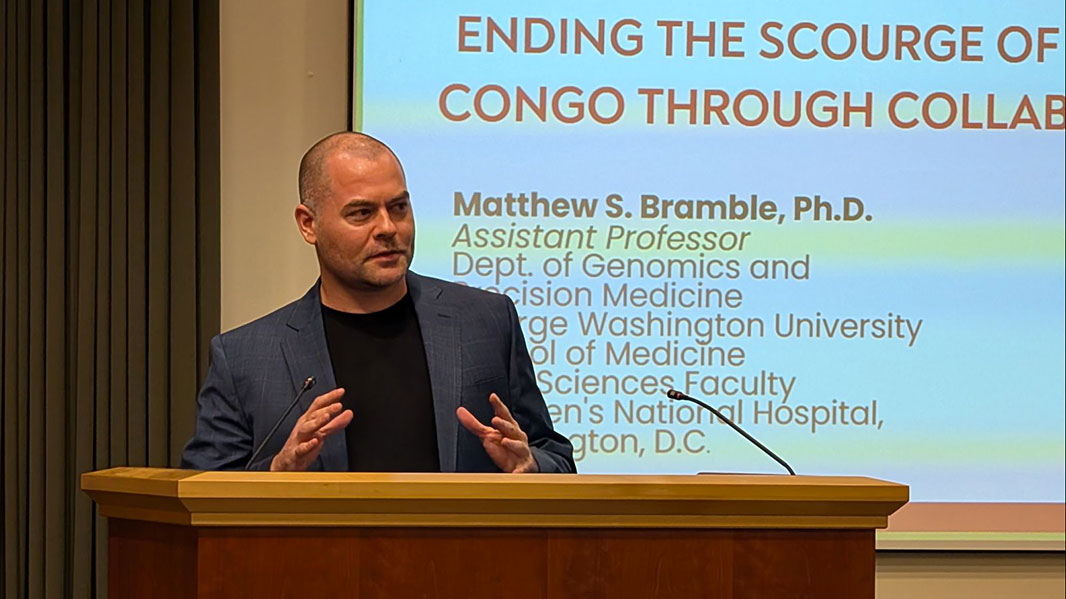
Matthew Bramble, PhD, assistant professor and data sciences faculty member at Children’s National, delivered the Global Health Sponsored Lecture on ending the scourge of Konzo in the Democratic Republic of Congo. His work in this isolated environment — where populations depend heavily on cassava — could unlock information relevant far beyond Konzo itself, potentially shedding light on diseases like ALS.
The joint Lecture on AI featured two complementary perspectives on machine learning’s growing role in care delivery. Hooman Rashidi, MD, MS, associate dean of AI in medicine, professor and endowed chair of Lombardi-Shinozuka Experimental Pathology Research at the University of Pittsburgh School of Medicine proposed a mixed approach with a human in the loop as the responsible path forward. Nam Tran, PhD, MS, professor of pathology, associate dean of biobanking and medical director of Point of Care Testing at the University of Pittsburgh, highlighted AI’s potential in austere settings, field medicine, military contexts and disaster response, where speed and limited resources put a premium on intelligent decision support.
What the abstracts reveal
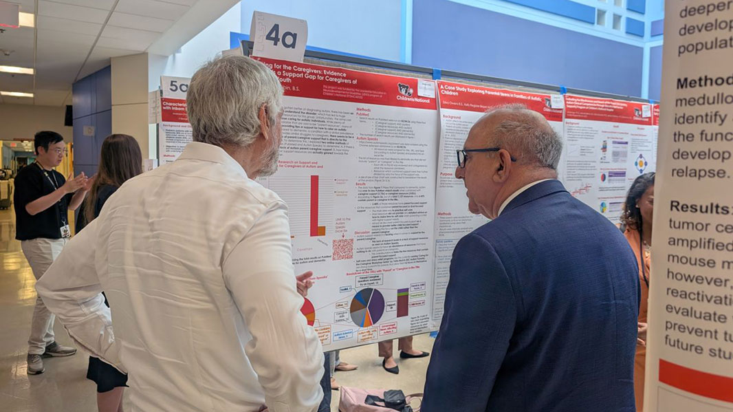
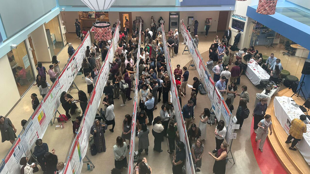
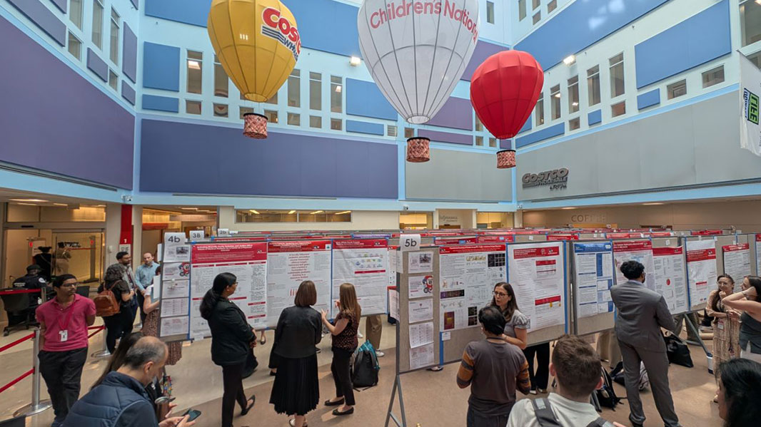
This year’s poster sessions drew more than 450 abstracts spanning 46 research categories — a record number that reflects both the depth of work underway at Children’s National and the range of challenges the institution has committed to tackling. Oncology, neuroscience and neuroprotection, cancer immunology and bone marrow transplantation represented some of the largest clusters of work, with investigators exploring everything from gene therapy for beta-thalassemia to advanced neuroimaging frameworks for fetal and neonatal populations. Digital innovation, data science and AI also drew a strong showing, with studies applying machine learning to EEG-based cognitive assessment, predictive modeling and clinical decision support, reflecting the institution’s growing investment in responsible, data-driven care.
Equally notable was how questions of access and reach have woven themselves into categories that might not have carried that framing in prior years. Research on firearm violence, pathways to mental health care and the downstream effects of social conditions on pediatric outcomes appeared across multiple categories. Community research submissions highlighted school-based telehealth programs and outreach initiatives that extend Children’s National’s reach into underserved communities across the Washington region. Psychology and wellness, quality improvement and treatment compliance rounds out a body of work that asks not just what works scientifically, but who benefits and whether they can actually access what works.
Global and climate health emerged as a distinct and growing area, with submissions connecting environmental conditions to pediatric disease burden and mortality. Taken together, this year’s abstracts capture a field that is translating molecular discoveries into clinical applications, integrating technology with intention and making sure research translates to care for children who need it most.
This year’s honorees
REI Week concluded Thursday with an awards ceremony honoring outstanding contributions across research, education and innovation.
Award winners:
POSTER SESSION AWARDS
Basic & Translational Research
Faculty: Yingshi Ouyang, PhD “Opioid Receptor Expression in the Human Placenta and Placental Transcriptomic Alterations in Opioid Use Disorders”
Postdocs/Fellows/Residents: Margaret Hines, PhD “Understanding the Role of Cranial Mesenchyme in Neural Tube Closure”
Postdocs/Fellows/Residents: Khatereh Khorsandi, PhD “Reprogramming the Immunosuppressive Microenvironment to Enhance CAR T‑Cell Therapy in Diffuse Intrinsic Pontine Glioma”
Staff: Zara Hasnani “Evaluation of Virus‑Specific T Cell Immunity in Pediatric Inflammatory Bowel Disease Patients on Biologic Therapy”
Graduate Students: Elton VanNoy “Modulating DNA Methylation During CAR T Manufacturing to Enhance Immunotherapy for Pediatric Glioma”
Graduate Students: Woudasie Admasu “Identification of Druggable Host Factors to Prevent RSV Infection Using CRISPR‑Cas9”
High School/Undergraduate Students: Akhil Chada “Designing of PepMLM‑Based Peptide Binders to Target Tumor‑Specific Splice Event‑Derived Proteoforms in Pediatric High‑Grade Gliomas”
Clinical Research
Faculty: Matthew Bramble, PhD “Pathways Involving Oxidative Damage Mitigation is Likely the Biological Risk Factor for the Development of Konzo”
Postdocs/Fellows/Residents: Maria Triantafyllou, MD “Plasma Metabolomic Signatures for Diagnosis and Risk Stratification of Pediatric Sepsis in the Emergency Department”
Staff: Pooneh Roshanitabrizi, PhD “Synthetic Dual‑Channel Color Doppler Echocardiography for Rheumatic Heart Disease Detection in Low‑Resource Settings”
Graduate Students: Jasmine Nguyen “The Relationship Between Cloacal Complexity and Early Vaginal Stenosis After Cloaca Repair”
Graduate Students: Maria Straker Brito, MD “High Airway Type‑III IFN Levels by Airway Epithelial Cells are Associated with Increased Pro‑Inflammatory Cytokines Production in the Airways”
Graduate Students: Artur Aharonyan, MS “Automating AMPs: An AI Pipeline for Generating 3D‑Printable BioAMP Plates in Unilateral Cleft Lip and Palate”
Graduate Students: Jaisimar Singh “Mechanical versus Non‑Mechanical Bowel Management in Children with Spina Bifida: A Cross‑Sectional Comparison of Patient‑Reported Symptoms”
High School/Undergraduate Students: Medha Pappula “Integrating Multimodal Clinical Data with Large Language Models to Predict Outcomes in Pediatric Metabolic and Bariatric Surgery”
High School/Undergraduate Students: Keertana Senthilkumar “Diagnostic Utility of Vascular Catheter Tip Cultures”
Community‑Based Research
Faculty: Katie Donnelly, MD, MPH “Evaluating the Propagation of Firearm Violence After an Incident Event”
Postdocs/Fellows/Residents: Brittany Fitzpatrick, MD, MPH “Walking Towards Equity: Enhancing Pediatric Pedestrian Safety Through Data‑Driven Solutions”
Postdocs/Fellows/Residents: Krithika Iyer, PhD “Predicting Early Cognitive Risk Using Ultra‑Low‑Field MRI Brain Volumetry and Demographic Measures in Low‑Resource Settings”
Staff: Megan Lau “A Longitudinal Examination of Social Support as a Mechanism of Change in Executive Function in a School‑Delivered Intervention for Adolescents with ADHD”
Graduate Students: Preeyanka Rao, MPH “Comparison of Asthma Utilizations Across Different Medicaid Insurance Plans in the District of Columbia”
High School/Undergraduate Students: Riya Mehta “Availability and Knowledge of Naloxone Distribution in an Urban Area”
Education, Training and Program Development
Faculty: Amy Wolfe, MD, MEd “A Missed Conversation: Spirituality as a Persistent Gap in PCCM Communication Training”
Postdocs/Fellows/Residents: Taylor Goodman, MD “The POCUS Pathway: A Novel Point‑of‑Care Ultrasound Longitudinal Curriculum for Pediatric Residents”
Postdocs/Fellows/Residents: Jennifer Bertollo, PhD “Online Educator Training for an Executive Function Intervention: Mixed Methods Educator Feedback and Impact on Adoption”
Staff: Tininka Rahman, MHA “Establishing a Community Engaged Research and Training (CERT) Hub at Children’s National”
Graduate Students: Novelle Leach “PCIT and SPACE Parenting Interventions: Are They Meeting the Needs of Young Children with Neurodevelopmental Disabilities and Their Families?”
Quality and Performance Improvement
Faculty: Jessica Lazerov, MD, MBA “Using LEAN Methodology to Improve Immunization Reconciliation and Vaccination Rates”
Postdocs/Fellows/Residents: Maya Gibson, MD “Evaluating Blood Product Utilization on ECMO Following Implementation of Restrictive Transfusion Strategies”
Staff: Abhijeet Parida, MS “HOPE4KIDS: AI‑Based Webtool for Neuro‑Oncology Segmentation and Volumetrics”
Graduate Students: Benjamin Upbin “Caring for the Caregivers: Evidence Reveals a Support Gap for Caregivers of Autistic Youth”
High School/Undergraduate Students: Safinabonu Juraeva, MPS “Diagnosing Primary Bottlenecks in the PICU‑to‑Floor Transfer Process to Improve Transfer Efficiency”
The conversations held during REI Week 2026 continue throughout the year. They carry forward into labs, clinics and classrooms, and ultimately into the lives of the children and families this institution exists to serve.
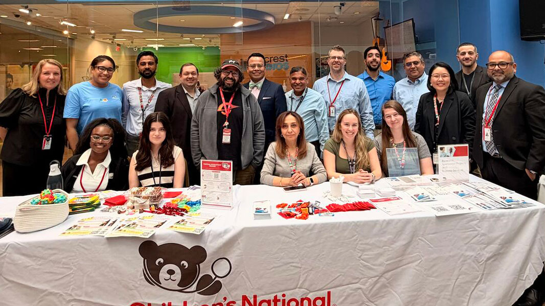
REI Week also highlighted the teams and programs that support research across its full lifecycle, with the Clinical Research Unit, Innovation and Technology Transfer Office and Research Sponsored Projects and Business Services maintaining a visible presence throughout the week to engage investigators and answer questions. Opportunities for connection extended beyond formal sessions through events such as the “global health mix & mingle”, sponsored by the Global Health Initiative, which brought participants together to engage around Children’s National’s work in local and global communities. Educational programming continued through Children’s Academy of Pediatric Educators sessions that explored how research and teaching intersect in practice, including discussions on the effectiveness of case-based teaching, pathways in medical education and the value of embracing failure as a driver of growth in academic medicine. Beyond the lecture halls, community engagement remained central, with a Safe Kids Worldwide event on Friday focused on fostering safety education and training for children and families. Supported by executive sponsors Nathan Kuppermann, MD, MPH; Catherine Bollard, MBChB, MD; Stella Ghattas, Esq; Elizabeth Wells, MD; and Cicely (CC) Brooks, DNP-ENL, MSN, RN, NE-BC, FAB, Children’s National continues to build the conditions where that kind of science can thrive.
REI Week 2027 takes place the week of April 5, and what happens between now and then matters just as much as what happens during that week. Every study advanced, every collaboration forged and every trainee who finds their footing in this community is part of the same long effort to reimagine what pediatric health can look like. That work doesn’t happen without investment in people, in ideas and in the infrastructure that turns discovery into care. If this week moved you, consider supporting what comes next.
https://innovationdistrict.childrensnational.org/wp-content/uploads/2026/04/REI-Week-2026-Costco-Atrium-feature.jpg300400Innovation Districthttps://innovationdistrict.childrensnational.org/wp-content/uploads/2025/09/InnovationDistrict_CN_WebHeader-1396px-1030x151.pngInnovation District2026-04-24 15:34:182026-04-24 15:37:00Bold research and novel collaborations reimagine a brighter future during REI Week 2026
Steven Hardy, PhD (left) and Dana Footer, PsyD (right), present Children’s National Hospital’s protocol for assessing psychological readiness for HSCT and gene therapy in sickle cell disease at the European Society for Blood and Marrow Transplantation (EBMT) Annual Meeting.
Hematopoietic stem cell transplant (HSCT) and gene therapy are changing the treatment landscape for sickle cell disease, offering the potential to reduce pain, prevent organ damage and improve quality of life. Alongside these clinical advances, it is essential to ensure patients and families are prepared for these transformative therapies from a psychological and social standpoint.
At Children’s National Hospital, psychologists have developed a structured approach to evaluating and supporting psychosocial readiness for HSCT and gene therapy. This includes addressing mental health concerns, fostering realistic expectations, considering motivations and activating social support networks. Ongoing monitoring after treatment helps ensure patients’ long-term psychological well-being.
This work was recently presented by Dana Footer, PsyD, and Steven Hardy, PhD, at the annual meeting of the European Society for Blood and Marrow Transplantation (EBMT) as an abstract titled A Protocol for Assessing Psychological Readiness for Hematopoietic Stem Cell Transplant and Gene Therapy in Sickle Cell Disease, highlighting Children’s National Hospital’s international leadership in integrating psychosocial care with cell and gene therapies.
Treating the whole patient
During the conference, the Children’s National psychologists within the Cancer and Blood Disorders Center provided concrete recommendations on how to evaluate readiness and use those results to guide individualized, team-based interventions, ensuring patients and families are fully supported throughout the treatment journey.
“Psychological readiness evaluations help the multidisciplinary care team understand patients’ mental health, relationships, identities and personal goals, which is essential for optimizing treatment outcomes,” said Dr. Hardy, director, Psychology and Patient Care Services in the Divisions of Hematology, Oncology, and BMT and co-author of the abstract.
This approach helps promote the patient voice, build confidence, strengthen disease self-management, and improve physical and psychosocial outcomes, ensuring care goes beyond treating the disease to supporting the whole person.
Patient and family feedback and real-world experiences continue to inform refinement of the readiness evaluations and interventions. “By listening to families who have undergone treatment talk about what they found most valuable about the preparation process and which aspects of treatment were surprising to them, we continue learning how best to support future patients,” said Dr. Footer, co-author and attending psychologist in the Divisions of Hematology, Oncology, and BMT.
Leading the field
Access to pediatric HSCT and gene therapy remains limited in the U.S., but Children’s National is a national leader. The embedded psychosocial care providers offer specialized mental health care before, during and after treatment.
By combining clinical innovation with psychosocial expertise, Children’s National is helping ensure these therapies improve not only health outcomes but also patients’ quality of life and ability to pursue their personal goals at every stage of life.
Researchers have uncovered how RNA splicing patterns, including CLK1 exon 4, shape pediatric brain tumors and may influence outcomes beyond DNA mutations.
Pediatric brain and other central nervous system tumors are the leading cause of cancer-related death in children. Over the past decade, DNA sequencing has helped doctors better classify these tumors and search for new treatments. However, gene mutations are only part of the story. A new study from researchers at Children’s National Hospital and Children’s Hospital of Philadelphia (CHOP) looks at another powerful layer of biology: how RNA is “spliced” inside tumor cells.
Splicing is the process cells use to cut and rearrange RNA before it becomes a protein. This allows one gene to create multiple versions of a protein. In healthy development, splicing is tightly controlled. In cancer, it can shift in ways that change how cells grow and survive.
A deeper dive
In a large-scale analysis of 729 pediatric central nervous system tumors, researchers mapped splicing patterns across many tumor types. They found that splicing patterns varied widely not only between tumor types, but also within them.
To better understand these differences, the team created a new measurement called the Splicing Burden Index. This tool measures how much a tumor’s splicing patterns differ from others in the group. Using this approach, the researchers identified tumor groups based on shared splicing patterns. Some of these splicing-defined groups were linked to patient outcomes, even after accounting for tumor type and other clinical factors. That means RNA splicing may provide important information beyond what we currently detect in the clinic.
“Splicing adds an entirely new layer of biology to how we understand these tumors,” said Jo Lynne Rokita, PhD, principal investigator of the Rokita Lab in the Center for Cancer and Immunology Research at Children’s National and senior author of the study. “When we look at tumors at the exon level, we see patterns that are not visible through DNA mutations or even overall gene expression alone.”
The team also found that overall splicing activity and spliceosome pathway signaling are not the same thing. Some tumors showed high activity in splicing-related pathways that were linked to worse survival, even if their overall splicing burden was not high. This suggests that splicing adds additional complexity to tumor biology.
A closer look
After mapping these patterns, the researchers focused on specific splicing events that might affect how proteins function. One event stood out in a gene called CDC-like kinase 1, or CLK1.
CLK1 helps regulate other splicing proteins. For CLK1 to work normally as a kinase, it must include a specific section called exon 4. When exon 4 is skipped, the protein loses its full activity.
The team found that exon 4 inclusion in CLK1 followed what is known as an oncofetal pattern. It is common in early brain development, usually decreases as the brain matures, and then appears again in many pediatric brain tumors. They found that higher levels of exon 4 inclusion were linked to improved patient outcomes in certain types of tumors, and these effects were different from simply measuring how much of the overall CLK1 gene was present. These findings highlight that looking at specific pieces of a gene can provide more meaningful insight than measuring total gene activity alone.
“CLK1 exon 4 is a clear example of why exon-level analysis matters,” said Ammar Naqvi, PhD, Principal Bioinformatics Scientist at CHOP and lead author of the study. “It is not just whether the gene is turned on or off. It is which version of the gene the tumor is using.”
What this means
To test whether this splicing event might influence tumor cells, researchers performed laboratory experiments in a pediatric high-grade glioma model. They used both a drug that targets CLK1 activity and a targeted approach to reduce inclusion of exon 4. In both cases, tumor cell growth decreased, and important cancer-related gene programs were disrupted. More work in additional models and in vivo systems will be needed to further confirm these findings. Still, the results suggest that exon-level splicing changes may help shape tumor behavior.
What comes next
This study represents one of the first systematic efforts to examine alternative splicing across many types of pediatric CNS tumors. By introducing the Splicing Burden Index and identifying splicing-defined tumor groups, the research offers a new framework for understanding how RNA regulation influences childhood brain cancer. For clinicians and families, the message is clear – DNA mutations are only one piece of tumor biology. RNA splicing adds another layer that may affect prognosis and, in the future, treatment strategies.
Read the full study “Alternative splicing in pediatric central nervous system tumors highlights oncofetal candidate CLK1 exon 4” in Neuro-Oncology Pediatrics.
This project was funded in part by the National Institutes of Health (R03OD036498), the anonymous private investors to the Children’s National Hospital Brain Tumor Institute, Children’s Brain Tumor Network, Chad Tough Foundation, CHOP Division of Neurosurgery, Mildred L. Roeckle Endowed Chair in Pathology at CHOP, and CureSearch for Children’s Cancer. This research utilized the Common Fund Cloud Workspace Pilot (1OT2OD030162-01) and the Kids First Data Research Center Cloud Credit Program (U2C HD109731 – 08S1).
https://innovationdistrict.childrensnational.org/wp-content/uploads/2026/03/RNA-feature.jpg300400Innovation Districthttps://innovationdistrict.childrensnational.org/wp-content/uploads/2025/09/InnovationDistrict_CN_WebHeader-1396px-1030x151.pngInnovation District2026-03-02 14:32:312026-03-04 09:42:03New insights into RNA splicing across pediatric brain tumors
The Children’s National team at the Kuwait Pediatric Stem Cell Transplant, Cellular & Gene Therapy Conference.
At the Kuwait Pediatric Stem Cell Transplant, Cellular & Gene Therapy Conference, David A. Jacobsohn, MD, MBA, chief of Blood and Marrow Transplantation at Children’s National Hospital, presented emerging approaches that are transforming the treatment of non-malignant pediatric diseases through allogeneic hematopoietic stem cell transplantation (HSCT). His presentation, Pediatric Stem Cell Transplant in Non-Malignant Disorders: Evolving Trends, highlighted sickle cell disease (SCD) as a key example of this progress, noting that decades of advances have made HSCT a highly effective curative option, with survival rates now exceeding 95% for children receiving matched sibling donor transplants. Newer reduced-intensity and non-myeloablative approaches are further lowering treatment-related toxicity while maintaining disease control, significantly decreasing hospitalization burden and improving quality of life.
The talk also addressed one of the field’s most pressing challenges, donor availability, and how the expansion of haploidentical (half-matched) donor strategies is opening curative treatment options to the vast majority of patients who previously lacked a suitable donor. In parallel, evolving conditioning regimens and immune-modulating therapies are helping reduce graft rejection and improve long-term outcomes.
Beyond SCD, the presentation highlighted national and international research efforts demonstrating the curative potential of HSCT for inherited immune disorders such as severe combined immunodeficiency (SCID) and chronic granulomatous disease (CGD). Data from large, multi-center collaborations show that earlier referral, tailored conditioning, and close collaboration between immunology and transplant teams are critical to optimizing survival and immune recovery.
Together, this work underscores a broader shift in pediatric transplantation, moving from high-risk rescue therapy to a more precise, accessible and patient-centered cure. It reinforces the role Children’s National plays in advancing innovative, life-changing treatments for children worldwide.
Kidney disease is a leading cause of mortality in SCD, underscoring the urgent need for earlier and more precise diagnostics.
“We’ve watched as silent kidney damage progresses year after year, often going undetected until significant harm has occurred,” said Andrew Campbell, MD, director of the Comprehensive Sickle Cell Disease Program at Children’s National and co-investigator of the study. “By identifying kidney disease earlier, we can intervene sooner with more effective disease-modifying therapies and curative treatments, potentially preventing or slowing progression to kidney failure.”
From discovery to impact
“We measure damaged mitochondria shed by the kidney in a few milliliters of urine so we can develop an early warning system for the onset of kidney damage,” said Marissa Howard, PhD, systems biologist and research fellow at George Mason University and principal investigator of the study. “Our partners at Children’s National are known for identifying innovative approaches to treat SCD. This exciting collaboration is collectively motivated by our ultimate goal of reducing the high mortality rate caused by Sickle Cell kidney failure in children affected by this disease.”
By translating cutting-edge science into clinical impact, Children’s National continues to advance next-generation diagnostics that improve outcomes for children with SCD and may extend to other diseases linked to mitochondrial dysfunction.
https://innovationdistrict.childrensnational.org/wp-content/uploads/2023/02/diseased-kidneys-feature.png300400Innovation Districthttps://innovationdistrict.childrensnational.org/wp-content/uploads/2025/09/InnovationDistrict_CN_WebHeader-1396px-1030x151.pngInnovation District2026-02-04 12:18:562026-02-04 12:21:53Advancing early detection for kidney disease in sickle cell

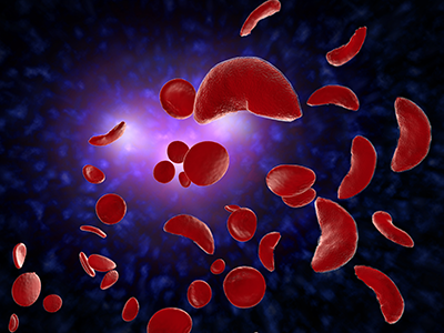
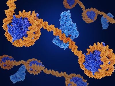
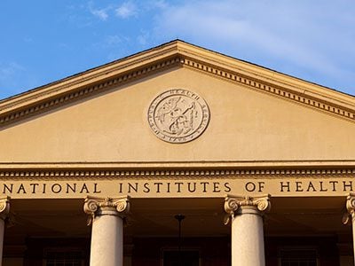
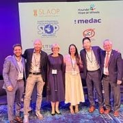
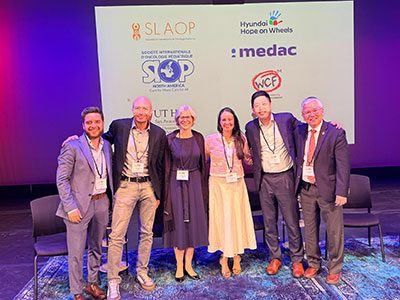
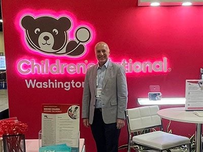

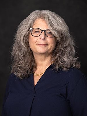
 Children’s National in Washington, D.C., was once again ranked as a top hospital in the nation by the U.S. News & World Report 2026-27 Best Children’s Hospitals