People with autism who struggle with challenges related to executive function are more likely to suffer poor mental health outcomes, according to findings from a large-scale longitudinal study published in The Journal of Child and Adolescent Psychiatry.
Lead author of the study, Lauren Kenworthy, PhD, division chief of Neuropsychology at Children’s National, recently appeared on Mind the Kids, a podcast hosted by the Association for Child and Adolescent Mental Health (ACAMH), to discuss the findings. Dr. Kenworthy touched on why interventions targeted to address executive function challenges, like Unstuck and On Target, may be critical tools to ensure better mental health outcomes for autistic youth and young adults.
What it means
Executive functions are higher order cognitive abilities that allow people to regulate their thoughts, feelings and behaviors to meet the demands of their environment. People with executive function challenges often struggle with several areas of self-regulation that can truly impact daily life, such as inhibition, flexibility and working memory.
“We’ve known for a long time now that executive functions are strongly linked to mental health, including depression, anxiety and aggression, for all of us,” Dr. Kenworthy told podcast host Clara Faria, PhD. “We’ve also known for a long time that executive functions and mental health are really challenging areas for a lot of people with autism.”
Despite the significance of executive function in autism, studies examining the trajectory and impact of these challenges long term have been difficult to complete in a comprehensive way. Small sample sizes and/or limited age ranges have prevented researchers from capturing a true notion of how executive function and mental health outcomes develop over time.
Why it matters
This study is the first large-scale longitudinal study of executive function and mental health in autism. It shows the lasting effect of executive function challenges on autistic people, especially drilling into specific subcategories of symptoms such as aggression, anxiety and depressive mood and finding that many of these symptoms only worsen over time.
Children’s National leads the way
Dr. Kenworthy and her study co-authors concluded from the findings that interventions targeting executive function skills, especially flexibility, in autistic children are critical to a child’s well-being throughout their lives. The right interventions may reduce the burden of both the executive function challenges themselves and their impact on a child’s mental health over time.
Unstuck and On Target, a set of tools created by Dr. Kenworthy and a team of researchers from Children’s National and Children’s Hospital Colorado, is one example of an evidence-based intervention that is ready to be deployed in schools and clinics.
“We developed executive function interventions designed to improve kids’ ability to be flexible, make plans and set goals,” Dr. Kenworthy notes in the podcast. “And we’ve designed them to be delivered in the public school system with fidelity by classroom teachers and special educators because every kid gets to go school. Some of their most important work occurs in school so it’s also the real-world setting where they need the help.”
https://innovationdistrict.childrensnational.org/wp-content/uploads/2026/06/kenworthy-podcast-feature.jpg300400Innovation Districthttps://innovationdistrict.childrensnational.org/wp-content/uploads/2025/09/InnovationDistrict_CN_WebHeader-1396px-1030x151.pngInnovation District2026-06-19 16:04:252026-06-19 16:08:06Executive function interventions may be critical to long-term mental health in autism
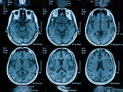
A new Children’s National study reveals that depression risk in pediatric epilepsy may be tied to how brain lesions interact with the brain’s default mode network — not just where those lesions are located.
Using focal cortical dysplasia (FCD), the most common cause of surgically treatable, drug-resistant epilepsy in children, investigators found that greater overlap between lesions and the brain’s default mode network (DMN) is associated with increased depressive symptoms, particularly during adolescence.
Dive deeper
FCD disrupts cortical development and functional connectivity, making it a powerful model for understanding how focal brain abnormalities affect distributed neural systems.
In this study, researchers analyzed children with FCD-related epilepsy who completed the Child Behavior Checklist, linking parent-reported psychiatric symptoms with advanced imaging. They mapped FCD lesions from MRI and measured how those lesions overlapped with major functional brain networks, then used statistical models accounting for age and gender to examine how network-level brain organization related to real-world psychiatric symptoms.
The results showed that greater overlap between FCD lesions and the default mode network was associated with higher depressive symptom scores, even after accounting for age and gender. Female sex was also associated with increased depressive symptoms. These associations were stronger in adolescents, suggesting that network maturation amplifies risk during this developmental stage.
“By anchoring depression risk to a specific brain network, we’re starting to connect the biology of epilepsy with the lived experience of patients in a much more direct way,” said Xiaotong Li, BA, first author of the study and clinical research coordinator with the Focal Cortical Dysplasia Research Program. “This kind of approach helps move the field toward identifying measurable markers that could eventually guide more personalized care.”
By contrast, overlap with the limbic network was not associated with depressive symptoms, and no meaningful relationship was found between network overlap and anxiety.
What this means
The findings support a shift from region-based to network-based models of pediatric epilepsy and its comorbidities.
“Rather than thinking of psychiatric symptoms as separate from epilepsy, this work suggests they may arise from the same underlying network disruptions that drive seizures,” said Nathan T. Cohen, MD, corresponding author and director of the Focal Cortical Dysplasia Research Program.
The default mode network is involved in self-referential processing and emotional regulation, aligning with broader evidence linking DMN dysfunction to depressive disorders.
The absence of a relationship with anxiety suggests that depression and anxiety in pediatric epilepsy may arise through distinct mechanisms. Overall, the results indicate that lesion–network colocalization contributes to depression vulnerability and that DMN involvement may serve as a neurobiological marker of risk, particularly in adolescence.
Why this matters
By showing that depression risk is tied to how lesions engage functional brain networks, this study provides a more precise framework for understanding psychiatric comorbidity in epilepsy.
The identification of DMN involvement as a potential marker of depressive symptoms opens the door to future work focused on individualized, network-informed diagnostics and therapies.
Together, these findings highlight how Children’s National is advancing a more integrated view of pediatric brain disease—one that connects imaging, behavior and clinical care to move toward more personalized treatment strategies.
Read the full study “Network Colocalization Correlates of Depression and Anxiety in Pediatric Focal Cortical Dysplasia-Related Epilepsy” in the Annals of Neurology here.
https://innovationdistrict.childrensnational.org/wp-content/uploads/2026/06/Brain-Scans-feature.jpg300400Innovation Districthttps://innovationdistrict.childrensnational.org/wp-content/uploads/2025/09/InnovationDistrict_CN_WebHeader-1396px-1030x151.pngInnovation District2026-06-09 13:23:142026-06-09 13:25:14Study links brain network interactions to depression risk in pediatric epilepsy
Neuroinflammation is a central feature of many pediatric neurologic conditions, yet its precise role has remained difficult to define. A recent study, published in the Journal of Neuroinflammation by researchers at Children’s National Hospital, applies high-sensitivity cerebrospinal fluid (CSF) proteomics to better understand this process across diseases, revealing distinct inflammatory signatures in post-hemorrhagic hydrocephalus (PHH) and anti-NMDA receptor encephalitis (NMDARE).
This collaborative effort brought together experts from the divisions of Critical Care Medicine, Neurosurgery, Neurology and Pathology and Laboratory Medicine, reflecting a multidisciplinary approach to complex pediatric brain disorders. The analysis showed that PHH is associated with a broad, sustained inflammatory response, while NMDARE demonstrates a more targeted immune profile.
Dive deeper
Neuroinflammation has been widely studied in adults, where CSF proteomics has helped identify biomarkers and therapeutic targets. In children, however, progress has been slower due to limited access to CSF samples and less sensitive analytical tools.
To address this gap, researchers used a high-sensitivity, multi-targeted proteomic platform to analyze CSF samples from children with PHH, NMDARE and brain tumor–associated hydrocephalus.
Their findings showed stark differences between conditions. PHH demonstrated a large number of differentially abundant proteins and enrichment of complement, coagulation and platelet-related pathways, pointing to a widespread inflammatory response. NMDARE, in contrast, exhibited a narrower profile, with fewer altered proteins and pathways centered on cytokine signaling and humoral immunity.
What this means
“These findings show that neuroinflammation is disease-specific rather than uniform across pediatric central nervous system (CNS) conditions,” says corresponding author Terry Dean, MD, critical care specialist at Children’s National.
In PHH, the persistence of complement and coagulation pathway activation over weeks suggests these mechanisms may be key drivers of disease progression and potential targets for intervention in a condition that currently lacks medical therapies.
In NMDARE, the identification of B‑cell–related proteins such as IGLC2, MZB1 and CD79B points to promising biomarker candidates. These markers may also extend beyond a single disease, with potential relevance across autoimmune CNS disorders.
How it worked
The study leveraged a pediatric CSF biorepository at Children’s National, enabling access to rare samples and longitudinal data collection.
Using advanced proteomic profiling, researchers analyzed hundreds of inflammation-associated proteins and identified disease-specific patterns through differential expression and pathway analysis.
This approach allowed the team to detect low-abundance inflammatory mediators that may have been missed by earlier techniques, providing a more detailed view of pediatric neuroinflammation.
Why this matters
Sometimes the most important advances come from clarifying how diseases differ at a molecular level. This study shows that pediatric neuroinflammation is not a single process, but a collection of distinct, disease-specific responses.
By identifying persistent inflammatory pathways in PHH and potential biomarkers in NMDARE, the work lays the foundation for more precise diagnostics and targeted therapies. It also highlights the importance of investing in pediatric research infrastructure, such as biorepositories, to enable discovery.
For clinicians and researchers, these insights move the field closer to tailoring treatment based on the underlying biology of each condition, an essential step toward improving outcomes for children with neurologic disease.
Read the full study “Characterizing CSF inflammatory proteomics in pediatric post-hemorrhagic hydrocephalus and Anti-NMDAR encephalitis” in the Journal of Neuroinflammation here.
Researchers at Children’s National Hospital are helping advance pediatric AI with a first-of-its-kind open-access brain tumor imaging dataset designed to improve radiology and precision medicine for children.
Artificial intelligence (AI) is moving quickly in medicine, and radiology is one of the easiest places to see that progress. But in pediatrics, the path forward is more complicated. Children are not just smaller versions of adult patients. They have different diseases, different biology and different care processes, which means AI has to be built with that reality in mind from the beginning.
That challenge is clear in a new effort focused on pediatric brain tumors. While rare, these tumors remain the most common solid tumors in children and the leading cause of cancer-related mortality in the pediatric population. Compared to adult brain tumors, they behave differently across biology, anatomy and clinical care, which makes diagnosis, monitoring and treatment response especially complex. AI has the potential to improve all of this. But progress has been slow, largely because the field has been working without the kind of large, standardized and accessible datasets that make AI actually work at scale.
Dive deeper
A new international effort, co-led by Marius George Linguraru, DPhil, MA, MSc, of Children’s National Hospital and Anahita Fathi Kazerooni, PhD of Children’s Hospital of Philadelphia, is helping change that. The Brain Tumor Segmentation in Pediatrics dataset, known as BraTS-PEDs, is the first large-scale, open-access benchmark dataset designed specifically for pediatric brain tumor segmentation and analysis. It brings together MRI data from multiple imaging sequences from 457 pediatric patients with high-grade gliomas, collected across several institutions and international research consortia.
Each case includes a full set of clinically relevant imaging sequences, including pre- and postcontrast T1-weighted, T2-weighted and T2-FLAIR MRI. Experts labeled different parts of each tumor using established pediatric neuro-oncology guidelines. They combined automated tools designed for pediatric cases with careful review and refinement by neuroradiologists. The result is a dataset that is not only large but consistent in a way the field has not had before.
Just as important, the dataset is structured to support real-world use. It is divided into training, validation and hidden testing subsets, allowing researchers to benchmark models in a reproducible way and evaluate how well those models generalize across institutions.
Why this matters
This kind of resource addresses one of the biggest barriers in pediatric AI. Without enough high-quality, standardized data, even the most promising algorithms struggle to translate into clinical use.
BraTS-PEDs changes that by giving researchers a shared foundation. It allows for consistent method comparison, supports model development across institutions and opens the door for integrating imaging with molecular and clinical data, which is where things start to move toward precision medicine in a meaningful way.
For pediatric radiology, that shift is critical. AI is not just about improving image analysis. It is about building tools that can help clinicians better understand disease, track how it evolves and make more informed decisions over time. Dr. Linguraru’s team has created public tools to provide open access to the volumetric analysis of pediatric brain tumors to doctors and researchers everywhere.
What comes next
At Children’s National, this work fits into a broader effort to rethink how AI is developed and deployed in pediatric care. Through its Division of AI Research, the focus is on creating a clinically grounded ecosystem where data, technology and clinical expertise are tightly connected. The goal is to move beyond isolated projects and build infrastructure that allows ideas to move from concept to application in a way that is scalable and sustainable. As Dr. Linguraru notes, “AI tools can play a key role in radiology, but radiologists must be able to trust in the systems’ design and receive adequate training.”
Radiology is already leading the way when it comes to AI in medicine, but pediatrics is still catching up in terms of data and infrastructure. Efforts like BraTS-PEDs are a step toward closing that gap. By creating shared, standardized resources, they make it possible to build and evaluate models in a way that is consistent, collaborative and clinically relevant. For children, that matters. Getting AI right in pediatrics is not just about innovation. It is about making sure the tools being developed actually reflect the patients they are meant to serve.
This research, Brain Tumor Segmentation in Pediatrics (BraTS-PEDs): A Multi-Institutional Benchmark Dataset for Pediatric Neuro-Oncology, was published in Radiology: Artificial Intelligence. Authors from Children’s National Hospital include Marius George Linguraru, DPhil, MA, MSc; Zhifan Jiang, PhD; Xinyang Liu, PhD; Brian Rood, MD; and Roger J. Packer, MD; in collaboration with colleagues from multiple institutions and international consortia working to advance artificial intelligence in pediatric neuro-oncology.
https://innovationdistrict.childrensnational.org/wp-content/uploads/2026/05/brain-tumors-CNRI.png385685Innovation Districthttps://innovationdistrict.childrensnational.org/wp-content/uploads/2025/09/InnovationDistrict_CN_WebHeader-1396px-1030x151.pngInnovation District2026-05-12 16:26:182026-05-12 16:28:16Advancing clinical deployment of artificial intelligence in pediatric radiology
The Neuromuscular Medicine Program at Children’s National Hospital has again been designated a Charcot‑Marie‑Tooth Association (CMTA) Center of Excellence, marking the second time the program has received this distinction. The designation recognizes the program’s sustained excellence in providing comprehensive, multidisciplinary care for children with Charcot‑Marie‑Tooth disease (CMT).
A core mission of the CMTA is to improve quality of life for individuals with CMT, a hereditary peripheral neuropathy most often identified in adolescence or early adulthood but also seen with onset in early childhood. Through its network of Centers of Excellence, CMTA ensures that patients and families have access to expert, coordinated care delivered by clinicians with deep experience in CMT.
Children’s National was first named a CMTA Center of Excellence five years ago following a rigorous review of clinical expertise, patient volume, and programmatic services. The renewed designation highlights the program’s consistency and growth during a period of rapid advancement in the field.
The Neuromuscular Medicine Program offers coordinated care across multiple specialties, including neurology, nutrition/diet, occupational therapy, orthopedic surgery, physiatry, physical therapy and social work/counseling, allowing for individualized, patient‑centered management across the continuum of care. CMTA Centers of Excellence play an increasingly important role as clinical trials and emerging therapies move forward, contributing both expert clinical insight and critical understanding of CMT’s natural history.
“It is a privilege to be recognized again as a CMTA Center of Excellence,” said Sarah Wright, DO, clinical director of the Neuromuscular Medicine Program at Children’s National. “This designation reflects our team’s continued commitment to delivering specialized, multidisciplinary care while supporting families and contributing to progress in neuromuscular research.”
https://innovationdistrict.childrensnational.org/wp-content/uploads/2026/05/cmta_logo-feature.jpg300400Innovation Districthttps://innovationdistrict.childrensnational.org/wp-content/uploads/2025/09/InnovationDistrict_CN_WebHeader-1396px-1030x151.pngInnovation District2026-05-07 11:35:392026-05-07 11:41:36Children’s National receives second CMTA Center of Excellence designation
Children’s National physician-scientists Chima Oluigbo, MD, Hasan Syed, MD, and Roger J. Packer, MD, welcomed the new NaviFUS focused ultrasound system alongside NaviFUS Corp. founder Hao‑Li Liu, PhD, Chief Executive Officer Arthur Lung, PhD, and Clinical Applications Manager David Moore.
Children’s National Hospital is advancing brain tumor care and research with a second-generation low-intensity focused ultrasound (LIFU) system donated by Taiwan-based NaviFUS Corp. This platform builds on existing capabilities and will support the development of more precise, less invasive treatments for children with brain tumors.
The system will expand clinical trials testing new therapies, including LIFU-based approaches. With two advanced systems and active first-in-human studies, Children’s National is now the only freestanding pediatric hospital in the world with this level of focused ultrasound capability dedicated to pediatric brain tumor research.
The big picture
Many childhood brain tumors are in areas where surgery is not possible, or medicines can’t reach. This makes drug delivery a major challenge. LIFU is a noninvasive technology under clinical investigation that can temporarily open the protective blood-brain barrier to help therapies reach tumors that are otherwise difficult or impossible to treat.
Children’s National is a leader in LIFU research, with an integrated program that moves discoveries from the laboratory into clinical trials. Preclinical studies in partnership with Virginia Tech explore whether LIFU can enhance the delivery of emerging immunotherapies, such as CAR-T and T-cell therapies. Clinically, investigators are leading first-in-human pediatric trials using LIFU to safely open the blood-brain barrier and deliver targeted treatments. Early results show strong safety and encouraging signs of benefit in children with high-grade gliomas, including diffuse intrinsic pontine glioma (DIPG).
The donation reflects NaviFUS’s recognition of Children’s National as a leader in focused ultrasound. A recent pilot study published in Neurosurgery supports the reliability of the NaviFUS system, showing that repeated LIFU-mediated blood-brain barrier opening is safe and feasible in adults with recurrent glioblastoma (rGBM) with encouraging results7.
“Our collaboration with Children’s National represents a pivotal step in expanding the clinical reach of our technology,” says Arthur Lung, PhD, CEO of NaviFUS Corp. “By providing this second-generation system, we aim to translate our successful findings in adult rGBM into the pediatric setting, offering a safer, more effective way to breach the blood-brain barrier and deliver hope to families facing the most challenging diagnoses.”
The patient impact
The NaviFUS system brings meaningful advances for patients and families. By integrating real-time neuronavigation — a computer-assisted, image-guided technology — with focused ultrasound, it delivers highly precise treatment without the need for a fixed frame. It supports repeated treatments within standard care, reducing the complexity of intracranial drug delivery. Its approach also allows for shorter procedures and may reduce sedation requirements, making treatment easier for patients.
With two advanced focused ultrasound systems now at Children’s National, more children may have access to personalized care and clinical trial options. It also gives care teams more flexibility to tailor therapies to each child’s needs.
Moving the Field Forward
The addition of a NaviFUS system is expected to speed up clinical trials and help bring novel treatments to patients sooner. This includes a new first-in-human pediatric LIFU trial starting this summer, as well as future studies combining LIFU with immunotherapies.
Focused ultrasound may also have broader uses in pediatric neurosurgery. Its precision nature could expand treatment options for conditions that currently have limited therapies, including epilepsy, movement disorders and rare genetic diseases.
“The next step is making focused ultrasound something we can do reliably and integrate into a real workflow,” says pediatric neurosurgeon Hasan Syed, MD, who co-directs the Focused Ultrasound Program and helped bring the new system to Children’s National. “It will strengthen the foundation for brain tumor studies and position us to evaluate other focused ultrasound applications in neurosurgery, as the evidence supports it.”
Beyond functionality, the NaviFUS system offers hope to children facing hard-to-treat tumors.
“This investment reflects our commitment to advancing new therapies for patients who have limited options,” says Roger J. Packer, MD, director of the Brain Tumor Institute and the Gilbert Family Neurofibromatosis Institute. “We are grateful to donors like NaviFUS for helping us move the field forward and bring better treatments closer to the children who need them most.”
https://innovationdistrict.childrensnational.org/wp-content/uploads/2026/05/NaviFUS-Group-Photo-feature.jpg300400Innovation Districthttps://innovationdistrict.childrensnational.org/wp-content/uploads/2025/09/InnovationDistrict_CN_WebHeader-1396px-1030x151.pngInnovation District2026-05-04 09:17:352026-05-04 09:18:48Advancing LIFU-enabled therapies for pediatric brain tumor care
The team of neurogeneticists, educators, and genetic counselors at the Genetics Summit directed by Kuntal Sen, MD.
As genetic testing and targeted therapies rapidly reshape care for children with neurologic conditions, a major gap has emerged between what medicine can do and how pediatric neurologists are trained to deliver it. Kuntal Sen, MD, a clinician-educator at Children’s National Hospital, dual-trained in neurology and genetics, has spent years working to close that gap.
His latest manuscript in Pediatric Neurology, “Emerging Topics in Neurogenomics: Summary from Inaugural CNS Genetics Summit”, brings together 26 authors from 19 institutions across the country and reflects five years of national, education-driven work to better prepare pediatric neurologists for the era of genomic medicine, a field that uses a patient’s genetic information to guide medical care. First authored by Dr. Sen and supported by collaborators including Jeffrey Strelzik, MD, program director of Child Neurology residency at Children’s National, the paper builds on the inaugural Child Neurology Society Genetics Summit held in 2025 in Charlotte, North Carolina.
The work examines some of the most important emerging areas in neurogenomics, including how to integrate genetic testing into everyday clinical practice, improve access and equity in testing, use rapid exome sequencing for critically ill children in the ICU and prepare for gene-targeted clinical trials. “This work is about making genetic testing a standard part of pediatric neurology care,” says Dr. Sen. “It should be viewed the same way we think about EEG, MRI or lumbar puncture, not as a specialty tool, but as a core part of how we diagnose and care for children with neurological symptoms.”
Dive deeper
The practice of child neurology has changed rapidly over the past two decades. Conditions once considered complex, degenerative or unexplained are now being molecularly defined through advances in genetic testing. Across epilepsy, autism, movement disorders and neuromuscular disease, clinicians can increasingly identify the exact cause of a child’s condition and, in some cases, offer targeted treatment. A national survey conducted by Dr. Sen in 2021 found that most pediatric neurology trainees across the U.S. did not feel comfortable ordering or interpreting genetic tests. Follow-up qualitative assessments with residency program directors and recent graduates confirmed the same challenge.
In response, Dr. Sen and collaborator Louis Dang, MD, PhD, of the University of Michigan developed a national genetics curriculum that has since been adopted across multiple residency programs. That effort later expanded into the Child Neurology Society Genetics Summit, an interactive national workshop that brought together neurogeneticists, medical educators and genetic counselors; designed to help clinicians apply genomics in real-world care settings.
The manuscript captures both the lessons from that summit and the larger effort behind it, using Kern’s curriculum development framework to create a structured, scalable model for improving genetics education across pediatric neurology.
What this means
For many families, navigating rare neurologic disease and getting answers can take years. Without timely access to genetic testing, patients often move through a long diagnostic odyssey without a clear explanation for their symptoms or connection to others facing the same condition. Larger academic medical centers may have the resources to keep pace with genomic advances, but smaller centers often face significant barriers.
By improving clinician confidence and making genetic tools more accessible across care settings, Dr. Sen hopes to ensure that advances like exome and genome sequencing and emerging gene therapies are not limited to a few specialized centers. “In rare disease, breakthroughs often happen not just through standardized research, but through shared conversations and collective learning,” says Dr. Sen. “That’s how we move faster for patients.”
Why this matters
Led by Children’s National, the project brought together experts from leading institutions including Children’s Hospital of Philadelphia, Stanford University, Mayo Clinic and the University of Alabama at Birmingham. This work reflects the hospital’s commitment to translating innovation into everyday practice and ensuring those advances reach children everywhere.
As genomic medicine continues to accelerate, Dr. Sen believes the future of pediatric neurology depends on building confidence across the entire workforce. “For the field of pediatric neurology to truly move forward, two things need to happen: every clinician needs to feel comfortable ordering and interpreting broad-based genetic testing in collaboration with genetics experts; and each clinician needs to go deep into a handful of disorders—driving natural history studies, biomarker development, and therapeutic advances—so the field evolves into a web of interconnected expertise,” Dr. Sen says. For his innovative contributions at the intersection of medical education and neurogenetics, Dr. Sen was recognized by the American Academy of Neurology with the prestigious A.B. Baker Teacher Award.
Read the full article “Emerging Topics In Neurogenomics: Summary from Inaugural CNS Genetics Summit” in Pediatric Neurology here.
https://innovationdistrict.childrensnational.org/wp-content/uploads/2026/04/Genetics-summit-feature.jpg300400Innovation Districthttps://innovationdistrict.childrensnational.org/wp-content/uploads/2025/09/InnovationDistrict_CN_WebHeader-1396px-1030x151.pngInnovation District2026-04-30 13:49:112026-04-30 13:52:05Building the future of pediatric neurogenomics through education and access
For five days in April, the hallways, auditoriums and virtual platforms of Children’s National Hospital became a space for the kind of honest and necessary conversations that move pediatric science forward. Research, Education & Innovation (REI) Week 2026, held April 13–17, brought together investigators, clinicians, trainees and partners across disciplines to grapple with the theme: reimagining a brighter future for children through bold research and novel collaborations. REI Week remains a cornerstone of the institution’s academic mission 16 years in and this year’s edition made clear that progress in pediatric health demands not just discovery, but clarity of purpose and the courage to push forward.
Making the case for pediatric-first development
Innovation Day opened the week at the Research & Innovation Campus with a panel discussion on drug and device development. Panelists were clear that making pediatric drug and device development a genuine priority is essential to delivering safe, effective care.
A media and social media panel pushed researchers toward another uncomfortable question: why should anyone outside this building care about what we do? Speakers explored how to frame research so it resonates beyond academic circles, making the case that storytelling and audience awareness are not soft skills but a part of the job.
A Shark Tank-style session showcased diverse artificial intelligence (AI) project proposals aimed at advancing pediatric care and research, with teams presenting bold ideas in a competitive format.
Standing up for science and innovation
Holden Thorp, PhD, editor-in-chief of Science, set a defining tone for the week in his Monday keynote, speaking frankly about the importance of resisting external pressures and standing up for scientific integrity. His message — that science must hold its ground even when it’s difficult — echoed throughout the days that followed.
Richard Childs, MD, assistant U.S. surgeon general and scientific director of the National Heart, Lung and Blood Institute, reinforced why sustained investment matters, highlighting how resources and infrastructure at the National Institutes of Health (NIH) enable the kinds of accomplishments that transform care. Kelly Gebo, MD, MPH, dean of the Milken Institute School of Public Health at George Washington University, grounded leadership in something simpler: “Listen before acting. Stay grounded in values. Adapt to change. Explore new funding. Communicate clearly. Lead with empathy.”
A trust problem, not a knowledge problem
Midweek programming zeroed in on research communication and didn’t flinch. Howard Bauchner, MD, vice chairman of pediatrics at the Boston University School of Medicine, delivered the Larrie Greenberg Grand Rounds Lecture with a call for a cultural shift: quality, not quantity, of publications must become the norm for the research enterprise to regain credibility.
The Jill Joseph Grand Rounds Lecture with Aaron Carroll, MD, MS, president and CEO of AcademyHealth, reframed how the field thinks about science dissemination. The problem, Dr. Carroll argued, is not that the public lacks knowledge, it is that they lack trust. He urged a shift toward solutions-based research and challenged researchers to understand the difference between relative and absolute risk, noting that headlines almost always reach relative figures. If science wants to grab attention responsibly, it has to understand how attention actually works.
Peter Hotez, MD, PhD, professor of pediatrics and molecular virology and microbiology at Baylor College of Medicine, co-director of the Texas Children’s Hospital Center for Vaccine Development and dean of the National School of Tropical Medicine, brought the stakes into sharp relief in his lecture on vaccines and immunizations in an era of anti-science, describing his personal and professional journey, including writing “Vaccines Did Not Cause Rachel’s Autism”, as part of an ongoing effort to counter misinformation and advocate for the families most affected by it.
Lauren Clark, PhD, RN, FAAN, professor and Shapiro Family Endowed Chair in Developmental Disability Studies at UCLA’s Joe C. Wen School of Nursing, closed the communication thread with a pointed observation: if researchers never leave their own scientific community, they are missing the boat. She emphasized that plain language is a strategy, not a compromise.
From global health to the clinic floor
Matthew Bramble, PhD, assistant professor and data sciences faculty member at Children’s National, delivered the Global Health Sponsored Lecture on ending the scourge of Konzo in the Democratic Republic of Congo. His work in this isolated environment — where populations depend heavily on cassava — could unlock information relevant far beyond Konzo itself, potentially shedding light on diseases like ALS.
The joint Lecture on AI featured two complementary perspectives on machine learning’s growing role in care delivery. Hooman Rashidi, MD, MS, associate dean of AI in medicine, professor and endowed chair of Lombardi-Shinozuka Experimental Pathology Research at the University of Pittsburgh School of Medicine proposed a mixed approach with a human in the loop as the responsible path forward. Nam Tran, PhD, MS, professor of pathology, associate dean of biobanking and medical director of Point of Care Testing at the University of Pittsburgh, highlighted AI’s potential in austere settings, field medicine, military contexts and disaster response, where speed and limited resources put a premium on intelligent decision support.
What the abstracts reveal
This year’s poster sessions drew more than 450 abstracts spanning 46 research categories — a record number that reflects both the depth of work underway at Children’s National and the range of challenges the institution has committed to tackling. Oncology, neuroscience and neuroprotection, cancer immunology and bone marrow transplantation represented some of the largest clusters of work, with investigators exploring everything from gene therapy for beta-thalassemia to advanced neuroimaging frameworks for fetal and neonatal populations. Digital innovation, data science and AI also drew a strong showing, with studies applying machine learning to EEG-based cognitive assessment, predictive modeling and clinical decision support, reflecting the institution’s growing investment in responsible, data-driven care.
Equally notable was how questions of access and reach have woven themselves into categories that might not have carried that framing in prior years. Research on firearm violence, pathways to mental health care and the downstream effects of social conditions on pediatric outcomes appeared across multiple categories. Community research submissions highlighted school-based telehealth programs and outreach initiatives that extend Children’s National’s reach into underserved communities across the Washington region. Psychology and wellness, quality improvement and treatment compliance rounds out a body of work that asks not just what works scientifically, but who benefits and whether they can actually access what works.
Global and climate health emerged as a distinct and growing area, with submissions connecting environmental conditions to pediatric disease burden and mortality. Taken together, this year’s abstracts capture a field that is translating molecular discoveries into clinical applications, integrating technology with intention and making sure research translates to care for children who need it most.
This year’s honorees
REI Week concluded Thursday with an awards ceremony honoring outstanding contributions across research, education and innovation.
Award winners:
POSTER SESSION AWARDS
Basic & Translational Research
Faculty: Yingshi Ouyang, PhD “Opioid Receptor Expression in the Human Placenta and Placental Transcriptomic Alterations in Opioid Use Disorders”
Postdocs/Fellows/Residents: Margaret Hines, PhD “Understanding the Role of Cranial Mesenchyme in Neural Tube Closure”
Postdocs/Fellows/Residents: Khatereh Khorsandi, PhD “Reprogramming the Immunosuppressive Microenvironment to Enhance CAR T‑Cell Therapy in Diffuse Intrinsic Pontine Glioma”
Staff: Zara Hasnani “Evaluation of Virus‑Specific T Cell Immunity in Pediatric Inflammatory Bowel Disease Patients on Biologic Therapy”
Graduate Students: Elton VanNoy “Modulating DNA Methylation During CAR T Manufacturing to Enhance Immunotherapy for Pediatric Glioma”
Graduate Students: Woudasie Admasu “Identification of Druggable Host Factors to Prevent RSV Infection Using CRISPR‑Cas9”
High School/Undergraduate Students: Akhil Chada “Designing of PepMLM‑Based Peptide Binders to Target Tumor‑Specific Splice Event‑Derived Proteoforms in Pediatric High‑Grade Gliomas”
Clinical Research
Faculty: Matthew Bramble, PhD “Pathways Involving Oxidative Damage Mitigation is Likely the Biological Risk Factor for the Development of Konzo”
Postdocs/Fellows/Residents: Maria Triantafyllou, MD “Plasma Metabolomic Signatures for Diagnosis and Risk Stratification of Pediatric Sepsis in the Emergency Department”
Staff: Pooneh Roshanitabrizi, PhD “Synthetic Dual‑Channel Color Doppler Echocardiography for Rheumatic Heart Disease Detection in Low‑Resource Settings”
Graduate Students: Jasmine Nguyen “The Relationship Between Cloacal Complexity and Early Vaginal Stenosis After Cloaca Repair”
Graduate Students: Maria Straker Brito, MD “High Airway Type‑III IFN Levels by Airway Epithelial Cells are Associated with Increased Pro‑Inflammatory Cytokines Production in the Airways”
Graduate Students: Artur Aharonyan, MS “Automating AMPs: An AI Pipeline for Generating 3D‑Printable BioAMP Plates in Unilateral Cleft Lip and Palate”
Graduate Students: Jaisimar Singh “Mechanical versus Non‑Mechanical Bowel Management in Children with Spina Bifida: A Cross‑Sectional Comparison of Patient‑Reported Symptoms”
High School/Undergraduate Students: Medha Pappula “Integrating Multimodal Clinical Data with Large Language Models to Predict Outcomes in Pediatric Metabolic and Bariatric Surgery”
High School/Undergraduate Students: Keertana Senthilkumar “Diagnostic Utility of Vascular Catheter Tip Cultures”
Community‑Based Research
Faculty: Katie Donnelly, MD, MPH “Evaluating the Propagation of Firearm Violence After an Incident Event”
Postdocs/Fellows/Residents: Brittany Fitzpatrick, MD, MPH “Walking Towards Equity: Enhancing Pediatric Pedestrian Safety Through Data‑Driven Solutions”
Postdocs/Fellows/Residents: Krithika Iyer, PhD “Predicting Early Cognitive Risk Using Ultra‑Low‑Field MRI Brain Volumetry and Demographic Measures in Low‑Resource Settings”
Staff: Megan Lau “A Longitudinal Examination of Social Support as a Mechanism of Change in Executive Function in a School‑Delivered Intervention for Adolescents with ADHD”
Graduate Students: Preeyanka Rao, MPH “Comparison of Asthma Utilizations Across Different Medicaid Insurance Plans in the District of Columbia”
High School/Undergraduate Students: Riya Mehta “Availability and Knowledge of Naloxone Distribution in an Urban Area”
Education, Training and Program Development
Faculty: Amy Wolfe, MD, MEd “A Missed Conversation: Spirituality as a Persistent Gap in PCCM Communication Training”
Postdocs/Fellows/Residents: Taylor Goodman, MD “The POCUS Pathway: A Novel Point‑of‑Care Ultrasound Longitudinal Curriculum for Pediatric Residents”
Postdocs/Fellows/Residents: Jennifer Bertollo, PhD “Online Educator Training for an Executive Function Intervention: Mixed Methods Educator Feedback and Impact on Adoption”
Staff: Tininka Rahman, MHA “Establishing a Community Engaged Research and Training (CERT) Hub at Children’s National”
Graduate Students: Novelle Leach “PCIT and SPACE Parenting Interventions: Are They Meeting the Needs of Young Children with Neurodevelopmental Disabilities and Their Families?”
Quality and Performance Improvement
Faculty: Jessica Lazerov, MD, MBA “Using LEAN Methodology to Improve Immunization Reconciliation and Vaccination Rates”
Postdocs/Fellows/Residents: Maya Gibson, MD “Evaluating Blood Product Utilization on ECMO Following Implementation of Restrictive Transfusion Strategies”
Staff: Abhijeet Parida, MS “HOPE4KIDS: AI‑Based Webtool for Neuro‑Oncology Segmentation and Volumetrics”
Graduate Students: Benjamin Upbin “Caring for the Caregivers: Evidence Reveals a Support Gap for Caregivers of Autistic Youth”
High School/Undergraduate Students: Safinabonu Juraeva, MPS “Diagnosing Primary Bottlenecks in the PICU‑to‑Floor Transfer Process to Improve Transfer Efficiency”
The conversations held during REI Week 2026 continue throughout the year. They carry forward into labs, clinics and classrooms, and ultimately into the lives of the children and families this institution exists to serve.
REI Week also highlighted the teams and programs that support research across its full lifecycle, with the Clinical Research Unit, Innovation and Technology Transfer Office and Research Sponsored Projects and Business Services maintaining a visible presence throughout the week to engage investigators and answer questions. Opportunities for connection extended beyond formal sessions through events such as the “global health mix & mingle”, sponsored by the Global Health Initiative, which brought participants together to engage around Children’s National’s work in local and global communities. Educational programming continued through Children’s Academy of Pediatric Educators sessions that explored how research and teaching intersect in practice, including discussions on the effectiveness of case-based teaching, pathways in medical education and the value of embracing failure as a driver of growth in academic medicine. Beyond the lecture halls, community engagement remained central, with a Safe Kids Worldwide event on Friday focused on fostering safety education and training for children and families. Supported by executive sponsors Nathan Kuppermann, MD, MPH; Catherine Bollard, MBChB, MD; Stella Ghattas, Esq; Elizabeth Wells, MD; and Cicely (CC) Brooks, DNP-ENL, MSN, RN, NE-BC, FAB, Children’s National continues to build the conditions where that kind of science can thrive.
REI Week 2027 takes place the week of April 5, and what happens between now and then matters just as much as what happens during that week. Every study advanced, every collaboration forged and every trainee who finds their footing in this community is part of the same long effort to reimagine what pediatric health can look like. That work doesn’t happen without investment in people, in ideas and in the infrastructure that turns discovery into care. If this week moved you, consider supporting what comes next.
https://innovationdistrict.childrensnational.org/wp-content/uploads/2026/04/REI-Week-2026-Costco-Atrium-feature.jpg300400Innovation Districthttps://innovationdistrict.childrensnational.org/wp-content/uploads/2025/09/InnovationDistrict_CN_WebHeader-1396px-1030x151.pngInnovation District2026-04-24 15:34:182026-04-24 15:37:00Bold research and novel collaborations reimagine a brighter future during REI Week 2026
“Leaky pipeline” in pediatric concussion care shows disparities in diagnosis and recovery.
Researchers uncovered a “leaky pipeline” in pediatric concussion care where disparities grow from diagnosis through recovery due to barriers facing patients and families.
Published in the Journal of Pediatrics, the study, led by Children’s Hospital of Philadelphia and co-led by Children’s National Hospital, drew on one of the nation’s largest pediatric concussion registries, spanning tens of thousands of patients, to examine how disparities emerge during the full spectrum of treatment from diagnosis, referral to specialty care and follow-up based on factors such as demographics, insurance status and a composite measure of factors important to child health and development called the Child Opportunity Index.
Why it matters
“By conceptualizing pediatric concussion care as a ‘leaky pipeline,’ this work provides a new framework for understanding and addressing inequities across acute injury care, informing future system-level solutions that can be applied well beyond head trauma,” said Sadiqa Kendi, MD, pediatric emergency physician who serves as associate chief of Academic Affairs and Research for the Division of Emergency Medicine at Children’s National Hospital, chief medical officer of Safe Kids Worldwide and co-author of the study.and co-author of the study.
Of more than 22,000 patients, steep drop-offs emerged at every stage based on patients’ age, race, insurance status and opportunity and disparities compounded as the care pathway progressed.
The big picture
“It’s important that every pediatric patient with head trauma, no matter how, where or when they first interact with the healthcare system, receives care that aligns with our best-practice evidence,” said study lead author Daniel J. Corwin, MD, MSCE, director of clinical and translational research in the Division of Emergency Medicine and associate director of the Minds Matter Concussion Program at CHOP. “By understanding where patients might fall away from the optimal care journey, we can develop strategies that ensure no patient is left behind.”
Building on these findings, researchers are advancing targeted solutions across two major studies. These include electronic health record (EHR)-based tools to standardize concussion diagnosis and risk stratification, as well as innovative patient management approaches that track recovery in real time and strengthen engagement beyond the clinic. Together, these efforts aim to address barriers as they emerge and reduce disparities across the care pathway.
This designation recognizes neurology programs committed to delivering high-quality, multidisciplinary care while advancing research and supporting families affected by Batten disease.
“On behalf of Children’s National, we are honored to receive an Affiliate Center designation from the Batten Disease Clinical Centers of Excellence Program,” said Laura Tochen, MD, neurologist and co-director of the Leukodystrophy and Myelin Disorders Program. “We look forward to working with our fellow Centers of Excellence to ensure the highest quality of support and care for Batten disease patients and collaborating on key research developments.”
Batten disease is a rare, inherited neurodegenerative disorder that primarily affects children. It causes progressive damage to the brain and nervous system, leading to symptoms such as seizures, vision loss, cognitive decline, movement difficulties, behavioral changes and loss of motor and communication abilities over time.
Because Batten disease affects multiple body systems and worsens over time, hospitals must take a multidisciplinary approach to care. No single specialist can manage the full range of medical, developmental and supportive needs. The Batten disease program at Children’s National includes specialists in neurology (movement disorders and epilepsy), genetics, palliative care, physical medicine and complex care pediatrics, ensuring comprehensive, coordinated care for patients and families.
“The continued growth of the Clinical Centers of Excellence Program reflects what is possible when leading institutions commit to working together,” said Ineka Whiteman, PhD, BDSRA Foundation’s head of Research and Medical Affairs. “The addition of Children’s National Hospital strengthens our ability to advance research, share expertise and improve outcomes for individuals living with Batten disease.”
https://innovationdistrict.childrensnational.org/wp-content/uploads/2026/03/BDSRA_CenterOfExcellenceLogo_Afifiliate_2024-Feature.jpg300400Innovation Districthttps://innovationdistrict.childrensnational.org/wp-content/uploads/2025/09/InnovationDistrict_CN_WebHeader-1396px-1030x151.pngInnovation District2026-03-24 12:27:092026-03-27 16:14:22Children’s National named Batten Disease Center of Excellence Affiliate Center
What if neurosurgeons could complete brain procedures with real-time continuous visualization and submillimeter robotic precision entirely inside the MRI scanner? That is the vision behind BrainBot, a first-of-its-kind MRI-compatible robotic platform developed at the Children’s National Hospital Sheikh Zayed Institute for Pediatric Surgical Innovation (SZI).
Designed specifically for image-guided, minimally invasive brain interventions, BrainBot addresses a longstanding limitation in the field: the absence of a fully robotic system capable of operating safely within the MRI environment.
Why BrainBot?
MRI imaging provides superior soft-tissue clarity and avoids ionizing radiation, a critical consideration in pediatric neurosurgery. Yet most stereotactic procedures still rely on manual frames, CT-based workflows or robotic systems that cannot function inside the MRI bore. These approaches often require moving patients between rooms, interrupting imaging continuity and prolonging anesthesia time.
MRI-compatible devices do exist, but they are manually adjusted and offer limited motion, making multi-target procedures such as epilepsy surgery inefficient and technically demanding.
“We identified a critical unmet need for a robotic platform that could operate entirely within the MRI scanner,” says Reza Monfaredi, PhD, principal investigator and lead inventor of BrainBot. In collaboration with co-leaders Chima Oluigbo, MD, neurosurgeon at Children’s National and clinical lead, and Kevin Cleary, PhD, associate director at SZI, the team set out to develop a system capable of delivering millimetric accuracy under continuous MRI guidance.
How does BrainBot move the field forward?
BrainBot is a fully robotic, scanner-agnostic system engineered for start-to-finish procedures inside the MRI suite. Its innovations include:
Real-time MRI guidance: Planning, targeting and confirmation occur without repositioning the patient.
Robotic system with four degrees of freedom: Allowing flexible, precise targeting across a spherical workspace.
Automatic needle driver with submillimeter accuracy: An air-powered automatic needle driver enables rotational and translational motion with approximately 0.5 mm precision under surgeon supervision.
Automatic path planning: Integrated proprietary software calculates optimal trajectories while accounting for vascular and eloquent structures.
Innovative custom 7-channel MRI head coil integration: Enhances image quality while maintaining robotic access, a capability not possible with standard diagnostic coils.
Modular head fixation system: Designed to accommodate patients across ages and anatomies while improving workflow efficiency.
Unlike existing systems that separate imaging and intervention, BrainBot unifies them into a single, continuous process. The platform is adaptable to brain biopsy, tumor ablation, epilepsy procedures, deep brain stimulation and precision drug delivery, with particular advantages in multi-target cases requiring numerous trajectories.
Backed by NIH support, advancing toward clinical translation
Supported by a $2 million NIH R01 grant in collaboration with Children’s National Research Institute and Cincinnati Children’s Hospital, the BrainBot team has achieved all major preclinical milestones. A renewal application is underway as the group prepares for first-in-human trials.
By integrating robotics, advanced imaging and automated planning into a single MRI-compatible platform, BrainBot represents a meaningful evolution in image-guided neurosurgery – one designed to enhance precision, reduce workflow inefficiencies and improve safety for patients of all ages.
The technical team from Children’s National includes Gang Li, PhD, Staff Scientist II, Atharva Paralikar, R&D Engineer I, Ayush Nankani, R&D Engineer II, Pavel Yarmolenko, PhD, Assistant Professor, and Nicholas Mouzakis, MRI Technologist. Subaward collaborators from Cincinnati Children’s Hospital are Chuck Dumoulin, PhD, and Wolfgang Loew.
https://innovationdistrict.childrensnational.org/wp-content/uploads/2026/03/brain-bot-feature.png300400Innovation Districthttps://innovationdistrict.childrensnational.org/wp-content/uploads/2025/09/InnovationDistrict_CN_WebHeader-1396px-1030x151.pngInnovation District2026-03-10 13:08:342026-03-10 13:09:47Redefining neurosurgery with BrainBot: MRI-compatible robot
Dravet syndrome is a rare, severe form of epilepsy that begins in infancy, often with prolonged seizures triggered by fever.
A study published in the New England Journal of Medicine found that children and teenagers with Dravet syndrome who were treated with the medication zorevunersen experienced reductions in seizure frequency, more seizure-free days and significant improvements in quality of life and overall functioning.
Why it matters
Dravet syndrome is a rare, severe form of epilepsy that begins in infancy, often with prolonged seizures triggered by fever. It is most commonly caused by mutations in the SCN1A gene and leads to frequent seizures, developmental delays and other neurological challenges. Treatment options remain limited for this lifelong condition.
“In this trial, beyond seizure reduction, we observed improvements in quality of life and overall functioning that were reported by both clinicians and caregivers,” said John Schreiber, MD, pediatric neurologist at Children’s National Hospital and co‑author of the study. “These outcomes are especially meaningful for individuals with Dravet syndrome and their families, given the broad and persistent impact of the disease.”
The big picture
Zorevunersen is an antisense oligonucleotide designed to target the SCN1A gene and increase production of the NaV1.1 protein, with the goal of addressing the underlying cause of the disease.
The study, supported by Stoke Therapeutics, enrolled 81 participants with Dravet syndrome between the ages 2 to 18 who were receiving anti-seizure medications into two Phase 1/2a open-label multicenter trials – MONARCH and ADMIRAL. Patients were put into two cohorts, a single-ascending-dose cohort or a multiple-ascending-dose cohort. Of the 81 participants, 75 who completed the Phase 1/2a trials and were eligible enrolled in open-label extension studies – LONGWING and SWALLOWTAIL – where treatment with zorevunersen continued.
The authors noted the median reductions in seizure frequency were largest in patients who received multiple initial doses of 70mg of zorevunersen in the Phase 1/2a trials. Continued treatment in the extension studies was associated with stabilized reductions in the frequency of convulsive seizures through 36 months.
What’s next
Zorevunersen is currently being further evaluated in an ongoing Phase 3 clinical study in patients with Dravet syndrome.
Researchers have uncovered how RNA splicing patterns, including CLK1 exon 4, shape pediatric brain tumors and may influence outcomes beyond DNA mutations.
Pediatric brain and other central nervous system tumors are the leading cause of cancer-related death in children. Over the past decade, DNA sequencing has helped doctors better classify these tumors and search for new treatments. However, gene mutations are only part of the story. A new study from researchers at Children’s National Hospital and Children’s Hospital of Philadelphia (CHOP) looks at another powerful layer of biology: how RNA is “spliced” inside tumor cells.
Splicing is the process cells use to cut and rearrange RNA before it becomes a protein. This allows one gene to create multiple versions of a protein. In healthy development, splicing is tightly controlled. In cancer, it can shift in ways that change how cells grow and survive.
A deeper dive
In a large-scale analysis of 729 pediatric central nervous system tumors, researchers mapped splicing patterns across many tumor types. They found that splicing patterns varied widely not only between tumor types, but also within them.
To better understand these differences, the team created a new measurement called the Splicing Burden Index. This tool measures how much a tumor’s splicing patterns differ from others in the group. Using this approach, the researchers identified tumor groups based on shared splicing patterns. Some of these splicing-defined groups were linked to patient outcomes, even after accounting for tumor type and other clinical factors. That means RNA splicing may provide important information beyond what we currently detect in the clinic.
“Splicing adds an entirely new layer of biology to how we understand these tumors,” said Jo Lynne Rokita, PhD, principal investigator of the Rokita Lab in the Center for Cancer and Immunology Research at Children’s National and senior author of the study. “When we look at tumors at the exon level, we see patterns that are not visible through DNA mutations or even overall gene expression alone.”
The team also found that overall splicing activity and spliceosome pathway signaling are not the same thing. Some tumors showed high activity in splicing-related pathways that were linked to worse survival, even if their overall splicing burden was not high. This suggests that splicing adds additional complexity to tumor biology.
A closer look
After mapping these patterns, the researchers focused on specific splicing events that might affect how proteins function. One event stood out in a gene called CDC-like kinase 1, or CLK1.
CLK1 helps regulate other splicing proteins. For CLK1 to work normally as a kinase, it must include a specific section called exon 4. When exon 4 is skipped, the protein loses its full activity.
The team found that exon 4 inclusion in CLK1 followed what is known as an oncofetal pattern. It is common in early brain development, usually decreases as the brain matures, and then appears again in many pediatric brain tumors. They found that higher levels of exon 4 inclusion were linked to improved patient outcomes in certain types of tumors, and these effects were different from simply measuring how much of the overall CLK1 gene was present. These findings highlight that looking at specific pieces of a gene can provide more meaningful insight than measuring total gene activity alone.
“CLK1 exon 4 is a clear example of why exon-level analysis matters,” said Ammar Naqvi, PhD, Principal Bioinformatics Scientist at CHOP and lead author of the study. “It is not just whether the gene is turned on or off. It is which version of the gene the tumor is using.”
What this means
To test whether this splicing event might influence tumor cells, researchers performed laboratory experiments in a pediatric high-grade glioma model. They used both a drug that targets CLK1 activity and a targeted approach to reduce inclusion of exon 4. In both cases, tumor cell growth decreased, and important cancer-related gene programs were disrupted. More work in additional models and in vivo systems will be needed to further confirm these findings. Still, the results suggest that exon-level splicing changes may help shape tumor behavior.
What comes next
This study represents one of the first systematic efforts to examine alternative splicing across many types of pediatric CNS tumors. By introducing the Splicing Burden Index and identifying splicing-defined tumor groups, the research offers a new framework for understanding how RNA regulation influences childhood brain cancer. For clinicians and families, the message is clear – DNA mutations are only one piece of tumor biology. RNA splicing adds another layer that may affect prognosis and, in the future, treatment strategies.
Read the full study “Alternative splicing in pediatric central nervous system tumors highlights oncofetal candidate CLK1 exon 4” in Neuro-Oncology Pediatrics.
This project was funded in part by the National Institutes of Health (R03OD036498), the anonymous private investors to the Children’s National Hospital Brain Tumor Institute, Children’s Brain Tumor Network, Chad Tough Foundation, CHOP Division of Neurosurgery, Mildred L. Roeckle Endowed Chair in Pathology at CHOP, and CureSearch for Children’s Cancer. This research utilized the Common Fund Cloud Workspace Pilot (1OT2OD030162-01) and the Kids First Data Research Center Cloud Credit Program (U2C HD109731 – 08S1).
https://innovationdistrict.childrensnational.org/wp-content/uploads/2026/03/RNA-feature.jpg300400Innovation Districthttps://innovationdistrict.childrensnational.org/wp-content/uploads/2025/09/InnovationDistrict_CN_WebHeader-1396px-1030x151.pngInnovation District2026-03-02 14:32:312026-03-04 09:42:03New insights into RNA splicing across pediatric brain tumors
With 13 million children under age 5 attending an early childhood education program in the United States, early childhood education providers are well positioned to identify early developmental concerns such as autism.
A study published in the Journal of Research in Childhood Education showed that a novel autism training program among early education providers was feasible and well-received. The program, facilitated by specialists at Children’s National Hospital and community agencies, also successfully improved the providers’ knowledge of autism and how to navigate resources to support children with autism.
What it means
Specialists at Children’s National hypothesized that with appropriate training delivered by specialists, providers at community agencies could also train early childhood education care providers to recognize autism in young children and help families access resources to support them.
With 13 million children under age 5 attending an early childhood education program in the United States, early childhood education providers are well positioned to identify early developmental concerns such as autism and support access to early intervention services. Increased knowledge of autism and resource navigation can help ensure quick and equitable access to care.
What’s been the hold-up in the field?
Early childhood education providers themselves have identified several barriers to their support of children with an increased likelihood of autism. These include:
Limited knowledge about autism
Hesitancy among providers to discuss developmental concerns with families
Limitations in knowing how best to engage parents in the diagnostic referral process
Moving the field forward
The study findings demonstrate a successful model that depends on and expands community-based partnerships between hospital specialists and child-serving community agencies. The findings also demonstrate that the program’s train-the-trainer model provides a sustainable approach that increases autism knowledge and improves resource navigation.
While the training was well-received and succeeded in meeting study goals, participants noted that some barriers still exist, and a system for continued training would be needed to maintain the program’s efficacy.
What is exciting about this work?
“It was exciting to co-develop this research with our community partners, who helped ensure that the work is relevant and timely,” says study lead author Serene Habayeb, PhD, a psychologist at Children’s National. “It has been wonderful to see the work sustained by community partners.”
As just one example, DC Public School’s Early Stages evaluation center is now providing regular training workshops with the materials developed through the project.
Children’s National leads the way
This is just one example of the many impactful behavioral health initiatives led by Children’s National. Partnerships created as part of this training program effort were leveraged to expand the reach of other hospital initiatives that aim to increase access to autism-related knowledge. This includes the work of ECHO (Extension of Community Health Outcomes) Autism, a program at Children’s National hosted through the Center for Autism Spectrum (CASD). ECHO Autism at Children’s National offers an innovative tele-mentoring model that creates virtual learning communities between multidisciplinary autism specialists and community professionals, including early childhood education providers. The early childhood education-focused ECHO at Children’s National has been highly successful, as shown by high attendance at training sessions and participant-reported gains in autism-related knowledge. The program’s success reflects both the need and interest of community professionals to improve care for children with autism or related developmental needs.
As a result of the identification of ongoing challenges and needs, a city-wide collaborative, the DC Autism Collaborative (DC-AC), was formed in 2020. More than 90 members from more than 45 different organizations regularly participate in the DC-AC. These members continue working toward increasing early and equitable access to high-quality autism care for all children.
A number of hospital teams are represented in the DC-AC collaborative, including CASD, the Divisions of Psychology and Behavioral Health, Neurodevelopmental Pediatrics, Primary Care, as well as the Community Mental Health CORE (Collaboration, Outreach, Research and Equity). The Community Mental Health CORE leads infrastructure building activities, develops enabling services to connect families to high quality care and launches innovative models to deliver direct services, all anchored in the mission of promoting equal access to health care for all children and families in Washington, D.C.
https://innovationdistrict.childrensnational.org/wp-content/uploads/2026/02/Autism-in-Blocks-feature.jpg300400Innovation Districthttps://innovationdistrict.childrensnational.org/wp-content/uploads/2025/09/InnovationDistrict_CN_WebHeader-1396px-1030x151.pngInnovation District2026-02-17 15:58:252026-04-15 17:20:23Training model improves autism knowledge among early childhood educators
Working together to study the genetic causes of Down syndrome are (from left) Meike van der Heijden, an assistant professor at the Fralin Biomedical Research Institute at VTC, and Kuangfu Hsiao, a principal investigator at the Center for Neuroscience Research at the Children’s National Research Institute. In addition, Erin Gloag, an assistant professor of biomedical sciences and pathobiology at Virginia Tech, and Andrea Hahn, an attending physician in infectious diseases at Children’s National Hospital, will focus on the underlying causes of persistent lung infections.
Researchers with Children’s National Hospital and Virginia Tech have joined to improve children’s health through new collaborative research projects focused on brain circuit changes in Down syndrome and persistent lung infections in cystic fibrosis.
Two cross-institutional research teams will launch the projects with joint Virginia Tech–Children’s National pilot awards, according to Catherine Bollard, MBChB, MD, senior vice president and chief research officer at Children’s National, and Michael Friedlander, executive director of the Fralin Biomedical Research Institute (FBRI) at VTC and Virginia Tech vice president for health sciences and technology.
The $100,000 each awards funded equally by Children’s National and FBRI were selected through a competitive process, with proposals reviewed by a committee of Virginia Tech and Children’s National researchers whose expertise aligned with each project area.
“These pilot awards are about moving promising ideas toward real-world impact — advancing discoveries that can ultimately improve health and care for children and families,” Bollard said.
“When we bring together the strengths of Virginia Tech and Children’s National, we can tackle complex problems in new ways,” Friedlander said. “This partnership is built to accelerate collaborative multi-disciplinary science and turn collaboration into real world breakthroughs.”
Meike van der Heijden, an assistant professor at the Fralin Biomedical Research Institute at VTC, and Kuangfu Hsiao, PhD, a principal investigator at the Center for Neuroscience Research at the Children’s National Research Institute, will study cerebellar function in trisomy 21, the genetic cause of Down syndrome.
Cerebellar differences are common in Down syndrome, but researchers don’t fully understand how those changes affect brain signaling and everyday skills like movement and speech.
The team will build on recent work in a trisomy 21 pre-clinical model and test whether intensive treadmill training can improve motor control, a practical, noninvasive approach with therapeutic potential.
Andrea Hahn, MD, MS, an Attending Physician in Infectious Diseases at Children’s National Hospital, and Erin Gloag, an assistant professor in the Department of Biomedical Sciences and Pathobiology at the Virginia-Maryland College of Veterinary Medicine, will explore how therapies that modulate the cystic fibrosis transmembrane conduction regulator (CFTR) are reshaping the approach to treating chronic lung infections by targeting the underlying cause of cystic fibrosis.
In cystic fibrosis, bacteria can settle in the airways and cause ongoing infections that are hard to clear, in part because they can form sticky biofilms that help them survive and resist treatment.
By pairing Children’s National strengths in long-term patient sampling and genomic analysis with Virginia Tech expertise in biofilm biology, the researchers aim to understand why these infections remain difficult to eliminate and identify new paths to improve long-term lung health.
Bollard and Friedlander said they look forward to following the progress of the projects, which reflect a shared commitment to advancing impactful, team-based research.
https://innovationdistrict.childrensnational.org/wp-content/uploads/2026/02/awardees-feature.jpg300400Innovation Districthttps://innovationdistrict.childrensnational.org/wp-content/uploads/2025/09/InnovationDistrict_CN_WebHeader-1396px-1030x151.pngInnovation District2026-02-12 11:00:252026-02-12 11:28:42Children’s National and Virginia Tech launch new pilot awards to advance children’s health research
Children’s National is transforming how doctors detect and treat focal cortical dysplasia, the leading cause of surgically treatable epilepsy in children.
https://innovationdistrict.childrensnational.org/wp-content/uploads/2026/02/NatRep-ID-Images-Neuro_400-x-300.jpg300400Innovation Districthttps://innovationdistrict.childrensnational.org/wp-content/uploads/2025/09/InnovationDistrict_CN_WebHeader-1396px-1030x151.pngInnovation District2026-02-06 15:17:572026-02-09 11:31:00Redefining the path forward for children with focal cortical dysplasia
The Divisions of Neurology and Neurosurgery at Children’s National Hospital are proud to host the 36th Annual Pediatric Neurology Update course. The focus will be on innovations in disorders of intracranial pressure and head pain.
Chairs:
Robert Keating, MD, Interim SVP, Neuroscience and Behavioral Medicine Center and Chief, Neurosurgery
Marc DiSabella, DO, Associate Chief, Neurology Operations and Director, Headache Program
The course attracts a national audience and brings together neuroscience clinicians and pediatricians in the Washington, D.C. and Mid-Atlantic region.
Guest speakers include:
Robert Avery, DO, pediatric neuro-ophthalmologist from Children’s Hospital of Philadelphia
Shawn Aylward, MD, pediatric neurologist from Nationwide Children’s Hospital
Abhaya Kulkarni, MD, PhD, professor of neurosurgery from Hospital for Sick Children, Toronto.
This year’s course highlights 4 major areas:
Primary and Secondary Disorders of Intracranial Pressure
Advances in Intracranial Pressure Monitoring
Neurodevelopmental, Psychiatric and Rehabilitation Needs of Patients Recovering from Disorders of Intracranial Pressure
Hydrocephalus and Current Approaches
We invite you to join us for presentations from experts in the field during this full-day, CME-accredited event on April 16, 2026. This is a hybrid event that will be held virtually and in-person at the Children’s National Research & Innovation Campus.
Children’s National is ranked one of the top 10 pediatric hospitals in the nation by U.S. News & World Report. Our faculty and staff are proud of the impact made on the lives of children and families in our community. Your participation in the U.S. News & World Report annual reputational survey validates the quality of care we provide and reflects the mutual respect and trust we share as healthcare professionals.
How to determine your voting eligibility
Voting for the U.S. News & World Report Best Children’s Hospitals rankings can be done only through Doximity.
To participate, physicians must:
Be board-certified and meet the eligibility criteria for the voting categories.
For child and adolescent psychologists, your account must be up to date with your specialty and subspecialty correctly marked.
Be a credential-verified member of Doximity (you must have an active and claimed Doximity profile).
Have all certifications and board documents currently up-to-date in your Doximity profile.
You have to claim your profile on Doximity.com to participate in the online survey. If you have not yet claimed your Doximity profile, go to Doximity.com, and click “Find My Profile.”
Once your profile has been claimed, you must confirm your email address and board certifications.
Verified Doximity members will receive an email inviting them to participate in the U.S. News survey.
For more information on how to claim your profile, visit Doximity.com
How to update and verify existing Doximity account information
Your Doximity profile must have up-to-date licenses, certifications and board documents.
Once you are logged in, your profile will automatically be in “Edit Mode.” You are able to add new items or edit existing information.
Update your Doximity profile and ensure your information is current.
Once registered, users wishing to participate in the online survey should:
Watch for an email from Doximity about the annual member survey.
Even if you don’t see the email, if you are a registered Doximity user, you can still vote by logging in to Doximity.com with your username and password during the voting period.
Once logged in, look for a U.S. News graphic or button on the homepage and click on it.
The survey asks users to name the hospitals that provide the best care in your respective specialty, without consideration to location or cost. Pediatric specialists will list 10 hospitals. The order in which you list the hospitals does not matter.
Please note: Children’s National Hospital is listed as “Children’s National Hospital Washington, DC” on the survey.
Visit Doximity’s FAQs if you have issues or questions about registration or claiming your profile.
How to cast your vote
In February 2026 when voting opens, all survey-eligible physicians will receive a notification on the Doximity app for Android or iOS. If you do not use the Doximity app, you will receive an email when voting opens.
Log in to your Doximity account at doximity.com or via the mobile app.
Click the Notifications icon or tap the “Submit your Nominations” button on the homepage. You can also search for “U.S. News Best Hospitals”
Select 10 hospitals in your respective specialty that you believe provide the best care in the United States.
Submit your vote
Having technical issues?
If you have difficulty registering with Doximity or completing the survey, please visit Doximity support for assistance.
Vote
The 2026 U.S. News & World Report Best Children’s Hospitals reputation voting will open in mid-February. Look for your Doximity notification to vote.
In a new retrospective study, researchers analyzed pediatric patients with myelin oligodendrocyte glycoprotein antibody–associated disease (MOGAD) to determine whether the presence of brain lesions affected clinical outcomes in those presenting with optic neuritis (ON).
In a new retrospective study published in Multiple Sclerosis and Related Disorders, researchers analyzed pediatric patients with myelin oligodendrocyte glycoprotein antibody–associated disease (MOGAD) to determine whether the presence of brain lesions affected clinical outcomes in those presenting with optic neuritis (ON). By comparing patients with ON and brain lesions to those with ON without brain lesions, the investigators found that brain lesions were often asymptomatic and were not associated with additional clinical disability.
The big picture
The study retrospectively looked at 32 pediatric patients with MOGAD being followed by Children’s National Hospital and Johns Hopkins Hospital between August 2010 and July 2024. Of these, 18 presented with ON without brain lesions and 14 with ON and brain lesions. Overall, 69% of patients were female and the median age at symptom onset was 12 years old, with a younger onset among those experiencing acute disseminated encephalomyelitis. Across the cohort, a total of 35 ON episodes were analyzed.
Features of ON, including severity of visual impairment, were largely consistent between groups, although individuals with concurrent brain lesions more frequently exhibited additional neurologic symptoms. Outcomes were favorable overall, with substantial recovery noted by one month and minimal disability at three months and beyond. Follow-up imaging demonstrated improvement in most brain lesions over time, suggesting that neither their presence nor persistence reliably predicts long-term disability or a more severe ON presentation.
The hold up in the field
“MOGAD is a rare disorder that has only been commercially testable since 2017, so we are still learning about its clinical phenotypes and long-term outcomes,” said Vivien Xie, MD, pediatric neuroimmunology fellow at Children’s National and lead author of the study. “In pediatrics, it can be especially difficult to conduct research on rare diseases because large cohort numbers are needed to draw meaningful conclusions.”
Moving the field forward
MOGAD encompasses a range of clinical phenotypes, and it remains unclear whether these phenotypes carry distinct prognostic implications in children.
“This study advances our understanding of prognosis in MOGAD and supports a more individualized approach to monitoring and treatment, while also helping clinicians more confidently counsel patients and families about the implications of brain lesions identified on initial MRI,” said Dr. Xie.
The Neuromuscular Medicine Program at Children’s National offers care through a robust, multidisciplinary clinic that brings together specialists in neurology, cardiology, pulmonology, endocrinology and bone health, physical and occupational therapy, nutrition and other key disciplines. This integrated model ensures that patients and families receive coordinated care that addresses the complex and evolving needs associated with Duchenne.
PPMD requires centers to reapply for certification every five years to ensure ongoing adherence to strict criteria, including specialist expertise, coordinated care models and comprehensive services. Recertification provides assurance of consistent, evidence-based care for individuals with Duchenne muscular dystrophy.
Introduced in 2025, the Clinical Research Designation distinguishes Certified Duchenne Care Centers that actively conduct Duchenne clinical trials and research. This designation highlights programs that contribute to reducing variability in care, strengthening clinical trial outcome measures and accelerating the development of emerging therapies.
“Recertification as a Certified Duchenne Care Center affirms Children’s National’s commitment to coordinated, evidence-based Duchenne care,” said Sarah Wright, DO, program director of the Neuromuscular Medicine Program. “The Clinical Research Designation builds on that foundation, reflecting our focus on advancing research, expanding access to clinical trials, and improving outcomes for patients and families.”
Through recertification and this new research designation, Children’s National continues to serve as a clinical and research resource within the national Duchenne care network while also improving the care of all patients with neuromuscular diagnoses.
https://innovationdistrict.childrensnational.org/wp-content/uploads/2026/01/Neuromuscular-Medicine-Team-feature.jpg300400Innovation Districthttps://innovationdistrict.childrensnational.org/wp-content/uploads/2025/09/InnovationDistrict_CN_WebHeader-1396px-1030x151.pngInnovation District2026-01-21 11:18:002026-01-21 11:19:05Children’s National recertified as PPMD Certified Duchenne Care Center with Clinical Research Designation

 Neuroinflammation is a central feature of many pediatric neurologic conditions, yet its precise role has remained difficult to define. A

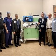
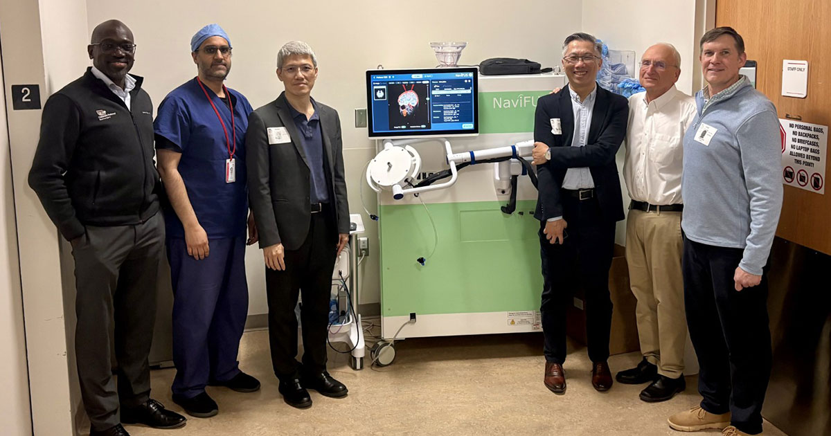
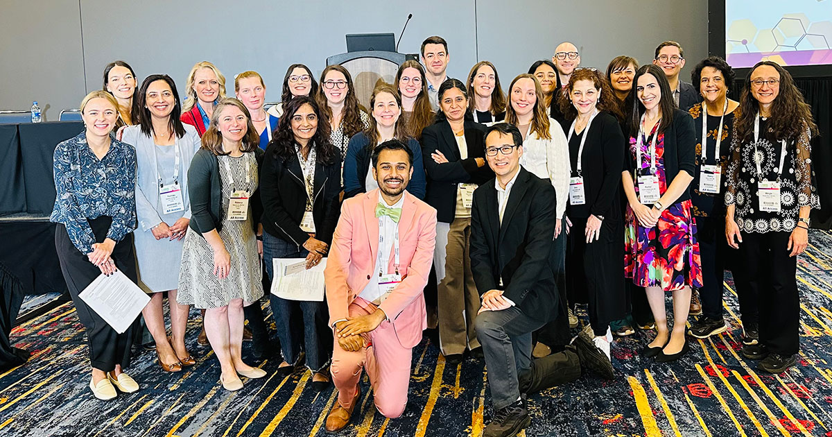

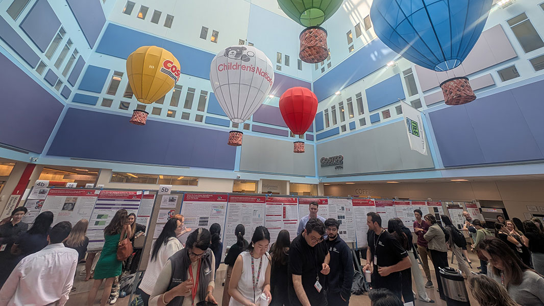
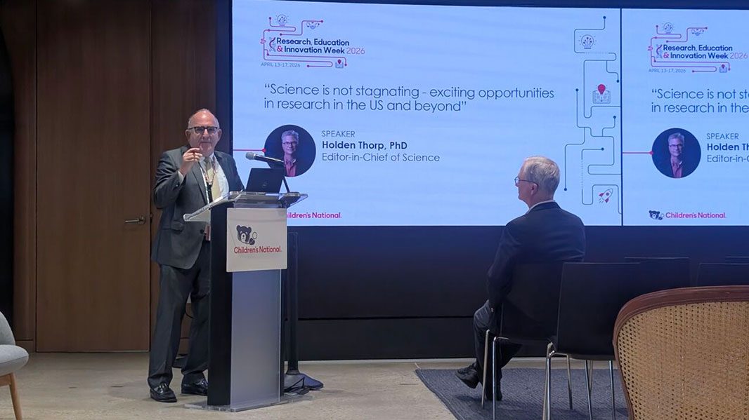
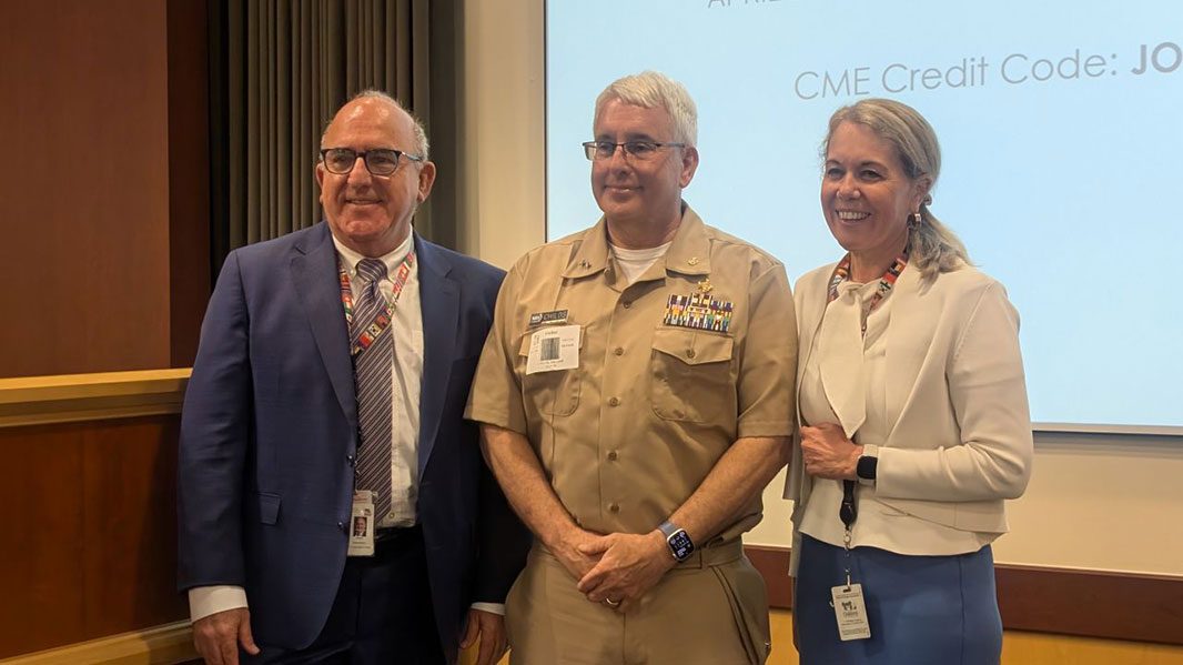
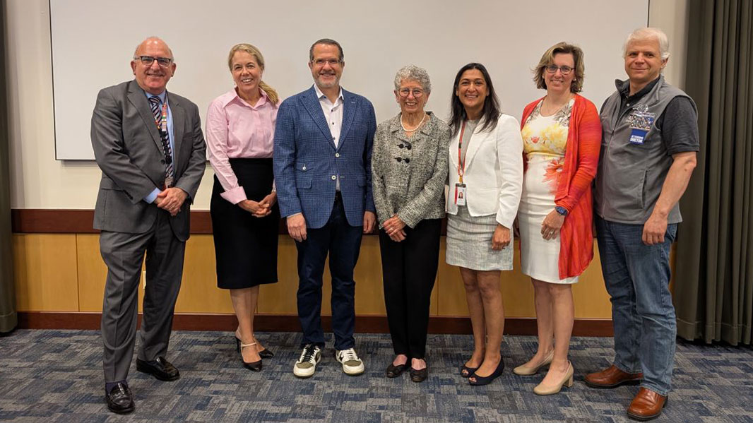
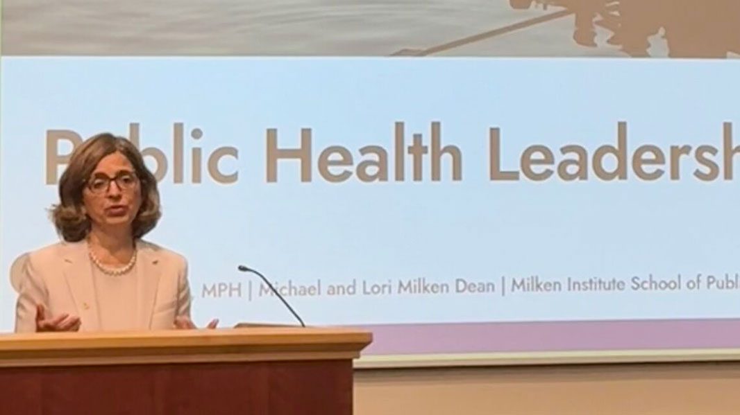
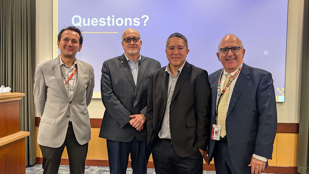
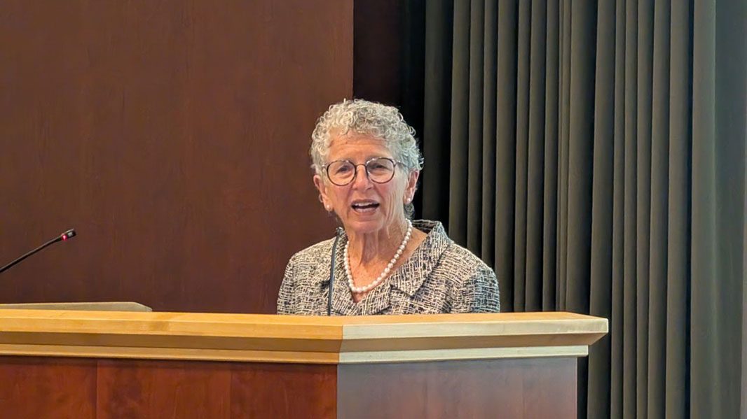
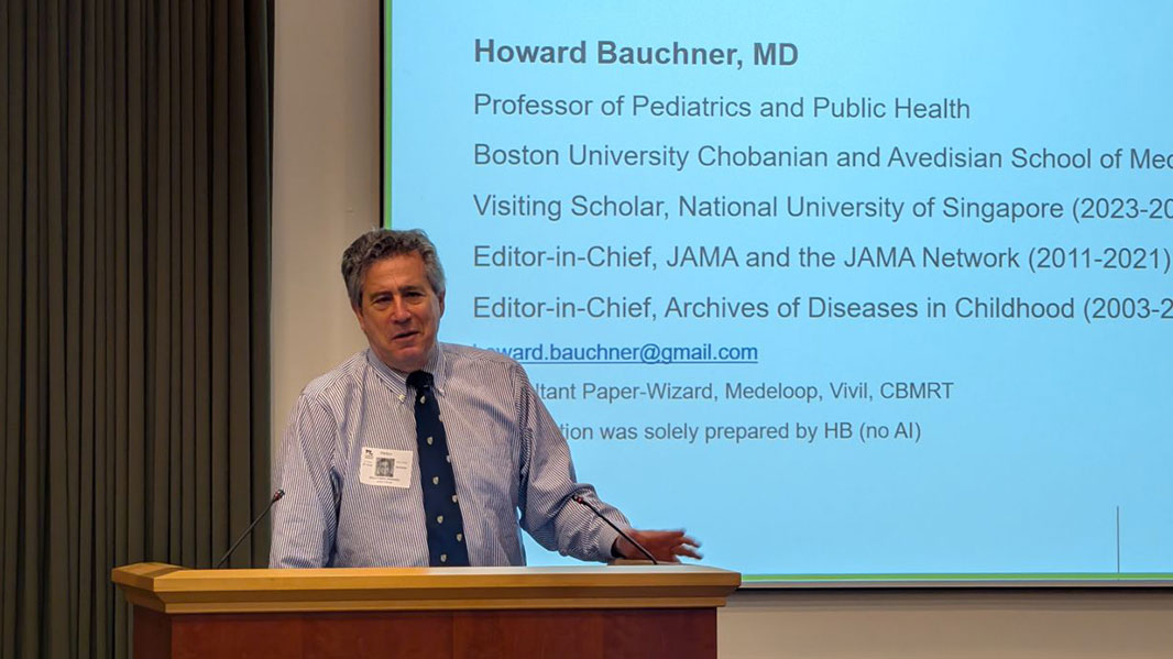
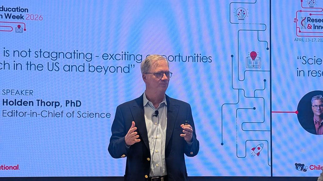
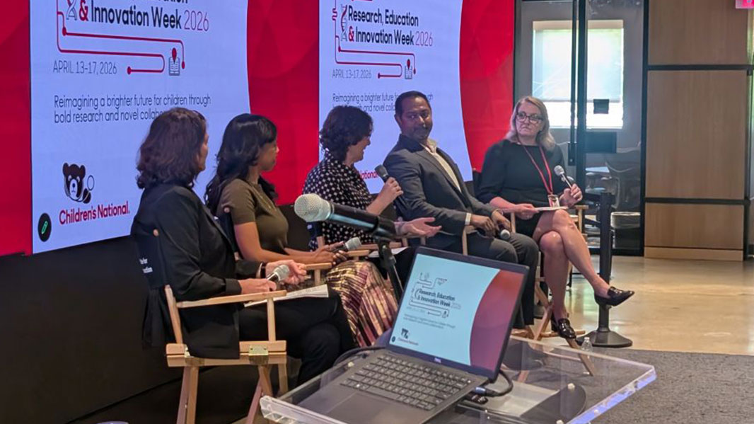
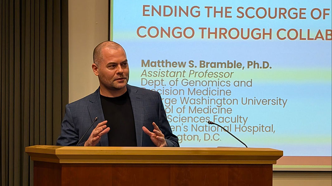
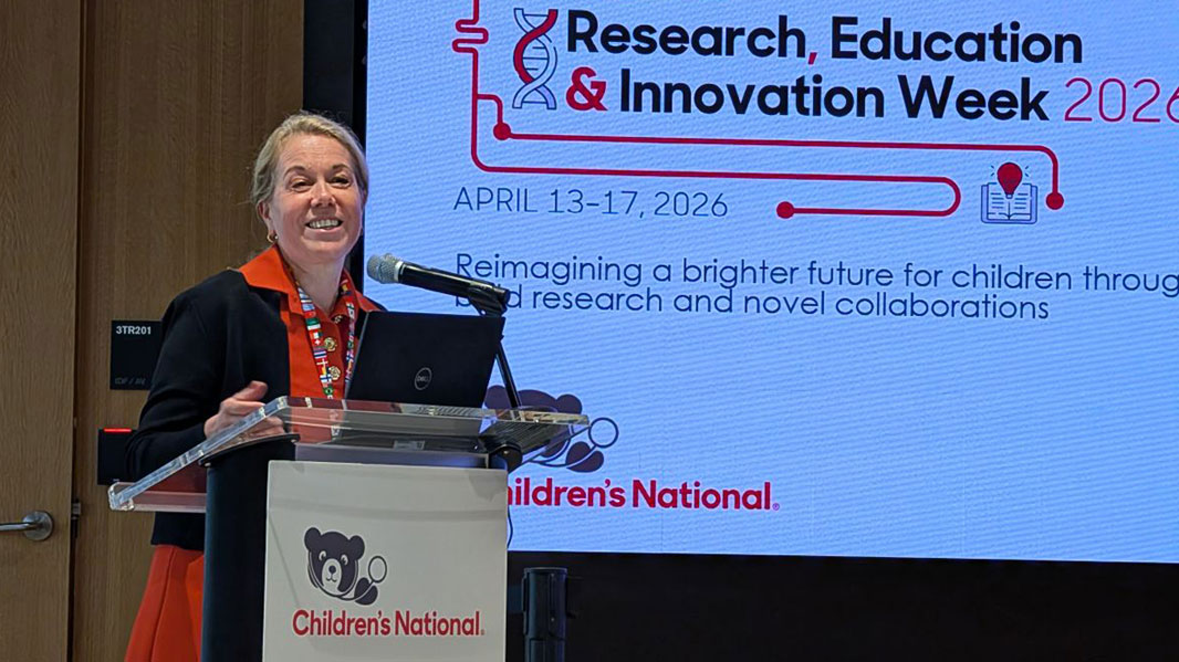

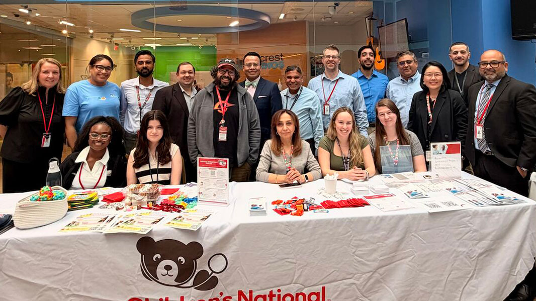
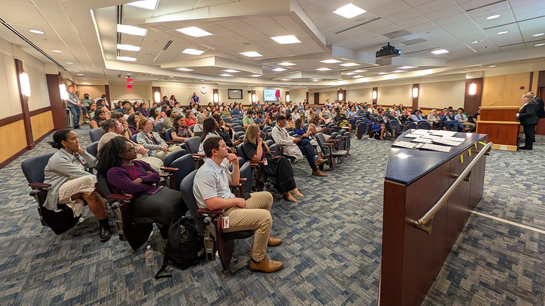
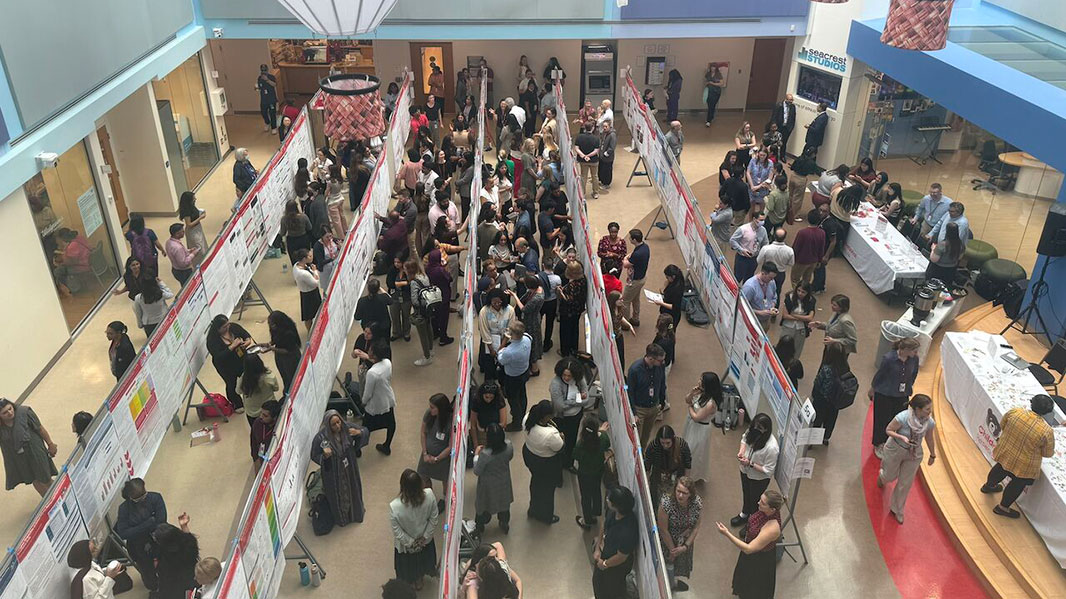
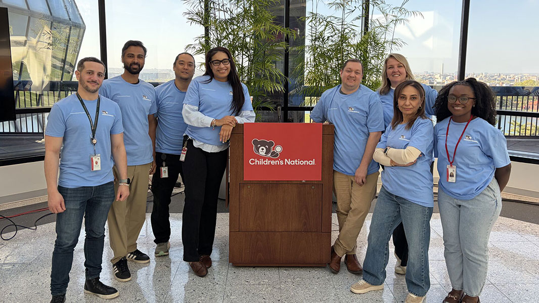
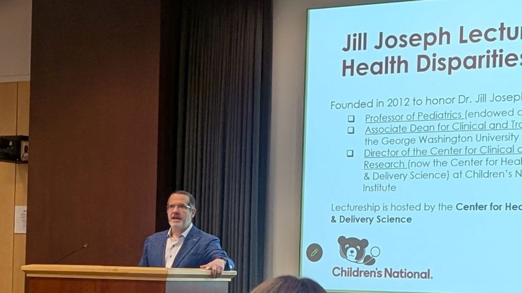
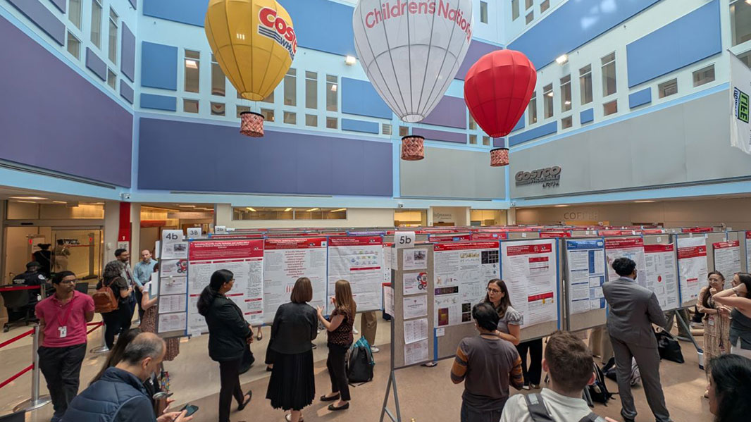
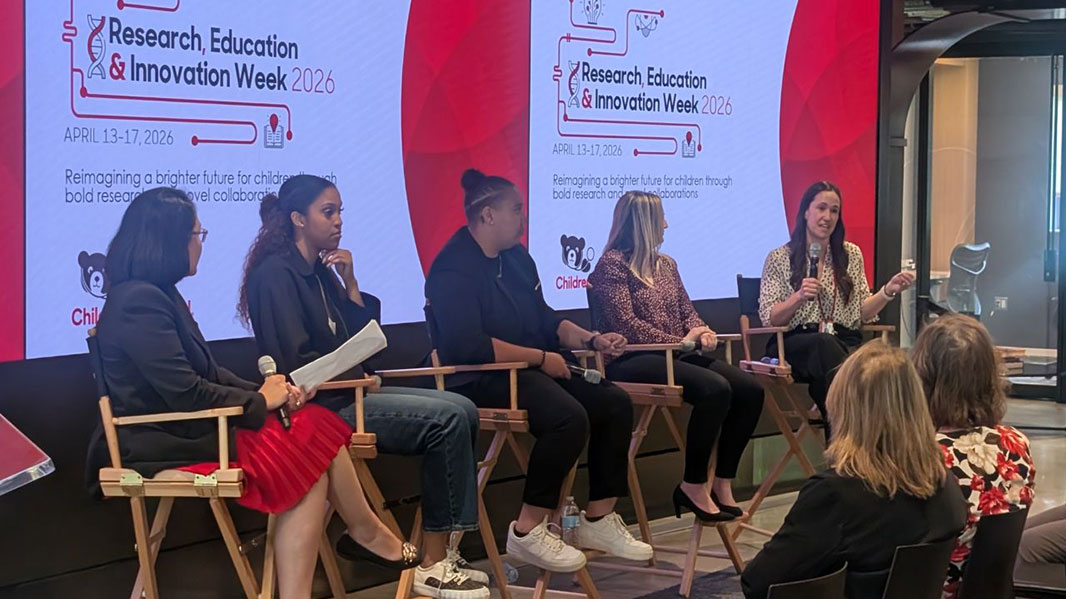
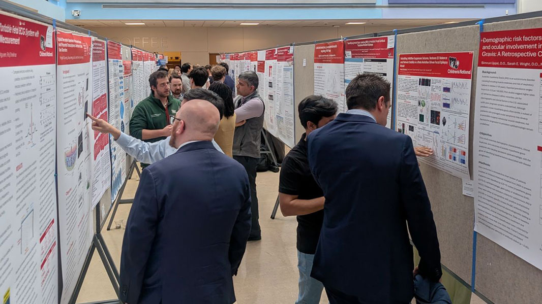
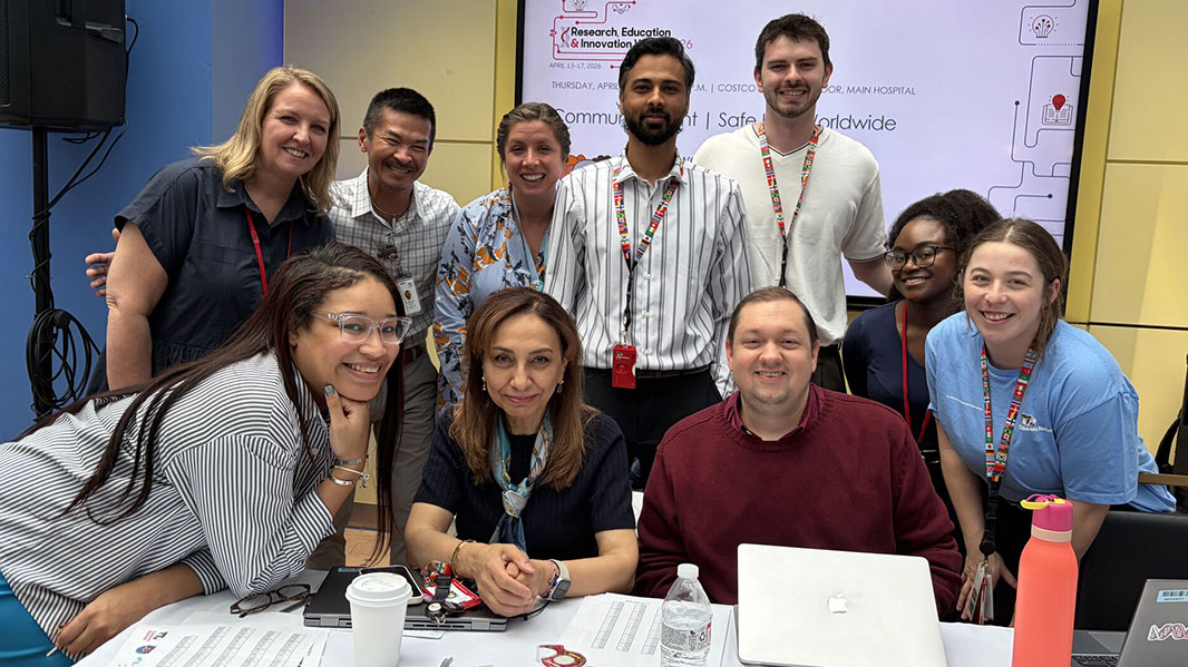
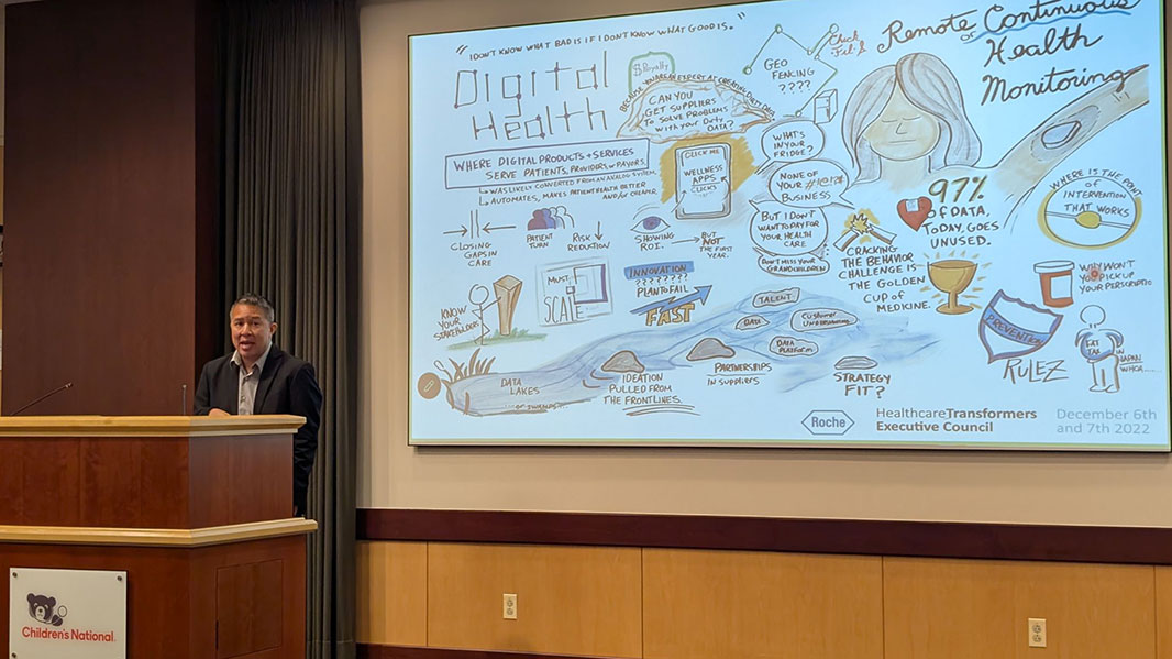
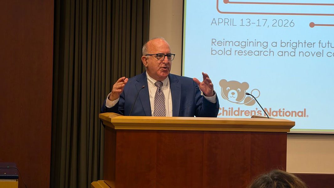

 The
The 


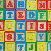


 The Divisions of Neurology and Neurosurgery at