Genetics & Rare Diseases

Andrew Dauber, M.D., MMSc, caps off research success with award and reception

Andrew Dauber

Unfortunately, we’ve been notified that the ENDO2020 conference has been canceled due to concerns of COVID-19. Because of this, we will not be hosting our reception in honor of Andrew Dauber, M.D., on Sunday, March 29.

We hope to see you at a future Endocrinology or Pediatric Endocrinology event.

Children’s National Hospital is incredibly proud of the work Dr. Dauber has done in the endocrinology community.

Andrew Dauber, M.D., MMSc, division chief of Endocrinology at Children’s National Hospital, will be awarded the 2020 Richard E. Weitzman Outstanding Early Career Investigator Award at ENDO 2020. The prestigious award will be presented at the annual meeting of the Endocrine Society in recognition of Dauber’s work in understanding the regulation of growth and puberty, and applying innovative genetic technologies to studying pediatric endocrinology. Dauber credits many collaborators throughout the world, as well as the team at Children’s National for the award.

With a five-year grant from the National Institutes of Health (NIH), Dauber and colleagues from the Cincinnati Children’s Hospital Medical Center, Boston Children’s Hospital and the Children’s Hospital of Philadelphia are using electronic health records to identify children who likely have rare genetic growth disorders. Using cutting-edge DNA sequencing technologies, including whole exome sequencing, the researchers are aiming to identify novel genetic causes of severe growth disorders. The first paper describing genetic findings in patients with high IGF-1 levels was published in Hormone Research in Paediatrics in December 2019.

Dauber and researchers at Cincinnati Children’s Hospital Medical Center are exploring how to treat patients with mutations in the PAPPA2 gene. In 2016, the group described the first patients with mutations in this gene who had decreased the bioavailability of IGF-1, stunting their growth and development. In their current phase of research, findings are emphasizing the importance of this gene in regulating IGF-1 bioavailability throughout childhood. The ultimate aim is to create therapies to increase IGF-1 bioavailability, thereby supporting healthy growth and development in children. Their first study to track PAPPA2 and intact IBGBP-3 concentrations throughout childhood was published in the European Journal of Endocrinology in January 2020.

Dauber is particularly interested in studying children with dominantly inherited forms of short stature. Along with collaborators in Cincinnati, he currently has an ongoing treatment trial using growth hormone in patients with Aggrecan gene mutations.  Dauber hopes to announce soon a new clinical trial for children with all forms of dominantly inherited short stature.

Study upon study has shown us that there are many factors that affect an individual’s height and growth. As these studies and the conversation around how to identify and address genomic anomalies become more prevalent, the team at Children’s National is increasingly interested in engaging with other centers around the country. In the coming months, the Children’s National Research & Innovation Campus will open on the grounds of the former Walter Reed Army Medical Center, which will serve as a one-of-a-kind pediatric research and innovation hub. A critical component to this campus is the co-location of Children’s National research with key partners and incubator space.

Working to improve the management of endocrine related conditions

Nadia Merchant

This past fall, Nadia Merchant, M.D., joined Children’s National Hospital as an endocrinologist in the Endocrinology and Diabetes Department. Dr. Merchant received her undergraduate and medical education at Weill Cornell Medical College in Qatar. She completed her pediatric residency at Wright State Boonshoft School of Medicine. She then completed her genetics residency and pediatric endocrine fellowship at Baylor College of Medicine/Texas Children’s Hospital.

Dr. Merchant was born with acromesomelic dysplasia, a rare genetic disorder, but that hasn’t stopped her from pursuing her medical career. While at Baylor College of Medicine, Dr. Merchant was very active in quality improvement projects, research and organizations that raise awareness of endocrine related conditions. For several years, she was a moderator at Baylor College of Medicine for “From Stress to Strength,” at a course for parents of children with genetic disorders and autism. Dr. Merchant also served as an endocrine fellow representative on the American Academy of Pediatrics Section on Endocrinology (SOEn) for the last two years and also served on the committee for a Bone and Mineral special interest group within the Pediatric Endocrine Society (PES). During medical school, she worked with Positive Exposure, an organization that uses visual arts to celebrate human diversity for individuals living with genetic, physical, behavioral and intellectual differences.

During the 2019 Endocrine Society Annual Meeting, Dr. Merchant won the Presidential Poster Award for her poster presentation: Assessing Metacarpal Cortical Thickness as a Tool to Evaluate Bone Density Compared to DXA in Osteogenesis Imperfecta a research project assessing whether hand film is an additional tool to detect low bone mineral density in children.

Dr. Nadia Merchant is currently one of the endocrinologists in the multidisciplinary bone health clinic at Children’s National, a clinic dedicated to addressing and improving bone health in children. Dr. Merchant also manages endocrine manifestations in children with rare genetic disorders.

The Endocrinology department at Children’s National is ranked among the best in the nation by “U.S. News & World Report”.

Making healthcare innovation for children a priority

Dr. Kurt Newman in front of the capitol building

Recently, Kurt Newman, M.D., president and CEO of Children’s National Hospital, authored an opinion piece for the popular political website, The Hill. In the article, he called upon stakeholders from across the landscape to address the significant innovation gap in children’s healthcare versus adults.

As Chair of the Board of Trustees of the Children’s Hospital Association,  Dr. Newman knows the importance of raising awareness among policy makers at the federal and state level about the healthcare needs of children. Dr. Newman believes that children’s health should be a national priority that is addressed comprehensively. With years of experience as a pediatric surgeon, he is concerned by the major inequities in the advancements of children’s medical devices and technologies versus those for adults. That’s why Children’s National is working to create collaborations, influence policies and facilitate changes that will accelerate the pace of pediatric healthcare innovation for the benefit of children everywhere. One way that the hospital is tackling this challenge is by developing the Children’s National Research & Innovation Campus, which will be the nation’s first innovation campus focused on pediatric research.

Children’s National welcomes Virginia Tech to its new campus

Children’s National Hospital and Virginia Tech create formal partnership that includes the launch of a Virginia Tech biomedical research facility within the new Children’s National Research & Innovation Campus.

Children’s National Hospital and Virginia Tech recently announced a formal partnership that will include the launch of a 12,000-square-foot Virginia Tech biomedical research facility within the new Children’s National Research & Innovation Campus. The campus is an expansion of Children’s National that is located on a nearly 12-acre portion of the former Walter Reed Army Medical Center in Washington, D.C. and is set to open its first phase in December 2020. This new collaboration brings together Virginia Tech, a top tier academic research institution, with Children’s National, a U.S. News and World Report top 10 children’s hospital, on what will be the nation’s first innovation campus focused on pediatric research.

Research & Innovation Campus

“Virginia Tech is an ideal partner to help us deliver on what we promised for the Children’s National Research & Innovation Campus – an ecosystem that enables us to accelerate the translation of potential breakthrough discoveries into new treatments and technologies,” says Kurt Newman, M.D., president and CEO, Children’s National. “Our clinical expertise combined with Virginia Tech’s leadership in engineering and technology, and its growing emphasis on biomedical research, will be a significant advance in developing much needed treatment and cures to save children’s lives.”

Earlier this year, Children’s National announced a collaboration with Johnson & Johnson Innovation LLC to launch JLABS @ Washington, DC at the Research & Innovation Campus. The JLABS @ Washington, DC site will be open to pharmaceutical, medical device, consumer and health technology companies that are aiming to advance the development of new drugs, medical devices, precision diagnostics and health technologies, including applications in pediatrics.

“We are proud to welcome Virginia Tech to our historic Walter Reed campus – a campus that is shaping up to host some of the top minds, talent and innovation incubators in the world,” says Washington, D.C. Mayor Muriel Bowser. “The new Children’s National Research & Innovation Campus will exemplify why D.C. is the capital of inclusive innovation – because we are a city committed to building the public and private partnerships necessary to drive discoveries, create jobs, promote economic growth and keep D.C. at the forefront of innovation and change.”

Faculty from the Children’s National Research Institute and the Fralin Biomedical Research Institute at Virginia Tech Carilion (VTC) have worked together for more than a decade, already resulting in shared research grants, collaborative publications and shared intellectual property. Together, the two institutions will now expand their collaborations to develop new drugs, medical devices, software applications and other novel treatments for cancer, rare diseases and other disorders.

“Joining with Children’s National in the nation’s capital positions Virginia Tech to improve the health and well-being of infants and children around the world,” says Virginia Tech President Tim Sands, Ph.D. “This partnership resonates with our land-grant mission to solve big problems and create new opportunities in Virginia and D.C. through education, technology and research.”

The partnership with Children’s National adds to Virginia Tech’s growing footprint in the Washington D.C. region, which includes plans for a new graduate campus in Alexandria, Va. with a human-centered approach to technological innovation. Sands said the proximity of the two locations – just across the Potomac – will enable researchers to leverage resources, and will also create opportunities with the Virginia Tech campus in Blacksburg, Va. and the Virginia Tech Carilion Health Science and Technology campus in Roanoke, Va.

Carilion Clinic and Children’s National have an existing collaboration for provision of certain specialized pediatric clinical services. The more formalized partnership between Virginia Tech and Children’s National will drive the already strong Virginia Tech-Carilion Clinic partnership, particularly for children’s health initiatives and facilitate collaborations between all three institutions in the pediatric research and clinical service domains.

Children’s National and Virginia Tech will engage in joint faculty recruiting, joint intellectual property, joint training of students and fellows, and collaborative research projects and programs according to Michael Friedlander, Ph.D., Virginia Tech’s vice president for health sciences and technology, and executive director of the Fralin Biomedical Research Institute at VTC.

“The expansion and formalization of our partnership with Children’s National is extremely timely and vital for pediatric research innovation and for translating these innovations into practice to prevent, treat and ultimately cure nervous system cancer in children,” says Friedlander, who has collaborated with Children’s National leaders and researchers for more than 20 years. “Both Virginia Tech and Children’s National have similar values and cultures with a firm commitment to discovery and innovation in the service of society.”

“Brain and other nervous system cancers are among the most common cancers in children (alongside leukemia),” says Friedlander. “With our strength in neurobiology including adult brain cancer research in both humans and companion animals at Virginia Tech and the strength of Children’s National research in pediatric cancer, developmental neuroscience and intellectual disabilities, this is a perfect match.”

The design of the Children’s National Research & Innovation Campus not only makes it conducive for the hospital to strengthen its prestigious partnerships with Virginia Tech and Johnson & Johnson, it also fosters synergies with federal agencies like the Biomedical Advanced Research and Development Authority, which will collaborate with JLABS @ Washington, DC to establish a specialized innovation zone to develop responses to health security threats. As more partners sign on, this convergence of key public and private institutions will accelerate discoveries and bring them to market faster for the benefit of children and adults.

“The Children’s National Research & Innovation Campus pairs an inspirational mission to find new treatments for childhood illness and disease with the ideal environment for early stage companies. I am confident the campus will be a magnet for big ideas and will be an economic boost for Washington DC and the region,” says Jeff Zients, who was appointed chair of the Children’s National Board of Directors effective October 1, 2019. As a CEO and the former director of President Obama’s National Economic Council, Zients says that “When you bring together business, academia, health care and government in the right setting, you create a hotbed for innovation.”

Ranked 7th in National Institutes of Health research funding among pediatric hospitals, Children’s National continues to foster collaborations as it prepares to open its first 158,000-square-foot phase of its Research & Innovation Campus. These key partnerships will enable the hospital to fulfill its mission of keeping children top of mind for healthcare innovation and research while also contributing to Washington D.C.’s thriving innovation economy.

Tailored T-cell therapies neutralize viruses that threaten kids with PID

t-cells

Tailored T-cells specially designed to combat a half dozen viruses are safe and may be effective in preventing and treating multiple viral infections, according to research led by Children’s National Hospital faculty.

Catherine Bollard, M.B.Ch.B., M.D., director of the Center for Cancer and Immunology Research at Children’s National and the study’s senior author, presented the teams’ findings Nov. 8, 2019, during a second-annual symposium jointly held by Children’s National and the National Institute of Allergy and Infectious Diseases (NIAID), part of the National Institutes of Health (NIH). Children’s National and NIAID formed a research partnership in 2017 to develop and conduct collaborative clinical research studies focused on young children with allergic, immunologic, infectious and inflammatory diseases. Each year, they co-host a symposium to exchange their latest research findings.

According to the NIH, more than 200 forms of primary immune deficiency diseases impact about 500,000 people in the U.S. These rare, genetic diseases so impair the person’s immune system that they experience repeated and sometimes rare infections that can be life threatening. After a hematopoietic stem cell transplantation, brand new stem cells can rebuild the person’s missing or impaired immune system. However, during the window in which the immune system rebuilds, patients can be vulnerable to a host of viral infections.

Because viral infections can be controlled by T-cells, the body’s infection-fighting white blood cells, the Children’s National first-in-humans Phase 1 dose escalation trial aimed to determine the safety of T-cells with antiviral activity against a half dozen opportunistic viruses: adenovirus, BK virus, cytomegalovirus (CMV), Epstein-Barr virus (EBV), Human Herpesvirus 6 and human parainfluenza-3 (HPIV3).

Eight patients received the hexa-valent, virus-specific T-cells after their stem cell transplants:

  • Three patients were treated for active CMV, and the T-cells resolved their viremia.
  • Two patients treated for active BK virus had complete symptom resolution, while one had hemorrhagic cystitis resolved but had fluctuating viral loads in their blood and urine.
  • Of two patients treated prophylactically, one developed EBV viremia that was treated with rituximab.

Two additional patients received the T-cell treatments under expanded access for emergency treatment, one for disseminated adenoviremia and the other for HPIV3 pneumonia. While these critically ill patients had partial clinical improvement, they were being treated with steroids which may have dampened their antiviral responses.

“These preliminary results show that hexaviral-specific, virus-specific T-cells are safe and may be effective in preventing and treating multiple viral infections,” says Michael Keller, M.D., a pediatric immunologist at Children’s National and the lead study author. “Of note, enzyme-linked immune absorbent spot assays showed evidence of antiviral T-cell activity by three months post infusion in three of four patients who could be evaluated and expansion was detectable in two patients.”

In addition to Drs. Bollard and Keller, additional study authors include Katherine Harris M.D.; Patrick J. Hanley Ph.D., assistant research professor in the Center for Cancer and Immunology; Allistair Abraham, M.D., a blood and marrow transplantation specialist; Blachy J. Dávila Saldaña, M.D., Division of Blood and Marrow Transplantation; Nan Zhang Ph.D.; Gelina Sani BS; Haili Lang MS; Richard Childs M.D.; and Richard Jones M.D.

###

Children’s National-NIAID 2019 symposium presentations

“Welcome and introduction”
H. Clifford Lane, M.D., director of NIAID’s Division of Clinical Research

“Lessons and benefits from collaboration between the NIH and a free-standing children’s hospital”
Marshall L. Summar, M.D., director, Rare Disease Institute, Children’s National

“The hereditary disorders of PropionylCoA and Cobalamin Metabolism – past, present and future”
Charles P. Venditti, M.D., Ph.D., National Human Genome Research Institute Collaboration

“The road(s) to genetic precision therapeutics in pediatric neuromuscular disease: opportunities and challenges”
Carsten G. Bönnemann, M.D., National Institute of Neurological Disorders and Stroke

“Genomic diagnostics in immunologic diseases”
Helen Su, M.D., Ph.D., National Institute of Allergy and Infectious Diseases

“Update on outcomes of gene therapy clinical trials for X-SCID and X-CGD and plans for future trials”
Harry Malech, M.D., National Institute of Allergy and Infectious Diseases

“Virus-specific T-cell therapies: broadening applicability for PID patients”
Catherine Bollard, M.D., Children’s National 

“Using genetic testing to guide therapeutic decisions in Primary Immune Deficiency Disease”
Vanessa Bundy, M.D., Ph.D., Children’s National 

Panel discussion moderated by Lisa M. Guay-Woodford, M.D.
Drs. Su, Malech, Bollard and Bundy
Morgan Similuk, S.C.M., NIAID
Maren Chamorro, Parent Advocate

“Underlying mechanisms of pediatric food allergy: focus on B cells
Adora Lin, M.D., Ph.D., Children’s National 

“Pediatric Lyme outcomes study – interim update”
Roberta L. DeBiasi, M.D., MS, Children’s National 

“Molecular drivers and opportunities in neuroimmune conditions of pediatric onset”
Elizabeth Wells, M.D., Children’s National 


$6M gift powers new PKD clinical and research activities

kidneys with cysts on them

PKD is a genetic disorder characterized by clusters of fluid-filled sacs (cysts) multiplying and interfering with the kidneys’ ability to filter waste from the blood.

When Lisa M. Guay-Woodford, M.D., McGehee Joyce Professor of Pediatrics at Children’s National Hospital, considers a brand-new gift, she likens it to 6 million gallons of “rocket fuel” that will power new research to better understand polycystic kidney disease.

Dr. Guay-Woodford received a $5.7 million dollar gift to support PKD clinical and research activities. PKD is a genetic disorder characterized by clusters of fluid-filled sacs (cysts) multiplying and interfering with the kidneys’ ability to filter waste from the blood. The kidneys’ smooth surface transforms to a bumpy texture as the essential organs grow oversized and riddled with cysts.

The extraordinary generosity got its start in an ordinary clinical visit.

Dr. Guay-Woodford saw a young patient in her clinic at Children’s National a few times in 2015. The child’s diagnosis sparked a voyage of discovery for the patient’s extended family and, ultimately, they attended a presentation she gave during a regional meeting about PKD. That led to a telephone conversation and in-person meeting as they invited her to describe “the white space” between what was being done at the time to better understand PKD and what could be done.

“It’s the power of the art and science of medicine. They come to see people like me because of the science. If we can convey to patients and families that who they are and their unique concerns are really important to researchers, that becomes a powerful connection,” she says. “The art plus the science equals hope. That is what these families are looking for: We give people the latest insights about their disease because information is power.”

The infusion of new funding will strengthen the global initiative’s four pillars:

  • Coordinated care for children and families impacted by renal cystic disease. The Inherited and Polycystic Kidney Disease (IPKD) program, launched September 2019, includes a cadre of experts working together as a team in the medical home so that “in a single, one-stop visit, Children’s National can address the myriad concerns they have,” she explains. A multi-disciplinary team that includes nephrologists, hepatologists and endocrinology experts meets weekly to ensure the Center of Excellence provides the highest-caliber patient care. The team includes genetic counselors to empower families with knowledge about genetic risks and testing opportunities. A nurse helps families navigate the maze of who to call about which issue. Psychologists help to ease anxiety. “There is stress. There is fear. There is pain that can be associated with this set of diseases. The good news is we can control their medical issues. The bad news is some children have difficulty coping. Our psychologists help children cope so they can be a child and do the normal things that children do,” she says.
  • Strengthening global databases to capture PKD variations. The team will expand its outreach to other centers located around the world – including Australia, Europe, India and Latin America – caring for patients with both the recessive and dominant forms of polycystic kidney disease, to better understand the variety of ways the disease can manifest in children. We really don’t know a lot about kids with the dominant form of the disease. How hard should we push to control their blood pressure, knowing that could ease symptoms? What are the ramifications of experiencing acute pain compared with chronic pain? How much do these pain flareups interfere with daily life and a child’s sense of self,” she asks. Capturing the nuances of the worldwide experience offers the power of harnessing even more data. And ensuring that teams collect data in a consistent way means each group would have the potential to extract the most useful information from database queries.
  • Filling a ‘desperate need’ for biomarkers. Developing clinical trials for new therapies requires having biomarkers that indicate the disease course. Such biomarkers have been instrumental in personalizing care for patients with other chronic conditions. “We are in desperate need for such biomarkers, and this new funding will underwrite pilot studies to identify and validate these disease markers. The first bite at the apple will leverage our imaging data to identify promising biomarkers,” she says.
  • Genetic mechanisms that trigger kidney disease. About 500,000 people in the U.S. have PKD. In many cases, children inherit a genetic mutation but, often, their genetic mutation develops spontaneously. Dr. Guay-Woodford’s research about the mechanisms that make certain inherited renal disorders lethal, such as autosomal recessive polycystic kidney disease, is recognized around the world. The fourth pillar of the new project provides funding to continue her lab’s research efforts to improve the mechanistic understanding of what triggers PKD.

Germline microsatellite genotypes differentiate children with medulloblastoma

pastel colored DNA strands

A new study suggests that medulloblastoma-specific germline microsatellite variations mark those at-risk for medulloblastoma development.

Brian Rood, M.D., oncologist and medical director at the Brain Tumor Institute, and Harold “Skip” Garner, Ph.D., associate vice provost for research development at Edward Via College of Osteopathic Medicine, published a report in the Society for Neuro-Oncology’s Neuro-Oncology Journal about using a novel approach to identify specific markers in germline (non-tumor) DNA called microsatellites that can differentiate children who have the brain tumor medulloblastoma (MB) from those who don’t.

“Ultimately, the best way to save children from brain tumors and prevent them from bearing long-term side effects from treatment is to prevent those tumors from occurring in the first place,” says Dr. Rood. “New advancements hold the potential to finally realize the dream of cancer prevention, but we must first identify those children at-risk.”

While analyzing germline sequencing data from a training set of 120 MB subjects and 425 controls, the doctors identified 139 individual microsatellites whose genotypes differ significantly between the groups. Using a genetic algorithm, they were able to construct a subset of 43 microsatellites that distinguish MB subjects from controls with a sensitivity and specificity of 92% and 88% respectively.

“We made discoveries in an untapped part of the human genome, enabled by unique bioinformatics data mining approaches combined with clinical insight,” said Dr. Garner. “Our findings establish new genomic directions that can lead to high accuracy diagnostics for predicting susceptibility to medulloblastoma.”

What the doctors discovered and demonstrated in the study was that MB-specific germline microsatellite variations mark those at risk for MB development and suggest that other mechanisms of cancer predisposition beyond heritable mutations exist for MB.

“This work is the first to demonstrate the ability of specific DNA sequences to differentiate children with cancer from their healthy counterparts,” added Dr. Rood.

Contributing Authors to this research study included:  Brian R. Rood, M.D., Harold R. Garner, Ph.D., Samuel Rivero-Hinojosa, Ph.D., and Nicholas Kinney, Ph.D.

Molecular gatekeepers that regulate calcium ions key to muscle function

mitochondria

Controlled entry of calcium ions into the mitochondria, the cell’s energy powerhouses, makes the difference between whether muscles grow strong or easily tire and perish from injury, according to research published in Cell Reports.

Calcium ions are essential to how muscles work effectively, playing a starring role in how and when muscles contract, tap energy stores to keep working and self-repair damage. Not only are calcium ions vital for the repair of injured muscle fibers, their controlled entry into the mitochondria, the cell’s energy powerhouses, spells the difference between whether muscles will be healthy or if they will easily tire and perish following an injury, according to research published Oct. 29, 2019, in Cell Reports.

“Lack of the protein mitochondrial calcium uptake1 (MICU1) lowers the activation threshold for calcium uptake mediated by the mitochondrial calcium uniporter in both, muscle fibers from an experimental model and fibroblast of  a patient lacking MICU1,” says Jyoti K. Jaiswal, MSc, Ph.D., a principal investigator in the Center for Genetic Medicine Research at Children’s National Hospital and one of the paper’s corresponding authors. “Missing MICU1 also tips the calcium ion balance in the mitochondria when muscles contract or are injured, leading to more pronounced muscle weakness and myofiber death.”

Five years ago, patients with a very rare disease linked to mutations in the mitochondrial gene MICU1 were described to suffer from a neuromuscular disease with signs of muscle weakness and damage that could not be fully explained.

To determine what was going awry, the multi-institutional research team used a comprehensive approach that included fibroblasts donated by a patient lacking MICU1 and an experimental model whose MICU1 gene was deleted in the muscles.

Loss of MICU1 in skeletal muscle fibers leads to less contractile force, increased fatigue and diminished capacity to repair damage to their cell membrane, called the sarcolemma. Just like human patients, the experimental model suffers more pronounced muscle weakness, increased numbers of dead myofibers, with greater loss of muscle mass in certain muscles, like the quadriceps and triceps, the research team writes.

“What was happening to the patient’s muscles was a big riddle that our research addressed,” Jaiswal adds. “Lacking this protein is not supposed to make the muscle fiber die, like we see in patients with this rare disease. The missing protein is just supposed to cause atrophy and weakness.”

Patients with this rare disease show early muscle weakness, fluctuating levels of fatigue and lethargy, muscle aches after exercise, and elevated creatine kinase in their bloodstream, an indication of cell damage due to physical stress.

“One by one, we investigated these specific features in experimental models that look normal and have normal body weight, but also show lost muscle mass in the quadriceps and triceps,” explains Adam Horn, Ph.D., the lead researcher in Jaiswal’s lab who conducted this study. “Our experimental model lacking MICU1 only in skeletal muscles responded to muscle deficits so similar to humans that it suggests that some of the symptoms we see in patients can be attributed to MICU1 loss in skeletal muscles.”

Future research will aim to explore the details of how the impact of MICU1 deficit in muscles may be addressed therapeutically and possible implications of lacking MICU1 or its paralog in other organs.

In addition to Jaiswal and Horn, Children’s National Hospital Center for Genetic Medicine Research co-authors include Marshall W. Hogarth and Davi A. Mazala. Additional co-authors include Lead Author Valentina Debattisti, Raghavendra Singh, Erin L. Seifert, Kai Ting Huang, and Senior Author György Hajnóczky, all from Thomas Jefferson University; and Rita Horvath, from Newcastle University.

Financial support for research described in this post was provided by the National Institutes of Health under award numbers R01AR55686, U54HD090257 and RO1 GM102724; National Institute of Arthritis and Musculoskeletal and Skin Diseases under award number T32AR056993; and Foundation Leducq.

$5M in federal funding to help patients with urea cycle disorders

Andrea Gropman

Andrea L. Gropman, M.D.: We have collected many years of longitudinal clinical data, but with this new funding now we can answer questions about these diseases that are meaningful on a day-to-day basis for patients with urea cycle disorders.

An international research consortium co-led by Andrea L. Gropman, M.D., at Children’s National Hospital has received $5 million in federal funding as part of an overall effort to better understand rare diseases and accelerate potential treatments to patients.

Urea cycle disorder, one such rare disease, is a hiccup in a series of biochemical reactions that transform nitrogen into a non-toxic compound, urea. The six enzymes and two carrier/transport molecules that accomplish this essential task reside primarily in the liver and, to a lesser degree, in other organs.

The majority of patients have the recessive form of the disorder, meaning it has skipped a generation. These kids inherit one copy of an abnormal gene from each parent, while the parents themselves were not affected, says Dr. Gropman, chief of the Division of Neurodevelopmental Pediatrics and Neurogenetics at Children’s National. Another more common version of the disease is carried on the X chromosome and affects boys more seriously that girls, given that boys have only one X chromosome.

Regardless of the type of urea cycle disorder, when the urea cycle breaks down, nitrogen converts into toxic ammonia that builds up in the body (hyperammonemia), particularly in the brain. As a result, the person may feel lethargic; if the ammonia in the bloodstream reaches the brain in high concentrations, the person can experience seizures, behavior changes and lapse into a coma.

Improvements in clinical care and the advent of effective medicines have transformed this once deadly disease into a more manageable chronic ailment.

“It’s gratifying that patients diagnosed with urea cycle disorder now are surviving, growing up, becoming young adults and starting families themselves. Twenty to 30 years ago, this never would have seemed conceivable,” Dr. Gropman says. “We have collected many years of longitudinal clinical data, but with this new funding now we can answer questions about these diseases that are meaningful on a day-to-day basis for patients with urea cycle disorders.”

In early October 2019, the National Institutes of Health (NIH) awarded the Urea Cycle Disorders Consortium for which Dr. Gropman is co-principal investigator a five-year grant. This is the fourth time that the international Consortium of physicians, scientists, neuropsychologists, nurses, genetic counselors and researchers has received NIH funding to study this group of conditions.

Dr. Gropman says the current urea cycle research program builds on a sturdy foundation built by previous principal investigators Mendel Tuchman, M.D., and Mark Batshaw, M.D., also funded by the NIH. While previous rounds of NIH funding powered research about patients’ long-term survival prospects and cognitive dysfunction, this next phase of research will explore patients’ long-term health.

Among the topics they will study:

Long-term organ damage. Magnetic resonance elastrography (MRE) is a state-of-the-art imaging technique that combines the sharp images from MRI with a visual map that shows body tissue stiffness. The research team will use MRE to look for early changes in the liver – before patients show any symptoms – that could be associated with long-term health impacts. Their aim is spot the earliest signs of potential liver dysfunction in order to intervene before the patient develops liver fibrosis.

Academic achievement. The research team will examine gaps in academic achievement for patients who appear to be underperforming to determine what is triggering the discrepancy between their potential and actual scholastics. If they uncover issues such as learning difficulties or mental health concerns like anxiety, there are opportunities to intervene to boost academic achievement.

“And if we find many of the patients meet the criteria for depression or anxiety disorders, there are potential opportunities to intervene.  It’s tricky: We need to balance their existing medications with any new ones to ensure that we don’t increase their hyperammonemia risk,” Dr. Gropman explains.

Neurologic complications. The researchers will tap continuous, bedside electroencephalogram, which measures the brain’s electrical activity, to detect silent seizures and otherwise undetectable changes in the brain in an effort to stave off epilepsy, a brain disorder that causes seizures.

“This is really the first time we will examine babies’ brains,” she adds. “Our previous imaging studies looked at kids and adults who were 6 years and older. Now, we’re lowering that age range down to infants. By tracking such images over time, the field has described the trajectory of what normal brain development should look like. We can use that as a background and comparison point.”

In the future, newborns may be screened for urea cycle disorder shortly after birth. Because it is not possible to diagnose it in the womb in cases where there is no family history, the team aims to better counsel families contemplating pregnancy about their possible risks.

Research described in this post was underwritten by the NIH through its Rare Diseases Clinical Research Network.

Preserving brain function by purposely inducing strokes

Bella when she was sick

Born to young parents, no prenatal testing had suggested any problems with Bella’s brain. But just a few hours after birth, Bella suffered her first seizure – one of many that would follow in the ensuing days. After brain imaging, her doctors in Iowa diagnosed her with hemimegalencephaly.

Strokes are neurologically devastating events, cutting off life-sustaining oxygen to regions of the brain. If these brain tissues are deprived of oxygen long enough, they die, leading to critical loss of function – and sometimes loss of life.

“As physicians, we’re taught to prevent or treat stroke. We’re never taught to inflict it,” says Taeun Chang, M.D., director of the Neonatal Neurology and Neonatal Neurocritical Care Program at Children’s National Hospital.

That’s why a treatment developed at Children’s National for a rare brain condition called hemimegalencephaly is so surprising, Dr. Chang explains. By inflicting controlled, targeted strokes, Children’s National physician-researchers have treated five newborns born with intractable seizures due to hemimegalencephaly before they’re eligible for epilepsy surgery, the standard of care. In the four surviving infants, the procedures drastically reduced or completely relieved the infants of hemimegalencephaly’s characteristic, uncontrollable seizures.

The most recent patient to receive this life-changing procedure is Bella, a 13-month-old from Iowa whose treatment at Children’s National began within her second week of life. Born to young parents, no prenatal testing had suggested any problems with Bella’s brain. But just a few hours after birth, Bella suffered her first seizure – one of many that would follow in the ensuing days. After brain imaging, her doctors in Iowa diagnosed her with hemimegalencephaly.

A congenital condition occurring in just a handful of children born worldwide each year, hemimegalencephaly is marked by one brain hemisphere growing strikingly larger and dysplastic than the other, Dr. Chang explains. This abnormal half of the brain is highly vascularized, rippled with blood vessels needed to support the seizing brain. The most conspicuous symptoms of hemimegalencephaly are the numerous seizures that it causes, sometimes several in the course of an hour, which also may prevent the normal half of the brain from developing and learning.

Prior studies suggest early surgery achieves better developmental outcomes with one study reporting as much as a drop of 10-20 IQ points with every month delay in epilepsy surgery.

The standard treatment for unilateral megalencephaly is a dramatic procedure called a hemispherectomy, in which surgeons remove and disconnect the affected half of the brain, allowing the remaining half to take over its neurological duties. However, Dr. Chang says, implementing this procedure in infants younger than 3 months of age is highly dangerous.  Excessive, potentially fatal blood loss is likely in infants younger than 3 months who have a highly vascularized brain in the setting of an immature coagulation system. That leaves their doctors with no choice but to wait until these infants are at least 3 months old, when they are more likely to survive the surgery.

However, five years ago, Dr. Chang and her colleagues came up with a different idea when a newborn continued to have several seizures per hour despite multiple IV seizure medications: Because strokes cause irreversible tissue death, it might be possible to effectively incapacitate the enlarged hemisphere from within by inflicting a stroke on purpose. At the very least, this “functional embolization” might buy time for a traditional hemispherectomy, and slow or halt ongoing brain damage until the infants are able to withstand surgery. Ideally, this procedure may be all some children need, knocking out the offending hemisphere completely so they’d never need a hemispherectomy, which has late complications, such as hydrocephalus.

A pediatrician friend of Bella’s paternal grandparents read a story on Children’s National website about Darcy, another baby who’d received functional embolization a year earlier and was doing well. She contacted Dr. Chang to see if the procedure would be appropriate for Bella.

Within days, Bella and her family headed to Washington, D.C., to prepare for functional embolization herself. Within the first weeks of life, Bella underwent three separate procedures, each three to four hours long. Under real-time fluoroscopic and angiographic guidance, interventional neuroradiologist Monica Pearl, M.D., threaded a micro-catheter up from the baby’s femoral artery through the complex network of blood vessels all the way to her brain. There, in targeted branches of her cerebral arteries, Dr. Pearl strategically placed liquid embolic agent to obstruct blood flow to the abnormal half of Bella’s brain.

Immediately after the first procedure, the team had to contend with the same consequences that come after any stroke: brain swelling that can cause bleeding and herniation, complicated further by the already enlarged hemisphere of Bella’s brain. Using neuroprotective strategies learned from treating hundreds of brain-injured newborns, the neonatal neurocritical care team and the neonatal intensive care unit (NICU) minimized the brain swelling and protected the normal half of the brain by tightly controlling the brain temperature, her sugar and electrolyte levels, her blood pressure and coagulation system.

As the brain tissue in the oversized hemisphere died, so did the seizures that had plagued Bella since birth. She has not had a seizure since she left Children’s National more than one year ago. Her adoptive parents report that Bella is hitting many of the typical developmental milestones for her age: She’s getting ready to walk, blowing kisses and saying a few words. Physical, speech and occupational therapy will keep her moving in the right direction, Dr. Chang says.

“We believe that Children’s National is the only place in the world that’s treating newborns in this way to preserve their futures,” Dr. Chang says. “We’re privileged to be able to care for Bella and other kids with this rare condition.”

Bella’s transfer and successful procedures required the support and collective efforts of many within the hospital organization including William D. Gaillard, M.D., and his surgical epilepsy team; interventional neuroradiology with Dr. Monica Pearl; Neurosurgery; Neonatology and the NICU; social work; and even approval from Robin Steinhorn, M.D., senior vice president of the Center for Hospital-Based Specialties, and David Wessel, M.D., executive vice president and Chief Medical Officer.

“While obvious credit goes to the medical team who saved Bella’s future and the neonatal intensive care nurses who provided exceptional, intensive, one-on-one care, Bella’s team of supporters extend to all levels within our hospital,” Dr. Chang adds.

Also read:

Born with hemimegalencephaly, Bella now has a bright future

bella's brain scans

PDF Version

Bella was born with a rare condition (hemimegalencephaly) in which one half of the brain developed abnormally, causing seizures. The textbook approach is to let babies grow big enough for a dramatic surgery. But Bella’s left hemisphere was triggering so many seizures each hour that waiting would mean her life would be defined by severe disability. Children’s National Hospital is believed to be the only center in the world that calms these seizures through controlled strokes.

Procedure one occurred five days after Bella came to Children’s National Hospital from Iowa, when she was 13 days old. The team first optimized control of her seizures and obtained special magnetic resonance images to plan their approach. They glued up the branches of the left posterior cerebral artery and branches of the left middle cerebral artery. Bella had a tiny bleed that was controlled immediately in the angio suite and afterwards in the Children’s National neonatal intensive care unit.

Procedure two occurred 10 days later when Bella was 23 days old. The team waited until brain swelling had subsided and brain tissue loss had occurred from the first procedure. This time, they glued up the remaining branches of the left posterior cerebral artery and some branches of the left anterior cerebral artery.

The third and final procedure was done nine days later when Bella was 29 days old.  This time the team glued and coiled, placing little wire coils where it was unsafe to use glue, getting at the remaining small and numerous branches that remained of the left anterior cerebral artery.

Also read:

Extracting actionable research data faster, with fewer hassles

Mihailo Kaplarevic

Mihailo Kaplarevic, Ph.D., the newly minted Chief Research Information Officer at Children’s National Hospital and Bioinformatics Division Chief at Children’s National Research Institute, will provide computational support, advice, informational guidance, expertise in big data and data analyses for researchers and clinicians.

Kaplarevic’s new job is much like the role he played most recently at the National Heart, Lung and Blood Institute (NHLBI), assembling a team of researchers and scientists skilled in computing and statistical analyses to assist as in-house experts for other researchers and scientists.

NHLBI was the first institute within the National Institutes of Health (NIH) family to set up a scientific information office. During his tenure, a half-dozen other NIH institutions followed, setting up the same entity to help bridge the enormous gap between basic and clinical science and everything related to IT.

“There is a difference compared with traditional IT support at Children’s National – which will remain in place and still do the same sort of things they have been doing so far,” he says of The Bear Institute for Health Innovation. “The difference is this office has experience in research because every single one of us was a researcher at a certain point in our career: We are published. We applied for grants. We lived the life of a typical scientist. On top of that, we’re coming from the computational world. That helps us bridge the gaps between research and clinical worlds and IT.”

Ultimately, he aims to foster groundbreaking science by recognizing the potential to enhance research projects by bringing expertise acquired over his career and powerful computing tools to help teams achieve their goals in a less expensive and more efficient way.

“I have lived the life of a typical scientist. I know exactly how painful and frustrating it can be to want to do something quickly and efficiently but be slowed by technological barriers,” he adds.

As just one example, his office will design the high-performance computing cluster for the hospital to help teams extract more useful clinical and research data with fewer headaches.

Right now, the hospital has three independent clinical systems storing patient data; all serve a different purpose. (And there are also a couple of research information systems, also used for different purposes.) Since databases are his expertise, he will be involved in consolidating data resources, finding the best way to infuse the project with the bigger-picture mission – especially for translational science – and creating meaningful, actionable reports.

“It’s not only about running fewer queries,” he explains. “One needs to know how to design the right question. One needs to know how to design that question in a way that the systems could understand. And, once you get the data back, it’s a big set of things that you need to further filter and carefully shape. Only then will you get the essence that has clinical or scientific value. It’s a long process.”

As he was introduced during a Children’s National Research Institute faculty meeting in late-September 2019, Kaplarevic joked that his move away from pure computer science into a health care and clinical research domain was triggered by his parents: “When my mom would introduce me, she would say ‘My son is a doctor, but not the kind of doctor who helps other people.’ ”

Some of that know-how will play out by applying tools and methodology to analyze big data to pluck out the wheat (useful data) from the chaff in an efficient and useful way. On projects that involve leveraging cloud computing for storing massive amounts of data, it could entail analyzing the data wisely to reduce its size when it comes back from the cloud – when the real storage costs come in. “You can save a lot of money by being smart about how you analyze data,” he says.

While he expects his first few months will be spent getting the lay of the land, understanding research project portfolios, key principal investigators and the pediatric hospital’s biggest users in the computational domain, he has ambitious longer-term goals.

“Three years from now, I would like this institution to say that the researchers are feeling confident that their research is not affected by limitations related to computer science in general. I would like this place to become a very attractive environment for up-and-coming researchers as well as for established researchers because we are offering cutting-edge technological efficiencies; we are following the trends; we are a secure place; and we foster science in the best possible way by making computational services accessible, affordable and reliable.”

Andrew Dauber, M.D., MMSc, awarded prestigious laureate award

Andrew Dauber

Unfortunately, we’ve been notified that the ENDO2020 conference has been canceled due to concerns of COVID-19. Because of this, we will not be hosting our reception in honor of Andrew Dauber, M.D., on Sunday, March 29.

We hope to see you at a future Endocrinology or Pediatric Endocrinology event.

Children’s National Hospital is incredibly proud of the work Dr. Dauber has done in the endocrinology community.

Andrew Dauber, M.D., MMSc, division chief of Endocrinology at Children’s National Hospital, will receive the 2020 Richard E. Weitzman Outstanding Early Career Investigator Award from The Endocrine Society. Given annually, the award was established in 1982 and honors the memory of the late Richard E. Weitzman, who had a brief but outstanding career studying neurohypophyseal hormone and cardiovascular-endocrine physiology – two seminal areas of modern endocrinology.

Dr. Dauber was selected as a recipient for the prestigious award for his contributions to understanding the regulation of growth and puberty, and his success at applying innovative genetic technologies to studying pediatric endocrinology.

“I feel extremely honored and humbled to be the recipient of the Richard E. Weitzman Outstanding Early Career Investigator Award from the Endocrine Society,” says Dr. Dauber. “I am so grateful to my many collaborators throughout the world as well as to my entire research team whose hard work and friendship are the basis for this award. I am excited to continue our work at Children’s National, an institution dedicated to innovation and team science.”

Dr. Dauber joined Children’s National in 2018 and specializes in studying and treating growth disorders. He has published over 75 studies examining genetic clues to endocrine disorders, with a focus on short stature and growth disorders.

The award will be presented at ENDO 2020, The Endocrine Society’s annual meeting, March 28-31, 2020, in San Francisco, California.

Getting to know Lee Beers, M.D., FAAP, future president-elect of AAP

Lee Beers

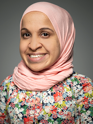
Lee Savio Beers, M.D., FAAP, Medical Director of Community Health and Advocacy at the Child Health Advocacy Institute (CHAI) at Children’s National Hospital carved out a Monday morning in late-September 2019, as she knew the American Academy of Pediatrics (AAP) would announce the results of its presidential election, first by telephone call, then by an email to all of its members.  Her husband blocked off the morning as well to wait with her for the results.  She soon got the call that she was elected by her peers to become AAP president-elect, beginning Jan. 1, 2020. Dr. Beers will then serve as AAP president in 2021 for a one-year term.

That day swept by in a rush, and then the next day she was back in clinic, caring for her patients, some of them teenagers whom she had taken care of since birth. Seeing children and families she had known for such a long time, some of whom had complex medical needs, was a perfect reminder of what originally motivated Dr. Beers to be considered as a candidate in the election.

“When we all work together – with our colleagues, other professionals, communities and families – we can make a real difference in the lives of children.  So many people have reached out to share their congratulations, and offer their support or help. There is a real sense of collaboration and commitment to child health,” Dr. Beers says.

That sense of excitement ripples through Children’s National.

“Dr. Beers has devoted her career to helping children. She has developed a national advocacy platform for children. I can think of no better selection for the president-elect role of the AAP. She will be of tremendous service to children within AAP national leadership,” says Kurt Newman, M.D., Children’s National Hospital President and CEO.

AAP comprises 67​,000 pediatricians, and its mission is to promote and safeguard the health and well-being of all children – from infancy to adulthood.

The daughter of a nuclear engineer and a schoolteacher, Dr. Beers knew by age 5 that she would become a doctor. Trained as a chemist, she entered the Emory University School of Medicine after graduation. After completing residency at the Naval Medical Center, she became the only pediatrician assigned to the Guantanamo Bay Naval Station.

That assignment to Cuba, occurring so early in her career, turned out to be a defining moment that shapes how she partners with families and other members of the team to provide comprehensive care.

“I was a brand-new physician, straight out of residency, and was the only pediatrician there so I was responsible for the health of all of the kids on the base. I didn’t know it would be this way at the time, but it was formative. It taught me to take a comprehensive public health approach to taking care of kids and their families,” she recalls.

On the isolated base, where she also ran the immunization clinic and the nursery, she quickly learned she had to judiciously use resources and work together as a team.

“It meant that I had to learn how to lead a multi-disciplinary team and think about how our health care systems support or get in the way of good care,” she says.

One common thread that unites her past and present is helping families build resiliency to shrug off adversity and stress.

“The base was a difficult and isolated place for some families and individuals, so I thought a lot about how to support them. One way is finding strong relationships where you are, which was important for patients and families miles away from their support systems. Another way is to find things you could do that were meaningful to you.”

Cuba sits where the Atlantic Ocean, Caribbean Sea and Gulf of Mexico meet. Dr. Beers learned how to scuba dive there – something she never would have done otherwise – finding it restful and restorative to appreciate the underwater beauty.

“I do think these lessons about resilience are universal. There are actually a lot of similarities between the families I take care of now, many of whom are in socioeconomically vulnerable situations, and military families when you think about the level of stress they are exposed to,” she adds.

Back stateside in 2001, Dr. Beers worked as a staff pediatrician at the National Naval Medical Center in Bethesda, Maryland, and Walter Reed Army Medical Center in Washington, D.C. In 2003, Dr. Beers joined Children’s National Hospital as a general pediatrician in the Goldberg Center for Community Pediatric Health. Currently, she oversees the DC Collaborative for Mental Health in Pediatric Primary Care, a public-private coalition that elevates the standards of mental health care for all children, and is Co-Director of the Early Childhood Innovation Network. She received the Academic Pediatric Association’s 2019 Public Policy and Advocacy Award.

As a candidate, Dr. Beers pledged to continue AAP’s advocacy and public policy efforts and to further enhance membership diversity and inclusion. Among her signature issues:

  • Partnering with patients, families, communities, mental health providers and pediatricians to co-design systems to bolster children’s resiliency and to alleviate growing pediatric mental health concerns
  • Tackling physician burnout by supporting pediatricians through office-based education and systems reforms
  • Expanding community-based prevention and treatment

“I am humbled and honored to have the support of my peers in taking on this newest leadership role,” says Dr. Beers. “AAP has been a part of my life since I first became a pediatrician, and my many leadership roles in the DC chapter and national AAP have given me a glimpse of the collective good that pediatricians can accomplish by working together toward common strategic goals.”

AAP isn’t just an integral part of her life, it’s where she met her future husband, Nathaniel Beers, M.D., MPA, FAAP, President of The HSC Health Care System. The couple’s children regularly attended AAP meetings with them when they were young.

Just take a glimpse at Lee Beers’ Twitter news feed. There’s a steady stream of images of her jogging before AAP meetings to amazing sunrises, jogging after AAP meetings to stellar sunsets and always, always, images of the entire family, once collectively costumed as The Incredibles.

“I really do believe that we have to set an example: If we are talking about supporting children and families in our work, we have to set that example in our own lives. That looks different for everyone, but as pediatricians and health professionals, we can model prioritizing our families while still being committed to our work,” she explains.

“Being together in the midst of the craziness is just part of what we do as a family. We travel a lot, and our kids have gone with us to AAP meetings since they were infants. My husband even brought our infant son to a meeting at the mayor’s office when he was on paternity leave. Recognizing that not everyone is in a position to be able to do things like that, it’s important for us to do it – to continue to change the conversation and make it normal to have your family to be part of your whole life, not have a separate work life and a separate family life.”

Understanding gut bacteria: forces for good (and sometimes evil)

gut bacteria

In a paper published Sept. 11, 2019, in PLOS ONE, a multi-institutional research team led by George Washington University (GW) faculty found 157 different types of organisms (eight phyla, 18 classes, 23 orders, 38 families, 59 genera and 109 species) living inside the guts of healthy volunteers.

Back in 2015, an interdisciplinary group of research scientists made their case during a business pitch competition: They want to create a subscription-based service, much like 23andMe, through which people could send in samples for detailed analyses. The researchers would crunch that big data fast, using a speedy algorithm, and would send the consumer a detailed report.

But rather than ancestry testing via cheek swab, the team sought to determine the plethora of diverse bacterial species that reside inside an individual’s gut in their ultimate aim to improve public health.

Hiroki Morizono, Ph.D., a member of that team, contributed detailed knowledge of Bacteroides, a key organism amid the diverse array of bacterial species that co-exist with humans, living inside our guts. These symbiotic bacteria convert the food we eat into elements that ensure their well-being as well as ours.

“Trillions of bacteria live in the gut. Bacteroides is one of the major bacterial species,” says Morizono, a principal investigator in the Center for Genetic Medicine Research at Children’s National in Washington, D.C. “In our guts they are usually good citizens. But if they enter our bloodstream, they turn evil; they’re in the wrong place. If you have a bacteroides infection, the mortality rate is 19%, and they resist most antibiotic treatments.”

The starting point for their project – as well as step one for better characterizing the relationship between gut bacteria and human disease – is taking an accurate census count of bacteria residing there.

In a paper published Sept. 11, 2019, in PLOS ONE, a multi-institutional research team led by George Washington University (GW) faculty did just that, finding 157 different types of organisms (eight phyla, 18 classes, 23 orders, 38 families, 59 genera and 109 species) living inside the guts of healthy volunteers.

The study participants were recruited through flyers on the GW Foggy Bottom campus and via emails.  They jotted down what they ate and drank daily, including the brand, type and portion size. They complemented that food journal by providing fecal samples from which DNA was extracted. Fifty fecal metagenomics samples randomly selected from the Human Microbiome Project Phase I research were used for comparison purposes.

“The gut microbiome inherently is really, really cool. In the process of gathering this data, we are building a knowledge base. In this paper, we’re saying that by looking at healthy people, we should be able to establish a baseline about what a normal, healthy gut microbiome should look like and how things may change under different conditions,” Morizono adds.

And they picked a really, really cool name for their bacteria abundance profile: GutFeelingKB.

“KB is knowledge base. Our idea, it’s a gut feeling. It’s a bad joke,” he admits. “Drosophila researchers have the best names for their genes. No other biology group can compete. We, at least, tried.”

Next, the team will continue to collect samples to build out their bacteria baseline, associate it with clinical data, and then will start looking at the health implications for patients.

“One thing we could use this for is to understand how the bacterial population in the gut changes after antibiotic treatment. It’s like watching a forest regrow after a massive fire,” he says. “With probiotics, can we do things to encourage the right bacteria to grow?”

In addition to Morizono, study co-authors include Lead Author Charles H. King, and co-authors Hiral Desai, Allison C. Sylvetsky, Jonathan LoTempio, Shant Ayanyan, Jill Carrie, Keith A. Crandall, Brian C. Fochtman, Lusine Gasparyan, Naila Gulzar, Najy Issa, Lopa Mishra, Shuyun Rao, Yao Ren, Vahan Simonyan, Krista Smith and Senior Author, Raja Mazumder, all of George Washington University; Paul Howell and Sharanjit VedBrat, of KamTek Inc.; Konstantinos Krampis, of City University of New York; Joseph R. Pisegna, of VA Greater Los Angeles Healthcare System; and Michael D. Yao, of Washington DC VA Medical Center.

Financial support for research described in this post was provided by the National Science Foundation under award number 1546491 and the National Institutes of Health National Center for Advancing Translational Sciences under award number UL1TR000075.

$2M from NIH to extract meaningful data from CRISPR screens

tube labeled "CRISPR"

Protein-coding genes comprise a mere 1% of DNA. While the other 99% of DNA was once derided as “junk,” it has become increasingly apparent that some non-coding genes enable essential cellular functions.

Wei Li, Ph.D., a principal investigator in the Center for Genetic Medicine Research at Children’s National in Washington, D.C., proposes to develop statistical and computational methods that sidestep existing hurdles that currently complicate genome-wide CRISPR/Cas9 screening. The National Institutes of Health has granted him $2.23 million in funding over five years to facilitate the systematic study of genes, non-coding elements and genetic interactions in various biological systems and disease types.

Right now, a large volume of screening data resides in the public domain, however it is difficult to compare data that is stored in one library with data stored at a different library. Over the course of the five-year project, Li aims to:

  • Improve functional gene identification from CRISPR screens.
  • Develop new analyses algorithms for screens targeting non-coding elements.
  • Study genetic interactions from CRISPR screens targeting gene pairs.

Ultimately, Li’s work will examine a range of disease types. Take cancer.

“There is abundant information already available in the public domain, like the Project Achilles  from the Broad Institute. However, no one is looking to see what is going in inside these tumors,” Li says. “Cancer is a disease of uncontrolled cell growth that makes tumors grow faster.”

Li and colleagues are going to ask which genes control this process by looking at genes that hit the brakes on cell growth as well as genes that pump the gas.

“You knock out one gene and then look: Does the cell grow faster or does it grow more slowly? If the cell grows more slowly, you know you are knocking out a gene that has the potential to stop tumor growth. If cells are growing faster, you know that you’re hitting genes that suppress cancer cell growth.”

In a nutshell, CRISPR (clustered regularly interspaced short palindromic repeats) screens knock out different genes and monitor changes in corresponding cell populations. When CRISPR first became popular, Li decided he wanted to do something with the technology. So, as a Postdoc at Harvard, he developed comprehensive computational algorithms for functional screens using CRISPR/Cas9.

To reach as many people as possible, he offered that MAGeCK/MAGeCK-VISPR software free to as many researchers as possible, providing source code and offering internet tutorials.

“So far, I think there are quite a lot of people using this. There have been more than 40,000 software downloads,” he adds. “It’s really exciting and revolutionary technology and, eventually, we hope the outcomes also will be exciting. We hope to find something really helpful for cancer patients.”

Research reported in this publication was supported by the National Human Genome Research Institute of the National Institutes of Health under award number R01HG010753.

Searching for the molecular underpinnings of asthma exacerbations

little boy using asthma inhaler

It’s long been known that colds, flu and other respiratory illnesses are major triggers for asthma exacerbations, says asthma expert Stephen J. Teach, M.D., MPH. Consequently, a significant body of research has focused on trying to figure out what’s happening on the cellular or molecular level as these illnesses progress to exacerbations.

People with asthma can be indistinguishable from people who don’t have this chronic airway disease – until they have an asthma attack, also known as an exacerbation. During these events, their airways become inflamed and swollen and produce an abundance of mucus, causing dangerous narrowing of the bronchial tubes that leads to coughing, wheezing and trouble breathing. These events are a major cause of morbidity and mortality, leading to the deaths of 10 U.S. residents every day, according to the Centers for Disease Control and Prevention.

It’s long been known that colds, flu and other respiratory illnesses are major triggers for asthma exacerbations, says Children’s National in Washington, D.C., asthma expert Stephen J. Teach, M.D., MPH. Consequently, a significant body of research has focused on trying to figure out what’s happening on the cellular or molecular level as these illnesses progress to exacerbations. Targeted searches have identified several different molecular pathways that appear to be key players in this phenomenon. However, Dr. Teach says researchers have been missing a complete and unbiased snapshot of all the important pathways in illness-triggered exacerbations and how they interrelate.

To develop this big picture view, Dr. Teach and  Inner-City Asthma Consortium colleagues recruited 208 children ages 6-17 years old with severe asthma – marked by the need for daily doses of inhaled corticosteroids, two hospitalizations or systemic corticosteroid treatments over the past year, and a high concentration of asthma-associated immune cells – from nine pediatric medical centers across the country, including Children’s National. (Inhaled corticosteroids are a class of medicine that calms inflamed airways.) The researchers collected samples of nasal secretions and blood from these patients at baseline, when all of them were healthy.

Then, they waited for these children to show symptoms of respiratory illnesses. Within six days of cold symptoms, the researchers took two more samples of nasal secretions and blood. They also administered breathing tests to determine whether these respiratory illnesses led to asthma exacerbations and recorded whether these patients were treated with systemic corticosteroids to stem the associated respiratory inflammation.

The researchers examined nasal fluid samples for evidence of viral infection during illness and used analytical methods to identify the causative virus. They analyzed all the samples they collected for changes in concentrations of various immune cells. They also looked globally in these samples for changes in gene expression compared with baseline and between the two collection periods during respiratory illness.

Together, this information told the molecular story about what took place after these children got sick and after some of them developed exacerbations. Of the 208 patients recruited, 106 got respiratory illnesses during the six-month study period, leading to a total of 154 illness events. Of those, 47 caused exacerbations, and 107 didn’t.

About half the exacerbations appeared to have been triggered by a rhinovirus, a cause of common colds, the research team reports in a study published online April 8, 2019, in Nature Immunology. The other children’s cold-like symptoms could have been triggered by pollution, allergens or other irritants.

In most exacerbations, virally triggered or not, the researchers saw early activation of a network of genes that appeared to be associated with SMAD3, a signaling molecule already known to be involved in airway inflammation. At the same time, genes that control a set of immune cells known as lymphocytes were turned down. However, as the exacerbation progressed and worsened, the researchers saw gene networks turned on that related to airway narrowing, mucus hypersecretion and activation of other immune cells.

Exacerbations triggered by viruses were associated with multiple inflammatory pathways, in contrast to those in which viruses weren’t found, which were associated with molecular pathways that affected cells in the airway lining.

The researchers validated these findings in 19 patients who each got respiratory illnesses at least twice during the study period but only developed an exacerbation during one of these episodes, finding the same upregulated and downregulated molecular pathways in these patients as in the study population as a whole. They also identified a set of molecular risk factors in patients at baseline – signatures of gene activation that appeared to put patients at risk for exacerbations when they got sick. When patients were treated with systemic corticosteroids during exacerbations, these medicines appeared to restore only some of the affected molecular pathways to normal, healthy levels. Other molecular pathways remained markedly changed.

Each finding could represent a new target for drugs that could prevent or more effectively treat exacerbations, keeping more patients with asthma healthy and out of the hospital.

“Our consortium study found increased gene expression of enzymes that produce molecules that contribute to narrowed airways and dilated blood vessels,” Dr. Teach adds. “This is especially intriguing because drugs that target kallikreins or bradykinin may help treat asthma attacks that aren’t caused by viruses.”

In addition to Dr. Teach, study co-authors include Lead Author Matthew C. Altman, University of Washington; Michelle A. Gill, Baomei Shao and Rebecca S. Gruchalla, all of University of Texas Southwestern Medical Center; Elizabeth Whalen and Scott Presnell of Benaroya Research Institute; Denise C. Babineau and Brett Jepson of Rho, Inc.; Andrew H. Liu, Children’s Hospital Colorado; George T. O’Connor, Boston University School of Medicine; Jacqueline A. Pongracic, Ann Robert H. Lurie Children’s Hospital of Chicago; Carolyn M. Kercsmar and Gurjit K. Khurana Hershey, , Cincinnati Children’s Hospital; Edward M. Zoratti and Christine C. Johnson, Henry Ford Health System; Meyer Kattan, Columbia University College of Physicians and Surgeons; Leonard B. Bacharier and Avraham Beigelman, Washington University, St. Louis; Steve M. Sigelman, Peter J. Gergen, Lisa M. Wheatley and Alkis Togias, National Institute of Allergy and Infectious Diseases; and James E. Gern, William W. Busse and Senior author Daniel J. Jackson, University of Wisconsin School of Medicine and Public Health.

Funding for research described in this post was provided by the National Institute of Allergy and Infectious Diseases under award numbers 1UM1AI114271 and UM2AI117870; CTSA under award numbers UL1TR000150, UL1TR001422 and 5UL1TR001425; the National Institutes of Health under award number UL1TR000451;  CTSI under award number 1UL1TR001430; CCTSI under award numbers UL1TR001082 and 5UM1AI114271; and NCATS under award numbers UL1 TR001876 and UL1TR002345.

“Liquid biopsies” could track diffuse midline gliomas

Test tube with DNA

A multi-institutional team led by researchers at Children’s National in Washington, D.C., developed and tested “liquid biopsy,” a measure of circulating tumor DNA in patients’ cerebrospinal fluid and blood plasma. They show that quantifying the amount of circulating tumor DNA possessing key mutations characteristic of diffuse midline gliomas could reliably predict the tumors’ response to radiotherapy.

Diffuse midline gliomas are rare, diagnosed in fewer than 800 Americans every year, the majority of whom are children. These cancers arise in the cellular “glue” that holds the brain and spinal cord’s neurons together, grow swiftly and have no cure. About half of patients with these cancers, including diffuse intrinsic pontine glioma, die within one year of diagnosis.

Clinical trials are increasingly investigating new treatments that could offer hope for patients and their families. Yet, thus far, there have been few ways to track the progression of these conditions, offering little insight on whether a treatment is hitting its intended goal.

To solve this problem, a multi-institutional team led by researchers at Children’s National in Washington, D.C., developed and tested “liquid biopsy,” a measure of circulating tumor DNA in patients’ cerebrospinal fluid and blood plasma. They show that quantifying the amount of circulating tumor DNA possessing key mutations characteristic of these cancers could reliably predict the tumors’ response to radiotherapy. The scientists published their results online Oct. 15, 2018, in Clinical Cancer Research.

“We heard from our clinician colleagues that many kids were coming in and their magnetic resonance imaging (MRI) suggested a particular type of tumor. But it was always problematic to identify the tumor’s molecular subtype,” says Javad Nazarian, Ph.D., MSC, a principal investigator in Children’s Center for Genetic Medicine Research. “Our colleagues wanted a more accurate measure than MRI to find the molecular subtype. That raised the question of whether we could actually look at their blood to determine the tumor subtype.”

Children’s liquid biopsy, which remains at the research phase, starts with a simple blood draw using the same type of needle as is used when people donate blood. When patients with brain tumors provide blood for other laboratory testing, a portion of it is used for the DNA detective work. Just as a criminal leaves behind fingerprints, tumors shed telltale clues in the blood. The team at Children’s National searches for the histone 3K27M (H3K27M), a mutation associated with worse clinical outcomes.

“With liquid biopsy, we were able to detect a few copies of tumor DNA that were hiding behind a million copies of healthy DNA,” Nazarian says. “The blood draw and liquid biopsy complement the MRI. The MRI gives the brain tumor’s ZIP code. Liquid biopsy gives you the demographics within that ZIP code.”

Working with collaborators around the nation, Children’s National continues to refine the technology to improve its accuracy.

Even though this research technique is in its infancy, the rapid, cheap and sensitive technology already is being used by people around the globe.

“People around the world are sending blood to us, looking for this particular mutation, H3K27M,” says Lindsay B. Kilburn, M.D., a neurooncologist, principal investigator at Children’s National for the Pacific Pediatric Neuro-Oncology Consortium, and study co-author. “In many countries or centers children to not have access to teams experienced in taking a biopsy of tumors in the brainstem, they can perform a simple blood draw and have that blood processed and analyzed by us. In only a few days, we can provide important molecular information on the tumor subtype previously only available to patients who had undergone a tumor biopsy.”

With that DNA finding, physicians can make more educated therapeutic decisions, including prescribing medications that could not have been given previously, Nazarian adds.

In addition to Nazarian and Dr. Kilburn, study co-authors include Eshini Panditharatna, Madhuri Kambhampati, Heather Gordish-Dressman, Ph.D., Suresh N. Magge, M.D., John S. Myseros, M.D., Eugene I. Hwang, M.D., and Roger J. Packer, M.D., all of Children’s National; Mariam S. Aboian, Nalin Gupta, Soonmee Cha, Michael Prados and Co-Senior Author Sabine Mueller, all of University of California, San Francisco; Cassie Kline, UCSF Benioff Children’s Hospital;  John R. Crawford, UC San Diego; Katherine E. Warren, National Cancer Institute; Winnie S. Liang and Michael E. Berens, Translational Genomics Research Institute; and Adam C. Resnick, Children’s Hospital of Philadelphia.

Financial support for the research described in the report was provided by the V Foundation for Cancer Research, Goldwin Foundation, Pediatric Brain Tumor Foundation, Smashing Walnuts Foundation, The Gabriella Miller Kids First Data Resource Center, Zickler Family Foundation, Clinical and Translational Science Institute at Children’s National under award 5UL1TR001876-03, Piedmont Community Foundation, Musella Foundation for Brain Tumor Research, Mathew Larson Foundation, The Lilabean Foundation for Pediatric Brain Cancer Research, The Childhood Brain Tumor Foundation, the National Institutes of Health and American Society of Neuroradiology.

Looking for atherosclerosis’ root cause

Cholesterol plaque in artery

A multi-institutional team led by research faculty at Children’s National in Washington, D.C., finds that extracellular vesicles derived from kids’ fat can play a pivotal role in ratcheting up risk for atherosclerotic cardiovascular disease well before any worrisome symptoms become visible.

According to the Centers for Disease Control and Prevention, about one in five U.S. kids aged 6 to 19 is obese, boosting their risk for a variety of other health problems now and later in life.

One of these is atherosclerosis, a term that translates literally as hardening of the arteries. Atherosclerosis causes blood vessels that carry oxygen-rich blood throughout the body to become inflamed. White blood cells called macrophages settle in the vessel wall, which becomes overloaded with cholesterol. A plaque forms that restricts blood flow. But it remains a mystery how fat cells residing in one place in the body can trigger mayhem in cells and tissues located far away.

Small, lipid-lined sacs called extracellular vesicles (EVs), released by cells into the bloodstream, are likely troublemakers since they enable intercellular communication. Now, a multi-institutional team led by research faculty at Children’s National in Washington, D.C., finds that EVs derived from kids’ fat can play a pivotal role in ratcheting up risk for atherosclerotic cardiovascular disease well before any worrisome symptoms become visible. What’s more, the team showed that EVs found in the body’s fat stores can disrupt disposal of cholesterol in a variety of kids, from lean to obese, the team reports online July 22, 2019, in the Journal of Translational Medicine.

“We found that seven specific small sequences of RNA (microRNA) carried within the extracellular vesicles from human fat tissue impaired the ability of white blood cells called macrophages to eliminate cholesterol,” says Robert J. Freishtat, M.D., MPH, senior scientist at the Center for Genetic Medicine Research at Children’s National and the study’s senior author. “Fat isn’t just tissue. It can be thought of as a metabolic organ capable of communicating with types of cells that predispose someone to develop atherosclerotic cardiovascular disease, the leading cause of death around the world.”

Research scientists and clinicians from Children’s National, the George Washington University, NYU Winthrop Hospital and the National Heart, Lung and Blood Institute collaborated to examine the relationship between the content of EVs and their effect on macrophage behavior. Their collaborative effort builds on previous research that found microRNA derived from fat cells becomes pathologically altered by obesity, a phenomenon reversed by weight-loss surgery.

Because heart disease can have its roots in adolescence, they enrolled 93 kids aged 12 to 19 with a range of body mass indices (BMIs), including the “lean” group, 15 youth whose BMI was lower than 22 and the “obese” group, 78 youths whose BMI was in the 99th percentile for their age. Their median age was 17. Seventy-one were young women. They collected visceral adipose tissue during abdominal surgeries and visited each other’s respective labs to perform the experiments.

“We were surprised to find that EVs could hobble the macrophage cholesterol outflow system in adolescents of any weight,” says Matthew D. Barberio, Ph.D., the study’s lead author, a former Children’s National scientist who now is an assistant professor at the George Washington University’s Milken Institute School of Public Health. “It’s still an open question whether young people who are healthy can tolerate obesity—or whether there are specific differences in fat tissue composition that up kids’ risk for heart disease.”

The team plans to build on the current findings to safeguard kids and adults against future cardiovascular risk.

“This study was a huge multi-disciplinary undertaking,” adds Allison B. Reiss, M.D., of NYU Winthrop Hospital and the study’s corresponding author. “Ultimately, we hope to learn which properties belonging to adipose tissue EVs make them friendly or unfriendly to the heart, and we hope that gaining that knowledge will help us decrease morbidity and mortality from heart disease across the lifespan.”

In addition to Dr. Freishtat, additional study co-authors include Samuel B. Epstein, Madeleine Goldberg, Sarah C. Ferrante, and Evan P. Nadler, M.D., director of the Bariatric Surgery Program, all of Children’s National’s Center for Genetic Medicine Research; Lead Author, Matthew D. Barberio, of Millken Institute School of Public Health at the George Washington University; Lora J. Kasselman, Heather A. Renna, Joshua DeLeon, Iryna Voloshyna, Ashley Barlev, Michael Salama and Allison B. Reiss, all of NYU Winthrop Hospital; and Martin P. Playford and Nehal Mehta, of the National Heart, Lung and Blood Institute.

Financial support for research described in this post was provided by the National Institutes of Health National Center for Advancing Translational Sciences under award number UL1TR000075, the National Heart, Lung and Blood Institute under award number Z1AHL-06193-4, the American Heart Association under award number 17POST33670787, the Clark Charitable Foundation, the Elizabeth Daniel Research Fund, and Robert Buescher.

Fighting lethal cancer with a one-two punch

The immune system is the ultimate yin and yang, explains Anthony D. Sandler, M.D., senior vice president and surgeon-in-chief of the Joseph E. Robert Jr. Center for Surgical Care at Children’s National in Washington, D.C. With an ineffective immune system, infections such as the flu or diarrheal illness can run unchecked, causing devastating destruction. But on the other hand, excess immune activity leads to autoimmune diseases, such as lupus or multiple sclerosis. Thus, the immune system has “checks and balances” to stay controlled.

Cancer takes advantage of “the checks and balances,” harnessing the natural brakes that the immune system puts in place to avoid overactivity. As the cancer advances, molecular signals from tumor cells themselves turn on these natural checkpoints, allowing cancers to evade immune attack.

Several years ago, a breakthrough in pharmaceutical science led to a new class of drugs called checkpoint inhibitors. These medicines take those proverbial brakes off the immune system, allowing it to vigorously attack malignancies. However, Dr. Sandler says, these drugs have not worked uniformly and in some cancers, they barely work at all against the cancer.

One of these non-responders is high risk neuroblastoma, a common solid tumor found outside the skull in children. About 800 U.S. children are diagnosed with this cancer every year. And kids who have the high-risk form of neuroblastoma have poor prognoses, regardless of which treatments doctors use.

However, new research could lead to promising ways to fight high-risk neuroblastoma by enabling the immune system to recognize these tumors and spark an immune response. Dr. Sandler and colleagues recently reported on these results in the Jan. 29, 2018, PLOS Medicine using an experimental model of the disease.

The researchers created this model by injecting the preclinical models with cancer cells from an experimental version of neuroblastoma. The researchers then waited several days for the tumors to grow. Samples of these tumors showed that they expressed a protein on their cell surfaces known as PD-L1, a protein that is also expressed in many other types of human cancers to evade immune system detection.

To thwart this protective feature, the researchers made a cancer vaccine by removing cells from the experimental model’s tumors and selectively turning off a gene known as Id2. Then, they irradiated them, a treatment that made these cells visible to the immune system but blocked the cells from dividing to avoid new tumors from developing.

They delivered these cells back to the experimental models, along with two different checkpoint inhibitor drugs – antibodies for proteins known as CLTA-4 and PD-L1 – over the course of three treatments, delivered every three days. Although most checkpoint inhibitors are administered over months to years, this treatment was short-term for the experimental models, Dr. Sandler explains. The preclinical models were completely finished with cancer treatment after just three doses.

Over the next few weeks, the researchers witnessed an astounding turnaround: While experimental models that hadn’t received any treatment uniformly died within 20 days, those that received the combined vaccine and checkpoint inhibitors were all cured of their disease. Furthermore, when the researchers challenged these preclinical models with new cancer cells six months later, no new tumors developed. In essence, Dr. Sandler says, the preclinical models had become immune to neuroblastoma.

Further studies on human patient tumors suggest that this could prove to be a promising treatment for children with high-risk neuroblastoma. The patient samples examined show that while tumors with a low risk profile are typically infiltrated with numerous immune cells, tumors that are high-risk are generally barren of immune cells. That means they’re unlikely to respond to checkpoint inhibiting drugs alone, which require a significant immune presence in the tumor microenvironment. However, Dr. Sandler says, activating an immune response with a custom-made vaccine from tumor cells could spur the immune response necessary to make these stubborn cancers respond to checkpoint inhibitors.

Dr. Sandler cautions that the exact vaccine treatment used in the study won’t be feasible for people. The protocol to make the tumor cells immunogenic is cumbersome and may not be applicable to gene targeting in human patients. However, he and his team are currently working on developing more feasible methods for crafting cancer vaccines for kids. They also have discovered a new immune checkpoint molecule that could make this approach even more effective.

“By letting immune cells do all the work we may eventually be able to provide hope for patients where there was little before,” Dr. Sandler says.

In addition to Dr. Sandler, study co-authors include Priya Srinivasan, Xiaofang Wu, Mousumi Basu and Christopher Rossi, all of the Joseph E. Robert Jr. Center for Surgical Care and The Sheikh Zayed Institute for Pediatric Surgical Innovation (SZI), at Children’s National in Washington, D.C.

Financial support for research described in this post was provided by the EVAN Foundation, the Catherine Blair foundation, the Michael Sandler Research Fund and SZI.

ID-KD vaccine induced T-cell cytotoxicity

Mechanism of Id2kd Neuro2a vaccination combined with α-CTLA-4 and α-PD-L1 immunotherapy in a neuroblastoma model. During a vaccine priming phase, CTLA-4 blockade enhances activation and proliferation of T-cells that express programmed cell death 1 (PD1) and migrate to the tumor. Programmed cell death-ligand 1 (PD-L1) is up-regulated on the tumor cells, inducing adaptive resistance. Blocking PD-L1 allows for enhanced cytotoxic effector function of the CD8+ tumor-infiltrating lymphocytes. Artist: Olivia Abbate