Fewer than half of California pharmacies provide correct drug disposal info
Fewer than half of California pharmacies provided correct prescription drug disposal details, a percentage that dropped if “secret shoppers” made their call on a weekend, according to a brief research report published online Dec. 31, 2019, in Annals of Internal Medicine.
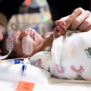
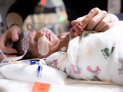
The callers pretended to be well-meaning parents who were trying to safely dispose of unneeded antibiotics and opioid-based prescription painkillers after their child’s surgery. Fewer than half of the California pharmacies they called provided correct prescription drug disposal details, a percentage that dropped sharply if the “secret shoppers” made their call on a weekend, according to a brief research report published online Dec. 31, 2019, in Annals of Internal Medicine.
“The Food and Drug Administration advises consumers about how to safely dispose of unneeded medicines and, because pharmacists can play an integral role in this conversation, the American Pharmacists Association says prescription medication disposal should follow FDA guidelines,” says Rachel E. Selekman, M.D., MAS, a pediatric urologist at Children’s National Hospital and the study’s first author. “We found very few California pharmacies permitted take-back of unneeded medications. There was also a striking difference in the accuracy and completeness of drug disposal information depending on whether they answered the call on a weekday or a weekend. That suggests room for improvement,” Dr. Selekman says.
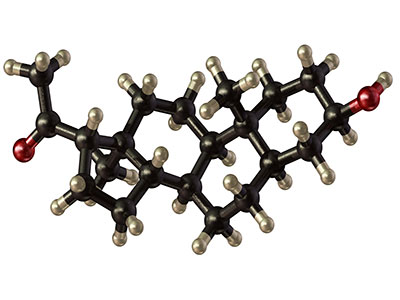
The multi-institutional research team, led by Primary Investigator and senior author Hillary L. Copp, M.D., MS, at University of California, San Francisco, identified licensed pharmacies located in urban and rural settings in California. That state that accounts for 10% of all U.S. pharmacies. They wrote a script that guided four male and two female “secret shoppers” to ask about what to do about leftover antibiotics (sulfamethoxazole-trimethoprim tablets) and a liquid opioid-based painkiller (hydrocodone-acetaminophen). From late-February to late-April 2018, they called 898 pharmacies from 8 a.m. to 8 p.m., asking about the correct way to dispose of these medicines.
According to the FDA, consumers should mix most unused medicines with an unappealing substance, like kitty litter, place it in a sealed container and toss the container in the trash. Medicines that can be harmful to others, like opioids, should be flushed down the sink or toilet. Many pharmacies have programs or kiosks to handle unused prescription medicines.
Of the pharmacies surveyed in California:
- 47% provided correct information about disposing of antibiotics
- 29% provided correct information about how to dispose of both antibiotics and opioids
- 19% provided correct information about how to dispose of opioids
- 49% provided correct antibiotic disposal information and 20% provided correct opioid disposal information on weekday calls
- 15% provided correct antibiotic disposal information and 7% provided correct opioid disposal information on weekend calls
Asked specifically about drug take-back programs, just 11% said their pharmacy had one that could be used to dispose of antibiotics or opioids.
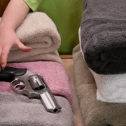
“Unused prescription medications can be misused by others and can result in accidental childhood poisonings,” Dr. Selekman adds. “The bottom line is that we often talk about how to address the problem of too many unused medications lingering in homes. There are many reasons this is a problem, but part of the problem is nobody knows what to do if they have too many prescription medicines. Because of this research, we have discovered that pharmacies don’t uniformly provide accurate information to our patients. Patients, families and health care professionals who advise families should work together to help improve and expand safe disposal options for these powerful medications.”
In addition to Drs. Selekman and Copp, the research team includes co-authors Thomas W. Gaither, M.D., MAS, Zachary Kornberg, BA, and Aron Liaw, M.D., all of whom were at the University of California, San Francisco, School of Medicine, Division of Pediatric Urology at the time the study was performed.