Extracting actionable research data faster, with fewer hassles

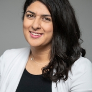
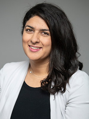
Mihailo Kaplarevic, Ph.D., the newly minted Chief Research Information Officer at Children’s National Hospital and Bioinformatics Division Chief at Children’s National Research Institute, will provide computational support, advice, informational guidance, expertise in big data and data analyses for researchers and clinicians.
Kaplarevic’s new job is much like the role he played most recently at the National Heart, Lung and Blood Institute (NHLBI), assembling a team of researchers and scientists skilled in computing and statistical analyses to assist as in-house experts for other researchers and scientists.
NHLBI was the first institute within the National Institutes of Health (NIH) family to set up a scientific information office. During his tenure, a half-dozen other NIH institutions followed, setting up the same entity to help bridge the enormous gap between basic and clinical science and everything related to IT.
“There is a difference compared with traditional IT support at Children’s National – which will remain in place and still do the same sort of things they have been doing so far,” he says of The Bear Institute for Health Innovation. “The difference is this office has experience in research because every single one of us was a researcher at a certain point in our career: We are published. We applied for grants. We lived the life of a typical scientist. On top of that, we’re coming from the computational world. That helps us bridge the gaps between research and clinical worlds and IT.”
Ultimately, he aims to foster groundbreaking science by recognizing the potential to enhance research projects by bringing expertise acquired over his career and powerful computing tools to help teams achieve their goals in a less expensive and more efficient way.
“I have lived the life of a typical scientist. I know exactly how painful and frustrating it can be to want to do something quickly and efficiently but be slowed by technological barriers,” he adds.
As just one example, his office will design the high-performance computing cluster for the hospital to help teams extract more useful clinical and research data with fewer headaches.
Right now, the hospital has three independent clinical systems storing patient data; all serve a different purpose. (And there are also a couple of research information systems, also used for different purposes.) Since databases are his expertise, he will be involved in consolidating data resources, finding the best way to infuse the project with the bigger-picture mission – especially for translational science – and creating meaningful, actionable reports.
“It’s not only about running fewer queries,” he explains. “One needs to know how to design the right question. One needs to know how to design that question in a way that the systems could understand. And, once you get the data back, it’s a big set of things that you need to further filter and carefully shape. Only then will you get the essence that has clinical or scientific value. It’s a long process.”
As he was introduced during a Children’s National Research Institute faculty meeting in late-September 2019, Kaplarevic joked that his move away from pure computer science into a health care and clinical research domain was triggered by his parents: “When my mom would introduce me, she would say ‘My son is a doctor, but not the kind of doctor who helps other people.’ ”
Some of that know-how will play out by applying tools and methodology to analyze big data to pluck out the wheat (useful data) from the chaff in an efficient and useful way. On projects that involve leveraging cloud computing for storing massive amounts of data, it could entail analyzing the data wisely to reduce its size when it comes back from the cloud – when the real storage costs come in. “You can save a lot of money by being smart about how you analyze data,” he says.
While he expects his first few months will be spent getting the lay of the land, understanding research project portfolios, key principal investigators and the pediatric hospital’s biggest users in the computational domain, he has ambitious longer-term goals.
“Three years from now, I would like this institution to say that the researchers are feeling confident that their research is not affected by limitations related to computer science in general. I would like this place to become a very attractive environment for up-and-coming researchers as well as for established researchers because we are offering cutting-edge technological efficiencies; we are following the trends; we are a secure place; and we foster science in the best possible way by making computational services accessible, affordable and reliable.”