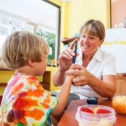
Public Health

To understand the preterm brain, start with the fetal brain

Nickie Andescavage

“My best advice to future clinician-scientists is to stay curious and open-minded; I doubt I could have predicted my current research interest or described the path between the study of early oligodendrocyte maturation to in vivo placental development, but each experience along the way – both academic and clinical – has led me to where I am today,” Nickie Andescavage, M.D., writes.

Too often, medical institutions erect an artificial boundary between caring for the developing fetus inside the womb and caring for the newborn whose critical brain development continues outside the womb.

“To improve neonatal outcomes, we must transform our current clinical paradigms to begin treatment in the intrauterine period and continue care through the perinatal transition through strong collaborations with obstetricians and fetal-medicine specialists,” writes Nickie Andescavage, M.D., an attending in Neonatal-Perinatal Medicine at Children’s National.

Dr. Andescavage’s commentary was published online March 25, 2019, in Pediatrics Research and accompanies recently published Children’s research about differences in placental development in the setting of placental insufficiency. Her commentary is part of a new effort by Nature Publishing Group to spotlight research contributions from early career investigators.

The placenta, an organ shared by a pregnant woman and the developing fetus, plays a critical but underappreciated role in the infant’s overall health. Under the mentorship of Catherine Limperopoulos, Ph.D., director of MRI Research of the Developing Brain, and Adré J. du Plessis, M.B.Ch.B., MPH, chief of the Division of Fetal and Transitional Medicine, Dr. Andescavage works with interdisciplinary research teams at Children’s National to help expand that evidence base.

While attending Cornell University as an undergraduate, Dr. Andescavage had an early interest in neuroscience and neurobehavior. As she continued her education by attending medical school at Columbia University, she corroborated an early instinct to work in pediatrics.

It wasn’t until the New Jersey native began pediatric residency at Children’s National that those complementary interests coalesced into a focus on brain autoregulation and autonomic function in full-term and preterm infants and imaging the brains of both groups. In normal, healthy babies the autonomic nervous system regulates heart rate, blood pressure, digestion, breathing and other involuntary activities. When these essential controls go awry, babies can struggle to survive and thrive.

“My best advice to future clinician-scientists is to stay curious and open-minded; I doubt I could have predicted my current research interest or described the path between the study of early oligodendrocyte maturation to in vivo placental development, but each experience along the way – both academic and clinical – has led me to where I am today,” Dr. Andescavage writes in the commentary.

Tamp down food allergy anxieties with this quiz


Asthma is associated with severe obstructive sleep apnea in children

Pulmonologists have often observed a link between asthma and the need for continuous positive airway pressure treatment (CPAP) among children with severe obstructive sleep apnea syndrome (OSAS).

Now, research published in the March 2019 issue of the journal Pediatric Pulmonology confirms the correlation.

Four-hundred eligible children with severe OSAS were included in a randomized, controlled study that took place at Children’s National Health System between September 2015 and June 2017. The mean age among study participants, ages 0 to 20, was 7.

Out of the 400 severe OSAS study participants, 113 children, about one-third, had asthma. Those with asthma were 29% more likely to require CPAP, compared to 14% of study participants without asthma. This association was independent of demographics, OSAS severity, obesity and a history of adenotonsillectomy, an operation to remove the tonsils.

“This is the first randomized, controlled study to test the association between asthma and CPAP among children with severe sleep apnea,” says Gustavo Nino, M.D., a corresponding study author, a pediatric pulmonologist and the director of sleep medicine at Children’s National Health System. “We’ve seen similar patterns in adults, but we needed to confirm the link in children to provide preventive screenings and personalized treatment.”

Dr. Nino mentions the goal now is to detect symptoms earlier, whether this occurs at an annual wellness exam with a pediatrician or at the first visit with a sleep medicine specialist.

“The next step for our research team, or for others interested in this topic, is to explore how these factors influence each other,” adds Dr. Nino. “Asthma itself is worse when you sleep. This leads us to wonder if obstructive sleep apnea exacerbates symptoms of asthma. Or could controlling asthma decrease the risk for CPAP therapy among children with severe obstructive sleep apnea?”

Until these questions have answers, Dr. Nino encourages pediatricians and specialists to keep the association in mind, especially since 7 million children nationwide have asthma, including 13,981 children in the District.

Parents should know that children who have severe obstructive sleep apnea and asthma are more likely to need extensive treatment, like CPAP, to maintain a positive flow of air to the nasal passages to keep the airway open.

Managing symptoms of asthma is also something parents can do at home, especially with the onset of spring asthma triggers, such as pollen, dust, dander, mold and smoke.

Kofi Essel, M.D., M.P.H. and Ankoor Shah, M.D., M.B.A., M.P.H., named among 40 Under 40 Leaders in Minority Health

Ankoor Shah and Kofi Essel

Ankoor Shah, M.D., M.B.A., M.P.H., and Kofi Essel, M.D., M.P.H., were named 40 Under 40 Leaders in Minority Health.

Two doctors from Children’s National Health System are among the recipients of the 40 Under 40 Leaders in Minority Health award by the National Minority Quality Forum (NMQF) for 2019. Kofi Essel, M.D., M.P.H., is a pediatrician, Ankoor Shah, M.D., M.B.A., M.P.H., is the medical director of the IMPACT DC Asthma Clinic and also a pediatrician at Children’s National.

Founded in 1998, the NMFQ is dedicated to ensuring that high-risk racial and ethnic populations and communities receive optimal health care. The 40 individuals selected for this award represent the next generation of thought leaders in reducing health disparities.

Dr. Kofi Essel is a pediatrician at the Children’s Health Center Anacostia.  His focus and research has been around health equity, obesity, food insecurity and nutrition.

“Hunger strikes so many of our families,” says Dr. Essel, “In D.C., we were number one in the nation for having the highest rate of food hardship in households with children.”

Dr. Essel is involved with many organizations and initiatives that raise awareness about hunger and how much of an issue it is.  He strives to be a partner for the families that he serves, many of whom are in the fight against obesity, and works alongside them to improve their overall health.

“It’s a huge honor to receive recognition from this national organization,” says Dr. Kofi Essel, “Ultimately, it allows us to have a bit more of a platform to continue to advance some of the great work we’re doing with health disparities.”

Dr. Ankoor Shah is the medical director for IMPACT DC asthma clinic and a pediatrician at the Children’s Health Center at THEARC.  His focus includes improving pediatric population health and reducing child health asthma disparities.

“Through the coordination of the best in class care at Children’s National with amazing on the ground community partners, we have been able to transform the lives of the most at-risk children with asthma” says Dr. Shah.

Dr. Shah collaborates with organizations to improve the outcomes of kids with asthma by targeting intervention in high-risk areas.

“This award is recognition of the great work we’re doing in terms of improving asthma health in high-risk child populations throughout the District of Columbia.”

Both Dr. Essel and Dr. Shah are from Arkansas, attended Emory University and they did their residency together at Children’s National.

Congratulations to these wonderful doctors and leaders for receiving this award.

The 40 Under 40 recipients received their awards at the 2019 NMQF Leadership Summit on Health Disparities and CBC Spring Health Braintrust Gala Dinner on April 9.

Community-based AIDS prevention organization recognizes Children’s National Health System

Thurlow Evans Tibbs, Jr. Award

Credit: Don Bon Photography

The Division of Adolescent and Young Adult Medicine at Children’s National Health System was honored with the Thurlow Evans Tibbs, Jr. Award for outstanding service in HIV prevention by Us Helping Us, People Into Living, a community-based AIDS service organization committed to reducing HIV infection in the African American community on April 2, 2019 in Washington, D.C.

“We are so honored to receive the Thurlow Evans Tibbs, Jr. Award from our colleagues and friends, Us Helping Us, People Into Living,” said Dr. Lawrence D’Angelo, director of the Youth Pride Clinic and Burgess Clinics, a division of Adolescent and Young Adult Medicine at Children’s National. “We have always considered Us Helping Us as an essential partner in this struggle and so it’s ingrained as a part of the community. It has been our honor to work with them, and an equal honor to be recognized in this way by them. We are so grateful and so proud!”

Dr. Lawrence D’Angelo and his team at the Burgess Clinic have cared for over 750 HIV infected youth, many who have gone on to live long and productive lives since the clinic opened its doors in 1988. The clinic started the Washington Area Consortium on HIV infection in Youth (WACHIVY) which later became MetroTeenAIDS Metro TeenAIDS, one of the largest and most successful prevention education programs for youth in the country. The Burgess Clinic is the home of over a dozen NIH and foundation grants that supported the diagnosis and treatment of HIV infected youth, accounting for over $55M in support.

“Receiving the Thurlow Evans Tibbs, Jr. Award from Us Helping Us, People Into Living is an honor beyond anything we could have hoped for,” says Dr. D’Angelo. “Our programs were started at the same time to meet similar needs and for years we have worked together and appreciated each other’s efforts in serving our community. D.C. could not have attained the progress it has made in the struggle against HIV without Us Helping Us, People Into Living and knowing that makes this award all the more special.”

Us Helping Us, People Into Living was incorporated in 1988 as a support group for HIV-positive black gay men.

Genetics 101: Rare diseases aren’t rare

The Rare Disease Institute staff on Rare Disease Day

Children’s National Health System is home to the Rare Disease Institute, the National Organization for Rare Disease’s first Center of Excellence, the largest clinical genetics program in the United States.

With the advent of DNA databanks, informatics, new technology, pediatric consortiums and global partnerships, clinical researchers have never been in a better position to diagnose and treat rare diseases. A rare disease is categorically defined as a condition that affects less than 200,000 people. However, 25 to 30 million Americans, about one in 10, have a rare disease.

Accelerations in genetic research and diagnostic criteria remain one of the most significant accomplishments in medicine, but these breakthroughs invite new challenges: How will researchers provide ongoing care and treatment for patients navigating a rare disease? How can doctors and researchers multiply themselves to ensure everyone has the latest information and resources they need? How can researchers use existing trials to augment other fields? How can we diagnose, catalogue and treat hundreds of new rare diseases each year, while accelerating the research and care of 7,000 existing rare conditions?

If these questions intrigue you, excite you and make you want to collaborate with scientific peers, welcome to the field of genetics. A common theme researchers and families talk about is that rare diseases affect a small proportion of the population, but have a huge impact.

On April 10, 1,200 international researchers, lawmakers, scientists and drug developers from 50 countries will meet in Oxon Hill, Md., 10 miles south of Washington, for a three-day summit, the World Orphan Drug Congress USA, to discuss how to unify efforts to enhance and maximize care for rare disease patients.

Here are eight themes to keep in mind:

    1. Rare diseases are chronic diseases. The human genome project has enabled the molecular mapping of 8,000 diseases with genetic underpinnings. Of these diseases, 600 diseases have therapies. A child born with a urea cycle disorder had a 5% chance of surviving the disease 40 years ago. Now the survival rate is 95%. Helping children survive is essential, but we need to think about the best treatments and standards for long-term care.
    2. Rare diseases are expensive. In Western Australia, according to the 2010 Western Australia Population Cohort, rare diseases account for less than 5% of hospital visits but for 10% of hospital costs. Similar data from Cleveland finds one-third of pediatric hospital visits have a genetic link but account for half of hospital costs.
    3. Rare diseases share common links. We’ve diagnosed 7,000 rare diseases but there are more to unravel. For example, breast cancer has over 30 molecular subtypes – some of which turn into rare diseases. By better understanding these molecular pathways, we may be able to inform common fields of medicine.
Marshall Summar's Rare Disease 101 presentation

Dr. Marshall Summar, a medical geneticist, speaks about the future of rare disease research and treatment at a Rare Disease 101 lecture hosted by the Rare Disease Congressional Caucus on Capitol Hill on Feb. 27. To sustain discoveries, Dr. Summar mentions a digital-first, flexible mindset is essential. Standard language and scalable, universal reference structures are required.

  1. Global partnerships create research repositories. Gold-standard research models – double blind, controlled studies with numerous participants – aren’t possible if five people in the world share the same disease. To increase the number of study participants, global partnerships and longitudinal registries are essential.
  2. Standard language helps. To avoid replicating existing research and to help teams quickly reference findings, we need to adopt standardized language to quantify measurements. Researchers from Berlin and Brazil may help inform the etiology of and future treatments for PKU, but they need to manage, store, access and share their collective findings, while remaining flexible.
  3. The science is here. The FDA is approving more drugs for rare diseases than ever before including gene therapy and micro organs, or Rare Diseases-on-chip models. The challenge with treating so many rare diseases isn’t developing new research, but creating therapies and studies to accommodate this patient volume. About 250 rare disease discoveries happen each year. At the current rate, it will take 2,000 years to treat them all.
  4. Progress is here. The Orphan Drug Act fast-tracked approval for rare disease treatments and therapies, and nearly half of all drugs coming in for FDA approval are for rare diseases. However, only 5% of rare diseases have FDA-approved drugs.
  5. We need to replicate geneticists. To provide optimal care, doctors need to standardize education models and use new forms of technology, such as artificial intelligence and deep learning, to share resources faster via patient education portals, resources for families, CME courses and virtual connections with pediatricians or families.

If you live in Washington, D.C., follow the genetics team and consider working with us as we move into a new home, the Children’s National Research and Innovation Campus, in 2020.

Pediatric Surgeon receives ACS/APSA Health Policy Scholarship

Jeffrey Lukish

Jeffrey Lukish, M.D., a pediatric surgeon at Children’s National Health System, has been named a 2019 American College of Surgeons (ACS) and American Pediatric Surgical Association (APSA) Health Policy Scholar for 2019.

The scholarship supports Dr. Lukish’s attendance at the Executive Leadership Program in Health Policy and Management at Brandeis University, which teaches knowledge and skills essential for participating in health care policy and equips health leaders with tools to create innovative and sustainable ways to improve health care service delivery. As a 2019 scholar, he will also provide health policy-related assistance to the ACS and the APSA as requested, and will have opportunities to build relationships with local, state and federal lawmakers.

Dr. Lukish is a nationally recognized expert in advanced minimally invasive surgery in infants and children, as well as pediatric surgical innovation. He has been voted a Baltimore Top Doctor by his peers for five of the last eight years. He holds academic appointments as a professor of surgery from the Uniformed Services University and associate professor of surgery at the George Washington University.

Dr. Lukish is a fellow of the American College of Surgeons and the American Academy of Pediatrics, and member of several prominent professional societies, including the American Pediatric Surgical Society, the Pediatric Cancer Oncology Group and the International Pediatric Endosurgery Group.  He has authored over 100 publications.

$2M NIH grant for treating disease linked to APOL1

Zhe Han

Children’s researcher Zhe Han, Ph.D., has received a $2 million award from the National Institutes of Health (NIH) to study new approaches to treat kidney disease linked to inheriting Apolipoprotein L1 (APOL1) risk alleles. These risk alleles are particularly common among persons of recent African descent, and African Americans are disproportionately affected by the increased risk in kidney disease associated with these risk alleles.

Han, an associate professor in Children’s Center for Genetic Medicine Research, has established a leading research program that uses the fruit fly Drosophila as a model system to study how genetic mutations lead to disease.

Drosophila is a very basic model, but studies in the fly have led to major breakthroughs in understanding fundamental biological processes that underlie health and disease in humans,” Han says. “Since coming to Children’s National five years ago, I have focused a significant part of my research studying particular fly cells called nephrocytes that carry out many of the important roles of human kidney glomeruli, units within the kidney where blood is cleaned. Working together with clinician colleagues here, we have demonstrated that these Drosophila cells can be used to very efficiently study different types of renal disease caused by genetic mutations.”

The APOL1 risk alleles are genetic variants, termed G1 and G2, found almost exclusively in people of African ancestry and can lead to a four-fold higher risk of end-stage kidney disease, the last of five stages of chronic kidney disease. Exactly how inheriting these risk alleles increases the risk of kidney disease remains an unanswered question and the focus of considerable research activity. Han’s laboratory has developed a Drosophila model of APOL1-linked renal disease by producing the G1 and G2 forms of APOL1 specifically in nephrocytes. This led to defects in fly renal cells that strikingly overlap with disease-associated changes in experimental model and human kidney cells expressing APOL1 risk alleles.

The new NIH award will fund large-scale screening and functional testing to identify new treatment targets and new drugs to treat kidney disease linked to APOL1. Using a genetic screening approach, Han’s lab will identify nephrocyte “modifier” genes that interact with APOL1 proteins and counter the toxic effects of risk-associated G1 and G2 variants.

The team also will identify nephrocyte genes that are turned on or off in the presence of APOL1 risk alleles, and confirm that such “downstream” APOL1-regulated genes are similarly affected in experimental model and human kidney cells. The potential of the newly identified “modifier” and “downstream” genes to serve as targets of novel therapeutic interventions will be experimentally tested in fly nephrocytes in vivo and in cultured mammalian kidney cells.

Finally, the Drosophila model will be used as a drug screening platform for in vivo evaluation of positive “hits” from a cell-based APOL1 drug screening study in order to identify compounds that are most effective with the fewest side effects.

“These types of studies can be most efficiently performed in Drosophila,” Han adds.  “They take advantage of the speed and low cost of the fly model system and the amazing array of well-established, sophisticated genetic tools available for the fly. Using this model to elucidate human disease mechanisms and to identify new effective therapies has truly become my research passion.”

ACC.19: A focus on pediatric cardiology

ACC19 attendees from Children's National

Dr. Gerard Martin, center, accepts an award before delivering the 2019 Dan G. McNamara Keynote lecture at ACC.19.

“Innovation meets tradition,” is how many attendees and journalists described the American College of Cardiology’s 68th Scientific Sessions (ACC.19), which took place March 16-18, 2019 in New Orleans, La.

Gerard Martin, M.D., F.A.A.P., F.A.C.C., F.A.H.A., a pediatric cardiologist and the medical director of Global Services at Children’s National, supported this narrative by referencing both themes in his 2019 Dan G. McNamara keynote lecture, entitled “Improved Outcomes in Congenital Heart Disease through Advocacy and Collaboration.” Dr. Martin highlighted advancements in the field of pediatric cardiology that took place over the past 15 years, while touting modern advancements – such as pulse oximetry screenings for critical congenital heart disease – that were a result of physician-led advocacy and collaboration.

Dr. Martin’s message was to continue to invest in research and technology that leads to medical breakthroughs, but to remember the power of partnerships, such as those formed by the National Pediatric Cardiology Quality Improvement Collaborative. These alliances, which generated shared protocols and infrastructure among health systems, improved interstage mortality rates between surgeries for babies born with hypoplastic left heart syndrome.

A dozen cardiologists and clinicians from the Children’s National Heart Institute also participated in CME panel discussions or delivered poster presentations to support future versions of this template, touching on early-stage innovations and multi-institution research collaborations. The themes among Children’s National Heart Institute faculty, presented to a diverse crowd of 12,000-plus professional attendees representing 108 countries, included:

Personalized guidelines:

  • Sarah Clauss, M.D., F.A.C.C., a cardiologist, presented “Unique Pediatric Differences from Adult Cholesterol Guidelines: Lipids and Preventive Cardiology,” before Charles Berul, M.D., division chief of cardiology and co-director of the Children’s National Heart Institute, presented “Unique Pediatric Differences from Adult Guidelines: Arrhythmias in Adults with Congenital Heart Disease,” in a joint symposium with the American Heart Association and the American College of Cardiology.
  • Berul, who specializes in electrophysiology, co-chaired a congenital heart disease pathway session, entitled “Rhythm and Blues: Electrophysiology Progress and Controversies in Congenital Heart Disease,” featuring components of pediatric electrophysiology, including heart block, surgical treatment of arrhythmias and sudden death risk.

Early detection:

  • Anita Krishnan, M.D., associate director of the echocardiography lab, presented “Identifying Socioeconomic and Geographic Barriers to Prenatal Detection of Hypoplastic Left Heart Syndrome and Transposition of the Great Arteries” as a moderated poster in Fetal Cardiology: Quickening Discoveries.
  • Jennifer Romanowicz, M.D., a cardiology fellow, and Russell Cross, M.D., director of cardiac MRI, presented the “Neonatal Supraventricular Tachycardia as a Presentation of Critical Aortic Coarctation” poster in FIT Clinical Decision Making: Congenital Heart Disease 2.
  • Pranava Sinha, M.D., a cardiac surgeon, presented the poster “Neuroprotective Effects of Vitamin D Supplementation in Children with Cyanotic Heart Defects: Insights from a Rodent Hypoxia Model” in Congenital Heart Disease: Therapy 2.

Coordinated care:

  • Ashraf Harahsheh, M.D., F.A.C.C., F.A.A.P., a cardiologist with a focus on hyperlipidemia and preventive cardiology, co-presented an update about BMI quality improvement (Q1) activity from the American College of Cardiology’s Adult Congenital and Pediatric Quality Network – BMI Q1 leadership panel.
  • Niti Dham, M.D., director of the cardio-oncology program, and Deepa Mokshagundam, M.D., cardiology fellow, presented the poster “Cardiac Changes in Pediatric Cancer Survivors” in Heart Failure and Cardiomyopathies: Clinical 3.
  • Nancy Klein, B.S.N., R.N., C.P.N., clinical program coordinator of the Washington Adult Congenital Heart program at Children’s National, presented the poster “Improving Completion of Advanced Directives in Adults with Congenital Heart Disease” in Risks and Rewards in Adult Congenital Heart Disease.

Innovation:

  • Jai Nahar, M.D., a cardiologist, moderated “Future Hub: Augmented Cardiovascular Practitioner: Giving Doctors and Patients a New Voice.” The session focused on technical aspects of artificial intelligence, such as language processing and conversational artificial intelligence, as well as how applications are used in patient-physician interactions.
  • Nahar also participated in a key event on the Heart-to-Heart stage, entitled “Rise of Intelligent Machines: The Potential of Artificial Intelligence in Cardiovascular Care.”

“While I enjoyed the significant representation of Children’s National faculty at the meeting and all of the presentations this year, one research finding that I found particularly compelling was Dr. Krishnan’s poster about geographical disparities in detecting congenital heart disease,” says Dr. Berul. “Her research finds obstetricians providing care to women in the lowest quartile of socioeconomic areas were twice as likely to miss a diagnosis for a critical congenital heart defect during a fetal ultrasound, compared to obstetricians providing care for women in the highest quartiles.”

Dr. Krishnan’s study was the collaborative effort of 21 centers in the United States and Canada, and investigated how socioeconomic and geographic factors affect prenatal detection of hypoplastic left heart syndrome and transposition of the great arteries.

“We studied over 1,800 patients, and chose these diseases because they require early stabilization by a specialized team at a tertiary care center,” says Dr. Krishnan, who led the research in conjunction with the Fetal Heart Society Research Collaborative. “We hope that by understanding what the barriers are, we can reduce disparities in care through education and community-based outreach.”

NUP160 genetic mutation linked to steroid-resistant nephrotic syndrome

DNA strands on teal background

Mutations in the NUP160 gene, which encodes one protein component of the nuclear pore complex nucleoporin 160 kD, are implicated in steroid-resistant nephrotic syndrome, an international team reports March 25, 2019, in the Journal of the American Society of Nephrology. Mutations in this gene have not been associated with steroid-resistant nephrotic syndrome previously.

“Our findings indicate that NUP160 should be included in the gene panel used to diagnose steroid-resistant nephrotic syndrome to identify additional patients with homozygous or compound-heterozygous NUP160 mutations,” says Zhe Han, Ph.D., an associate professor in the Center for Genetic Medicine Research at Children’s National and the study’s senior author.

The kidneys filter blood and ferry waste out of the body via urine. Nephrotic syndrome is a kidney disease caused by disruption of the glomerular filtration barrier, permitting a significant amount of protein to leak into the urine. While some types of nephrotic syndrome can be treated with steroids, the form of the disease that is triggered by genetic mutations does not respond to steroids.

The patient covered in the JASN article had experienced persistently high levels of protein in the urine (proteinuria) from the time she was 7. By age 10, she was admitted to a Shanghai hospital and underwent her first renal biopsy, which showed some kidney damage. Three years later, she had a second renal biopsy showing more pronounced kidney disease. Treatment with the steroid prednisone; cyclophosphamide, a chemotherapy drug; and tripterygium wilfordii glycoside, a traditional therapy, all failed. By age 15, the girl’s condition had worsened and she had end stage renal disease, the last of five stages of chronic kidney disease.

An older brother and older sister had steroid-resistant nephrotic syndrome as well and both died from end stage kidney disease before reaching 17. When she was 16, the girl was able to receive a kidney transplant that saved her life.

Han learned about the family while presenting research findings in China. An attendee of his session said that he suspected an unknown mutation might be responsible for steroid-resistant nephrotic syndrome in this family, and he invited Han to work in collaboration to solve the genetic mystery.

By conducting whole exome sequencing of surviving family members, the research team found that the mother and father each carry one mutated copy of NUP160 and one good copy. Their children inherited one mutated copy from either parent, the variant E803K from the father and the variant R1173X, which causes truncated proteins, from the mother. The woman (now 29) did not have any mutations in genes known to be associated with steroid-resistant nephrotic syndrome.

Some 50 different genes that serve vital roles – including encoding components of the slit diaphragm, actin cytoskeleton proteins and nucleoporins, building blocks of the nuclear pore complex – can trigger steroid-resistant nephrotic syndrome when mutated.

With dozens of possible suspects, they narrowed the list to six variant genes by analyzing minor allele frequency, mutation type, clinical characteristics and other factors.

The NUP160 gene is highly conserved from flies to humans. To prove that NUP160 was the true culprit, Dr. Han’s group silenced the Nup160 gene in nephrocytes, the filtration kidney cells in flies. Nephrocytes share molecular, cellular, structural and functional similarities with human podocytes. Without Nup160, nephrocytes had reduced nuclear volume, nuclear pore complex components were dispersed and nuclear lamin localization was irregular. Adult flies with silenced Nup160 lacked nephrocytes entirely and lived dramatically shorter lifespans.

Significantly, the dramatic structural and functional defects caused by silencing of fly Nup160 gene in nephrocytes could be completely rescued by expressing the wild-type human NUP160 gene, but not by expressing the human NUP160 gene carrying the E803K or R1173X mutation identified from the girl’s  family.

“This study identified new genetic mutations that could lead to steroid-resistant nephrotic syndrome,” Han notes. “In addition, it demonstrates a highly efficient Drosophila-based disease variant functional study system. We call it the ‘Gene Replacement’ system since it replaces a fly gene with a human gene. By comparing the function of the wild-type human gene versus mutant alleles from patients, we could determine exactly how a specific mutation affects the function of a human gene in the context of relevant tissues or cell types. Because of the low cost and high efficiency of the Drosophila system, we can quickly provide much-needed functional data for novel disease-causing genetic variants using this approach.”

In addition to Han, Children’s co-authors include Co-Lead Author Feng Zhao, Co-Lead Author Jun-yi Zhu, Adam Richman, Yulong Fu and Wen Huang, all of the Center for Genetic Medicine Research; Nan Chen and Xiaoxia Pan, Shanghai Jiaotong University School of Medicine; and Cuili Yi, Xiaohua Ding, Si Wang, Ping Wang, Xiaojing Nie, Jun Huang, Yonghui Yang and Zihua Yu, all of Fuzhou Dongfang Hospital.

Financial support for research described in this post was provided by the Nature Science Foundation of Fujian Province of China, under grant 2015J01407; National Nature Science Foundation of China, under grant 81270766; Key Project of Social Development of Fujian Province of China, under grant 2013Y0072; and the National Institutes of Health, under grants DK098410 and HL134940.

African American stakeholders help to perfect the APOLLO study

Nichole Jefferson and Patrick Gee

Nichole Jefferson and Patrick O. Gee

African Americans who either donated a kidney, received a kidney donation, are on dialysis awaiting a kidney transplant or have a close relative in one of those categories are helping to perfect a new study that aims to improve outcomes after kidney transplantation.

The study is called APOLLO, short for APOL1 Long-Term Kidney Transplantation Outcomes Network. Soon, the observational study will begin to enroll people who access transplant centers around the nation to genotype deceased and living African American kidney donors and transplant recipients to assess whether they carry a high-risk APOL1 gene variant.

The study’s Community Advisory Council – African American stakeholders who know the ins and outs of kidney donation, transplantation and dialysis because they’ve either given or  received an organ or are awaiting transplant – are opening the eyes of researchers about the unique views of patients and families.

Already, they’ve sensitized researchers that patients may not be at the same academic level as their clinicians, underscoring the importance of informed consent language that is understandable, approachable and respectful so people aren’t overwhelmed. They have encouraged the use of images and color to explain the apolipoprotein L1 (APOL1) gene. The APOL1 gene is found almost exclusively in people of recent African descent, however only 13 percent of these people carry the high-risk APOL1 variant that might cause kidney problems.

One issue arose early, during one of the group’s first monthly meetings, as they discussed when to tell patients and living donors about the APOLLO study. Someone suggested the day of the transplant.

“The Community Advisory Council told them that would not be appropriate. These conversations should occur well before the day of the transplant,” recalls Nichole Jefferson.

“The person is all ready to give a kidney. If you’re told the day of transplant ‘we’re going to include you in this study,’ that could possibly stop them from giving the organ,” Jefferson says. “We still remember the Tuskegee experiments. We still remember Henrietta Lacks. That is what we are trying to avoid.”

Patrick O. Gee, Ph.D., JLC, another Community Advisory Council member, adds that it’s important to consider “the mental state of the patient and the donor. As a patient, you know you are able to endure a five- to eight-hour surgery. The donor is the recipient’s hero. As the donor, you want to do what is right. But if you get this information; it’s going to cause doubt.”

Gee received his kidney transplant on April 21, 2017, and spent 33 days in the hospital undergoing four surgeries. His new kidney took 47 days to wake up, which he describes as a “very interesting journey.” Jefferson received her first transplant on June 12, 2008. Because that kidney is in failure, she is on the wait list for a new kidney.

“All I’ve ever known before APOLLO was diabetes and cardiovascular issues. Nobody had ever talked about genetics,” Gee adds. “When I tell people, I tread very light. I try to stay in my lane and not to come off as a researcher or a scientist. I just find out information and just share it with them.”

As he spoke during a church function, people began to search for information on their smart phones. He jotted down questions “above his pay grade” to refer to the study’s principal investigator. “When you start talking about genetics and a mutated gene, people really want to find out. That was probably one of the best things I liked about this committee: It allows you to learn, so you can pass it on.”

Jefferson’s encounters are more unstructured, informing people who she meets about her situation and kidney disease. When she traveled from her Des Moines, Iowa, home to Nebraska for a transplant evaluation, the nephrologist there was not aware of the APOL1 gene.

And during a meeting at the Mayo Clinic with a possible living donor, she asked if they would test for the APOL1 gene. “They stopped, looked at me and asked: ‘How do you know about that gene?’ Well, I’m a black woman with kidney failure.”

Patrick O. Gee received his kidney transplant on April 21, 2017, and spent 33 days in the hospital undergoing four surgeries. His new kidney took 47 days to wake up, which he describes as a “very interesting journey.”

About 100,000 U.S. children and adults await a kidney transplant. APOLLO study researchers believe that clarifying the role that the APOL1 gene plays in kidney-transplant failure could lead to fewer discarded kidneys, which could boost the number of available kidneys for patients awaiting transplant.

Gee advocates for other patients and families to volunteer to join the APOLLO Community Advisory Council. He’s still impressed that during the very first in-person gathering, all researchers were asked to leave the table. Only patients and families remained.

“They wanted to hear our voices. You rarely find that level of patient engagement. Normally, you sit there and listen to conversations that are over your head. They have definitely kept us engaged,” he says. “We have spoken the truth, and Dr. Kimmel is forever saying ‘who would want to listen to me about a genotype that doesn’t affect me? We want to hear your voice.’ ”

(Paul L. Kimmel, M.D., MACP, a program director at the National Institute of Diabetes and Digestive and Kidney Diseases, is one of the people overseeing the APOLLO study.)

Jefferson encourages other people personally impacted by kidney disease to participate in the APOLLO study.

“Something Dr. Kimmel always says is ‘You’re in the room.’ We’re in the room while it’s happening. It’s a line from Hamilton. That’s a good feeling,” she says. “I knew right off, these are not necessarily improvements I will see in my lifetime. I am OK with that. With kidney disease, we have not had advances in a long time. As long as my descendants don’t have to go through the same things I have gone through, I figure I have done my part. I have done my job.”

Kurt Newman, M.D., shares journey as a pediatric surgeon in TEDx Talk

Kurt Newman, M.D., president and chief executive officer of Children’s National, shares his poignant journey as a pediatric surgeon, offering a new perspective for approaching the most chronic and debilitating health conditions. In this independently-organized TEDx event, Dr. Newman also shares his passion for Children’s National and the need to increase pediatric innovations in medicine.

Coming together as a team for the good of the baby

Robin Steinhorn in the NICU

Children’s National has a new program to care for children who have severe bronchopulmonary dysplasia, a serious complication of preterm birth.

Around the 1-year-old’s crib is a tight circle of smiling adults, and at the foot of his bed is a menagerie of plush animals, each a different color and texture and shape to spark his curiosity and sharpen his intellect.

Gone are the days a newborn with extremely complex medical needs like Elijah would transfer from the neonatal intensive care unit (NICU) to the pediatric intensive care unit and transition through a couple of other hospital units by the time he was discharged. Gone are the days when he’d see a variety of new physician faces at every stop. And gone are the days he’d be confined to his room, divorced from the sights and sounds and scents of the outside world, stimulation that helps little baby’s neural networks grow stronger.

Children’s National has a new program designed to meet the unique needs of children like Elijah who have severe bronchopulmonary dysplasia (BPD), a common complication of preterm birth.

“It’s more forward-thinking – and I mean thinking for the future of each individual baby, and it’s allowing the baby to have one team and one location to take advantage of a deep knowledge of and relationship with that baby and family,” says Robin Steinhorn, M.D. Dr. Steinhorn is senior vice president of the Center for Hospital-Based Specialties and one of Children’s multidisciplinary team members who visited Elijah’s bed twice weekly during his lengthy hospitalization and who continues to see him regularly during outpatient visits.

“The pulmonologist, the neonatologist, the respiratory therapist, the physical therapist, the dietitian, the cardiologist – we all come as a team to work together for the good of the baby,” Dr. Steinhorn adds. “We stick with these babies through thick and thin. We will stick with that baby with this team and this location until they are ready to go home – and beyond.”

BPD, a serious lung condition, mostly affects extremely low birthweight preterm babies whose lungs were designed to continue developing inside the womb until the pregnancy reaches full term. Often born months before their due dates, these extremely vulnerable newborns have immature organs, including the lungs, which are not ready for the task of breathing air. Children’s program targets infants who experience respiratory failure from BPD. The respiratory support required for these infants ranges from oxygen delivered through a nasal cannula to mechanical ventilators.

Robin Steinhorn and Colleague

“It’s more forward-thinking – and I mean thinking for the future of each individual baby, and it’s allowing the baby to have one team and one location to take advantage of a deep knowledge of and relationship with that baby and family,” says Robin Steinhorn, M.D.

About 1 percent of all preterm births are extremely low birthweight, or less than 1,500 grams. Within that group, up to 40 percent will develop BPD. While they represent a small percentage of overall births, these very sick babies need comprehensive, focused care for the first few years of their lives. And some infants with severe BPD also have pulmonary hypertension which, at Children’s National, is co-managed by cardiology and pulmonary specialists.

Children’s BPD team not only focuses on the child’s survival and medical care, they focus on the neurodevelopmental and social care that a baby needs to thrive. From enhanced nutrition to occupational and physical therapy to a regular sleep cycle, the goal is to help these babies achieve their full potential.

“These babies are at tremendous risk for long-term developmental issues. Everything we do is geared to alleviate that,” adds John T. Berger III, M.D., director of Children’s Pulmonary Hypertension Program.

“Our NICU care is more focused, comprehensive and consistent,” agrees Mariam Said, M.D., a neonatologist on the team. “We’re also optimizing the timing of care and diagnostic testing that will directly impact health outcomes.”

Leaving no detail overlooked, the team also ensures that infants have age-appropriate developmental stimuli, like toys, and push for early mobility by getting children up and out of bed and into a chair or riding in a wagon.

“The standard approach is to keep the baby in a room with limited physical or occupational therapy and a lack of appropriate stimulation,” says Geovanny Perez, M.D., a pulmonologist on the team. “A normal baby interacts with their environment inside the home and outside the home. We aim to mimic that within the hospital environment.”

Dr. Steinhorn, who had long dreamed of creating this comprehensive team care approach adds that “it’s been so gratifying to see it adopted and embraced so quickly by Children’s NICU caregivers.”

Prescription for a healthy heart: pediatric-driven partnerships

Dr. Martin and a patient share a smile after a visit at Children’s National Health System.

For pediatric cardiologists, February, National Heart Month, is a special time. We share health tips in the hospital and talk about heart health with those looking for advice, especially with patients and families impacted by congenital heart disease (CHD). It’s also a time to look back at what’s worked well in the field, while accelerating advancements for CHD treatment.

To start, congenital heart disease, a structural abnormality of the heart or of the blood vessels surrounding it, is the most common birth defect and occurs in about one in every 100 live births, affecting 40,000 babies born in the U.S. each year. One million children and 1.4 million adults in the U.S. have CHD. Over the past 15 years, pediatric cardiologists have cut mortality rates for CHD in half. Gratefully, now instead of saving children’s lives, the emphasis is on improving them. The catalyst for this paradigm shift isn’t simply due to a medical breakthrough, but is also the result of collaboration and advocacy.

Pediatric cardiologists worked together with other stakeholders – nurses, neonatologists, parents, state and federal agencies – to implement newborn screening methods in hospitals, with the introduction pulse oximetry screenings for critical congenital heart defects (CCHD). The screening, which measures blood oxygen levels in newborns, focuses on screening babies for CCHD before they leave the hospital. The concept and a national protocol for screening began with a small project in 2002, was endorsed by medical associations by 2012 and required by all states in 2018. The impact of CCHD screening of newborns is remarkable. Data published in JAMA showed a 33 percent reduction in CCHD infant deaths associated with states that required CCHD screening.

The pulse oximetry screening’s impact on the number of lives saved goes beyond identifying newborns with CCHD. Worldwide, though the detection of secondary conditions, such as hypothermia, pneumonia, and sepsis, the pulse oximetry screening is estimated to save roughly 772,000 lives by 2030.

In addition to newborn screening recommendations for CCHD, a group of cardiologists, including myself, worked for the Joint Council on Congenital Heart Disease (JCCHD) to form and support the National Pediatric Cardiology Quality Improvement Collaborative (NPC-QIC). We developed measures to see how we could improve survival rates between surgeries for infants born with hypoplastic left heart syndrome (HLHS), one of the most common and severe forms of CCHD.

Babies born with HLHS require two heart surgeries within the baby’s first six months. Babies that survived the first operation had a significant mortality rate (15 percent) and frequent growth failure, while waiting for the second operation. Our focused aims were to both decrease the death rate and improve growth in these children. We analyzed data from medical centers, utilized quality improvement principals from the Institute for Health Care Improvement, talked with doctors and families, and invited teams from across the U.S. to partner with us to put quality and safety measures into place.

We emphasized the following points:

  1. Clear communication. Parents leaving the hospital received consistent messages about CHD, the type of surgery their baby had, next steps and how to care for their child at home.
  2. Improved nutrient intake. Parents received clear guidelines about how many calories babies needed to consume, were asked to weigh their baby each day, and taught how to augment feeding.
  3. Warning signs.Parents received a list of typical infant behaviors and HLHS red flags to watch out for, such as if a baby isn’t gaining a certain amount of weight. They received monitors to measure oxygen saturation levels at home. If oxygen saturation dropped significantly or if parents noticed a problem, they called their doctor immediately.

The implementation of these procedures reduced interstage mortality rates and the number of growth failures for HLHS patients. In 2008, six centers participated in the NPC-QIC pilot. By 2018, 65 medical centers in the U.S. and Canada used these methods. Similar to the pulse oximetry screening guidelines, this new method wasn’t the result of a medical breakthrough, but the result of shared learning and shared infrastructure.

Now, we’re referring more adult congenital heart patients to board-certified adult congenital heart disease (ACHD) specialists, a better fit than internists or pediatric cardiologists. Adults with congenital heart defects should have their heart examined at least once by a specialist and those with complex needs should meet with a specialist at least every two years. More than 300 board-certified ACHD specialists practice in the U.S. and the field is growing. The third ACHD board exam takes place this year.

Over the next few decades, I hope we’ll make even more progress with understanding, diagnosing and treating CHD.

Emerging research examines genetic clues for congenital heart defects, which were once thought to account for 8 percent of cases and may now account for 30 percent of conditions. We’re working with neurologists to examine the timing and pathway of potential oxygen inefficiencies that occur as the brain develops in utero, infancy, and after neonatal surgery. We’ve come a long way, but we continue looking at new frontiers and for innovative solutions.

Fortunately, as cardiologists, we’re good at fixing problems. We work with surgeons and medical teams to repair holes in hearts, or replace them, and reroute blood from an underdeveloped left ventricle to improve circulation. For almost every heart defect, we have evidence-based solutions. However, to continue to help children worldwide, it’s imperative that we don’t forget about what works well: good science, tracking data, sharing best practices, active listening, transparency and constant collaboration.

Gerard Martin, M.D., F.A.A.P., F.A.C.C., F.A.H.A., is a cardiologist and the medical director of global services at Children’s National Health System. Dr. Martin has practiced pediatric cardiology for 34 years and is the Dan G. McNamara keynote speaker at the American College of Cardiology’s 2019 Scientific Sessions. Follow Dr. Martin on Twitter @Gerard_MD.

This article first appeared on KevinMD.com.

$2 million NIH grant to study nephrotic syndrome

Zhe Han lab 2018

A Children’s researcher has received a $2 million grant from the National Institutes of Health (NIH) to study nephrotic syndrome in Drosophila, a basic model system that has revealed groundbreaking insights into human health. The award for Zhe Han, Ph.D., an associate professor in Children’s Center for Genetic Medicine Research, is believed to be the first ever NIH Research Project grant (R01)  to investigate glomerular kidney disease using Drosophila. Nephrotic syndrome is mostly caused by damage of glomeruli, so it is equivalent to glomerular kidney disease.

“Children’s National leads the world in using Drosophila to model human kidney diseases,” Han says.

In order to qualify for the five-year funding renewal, Han’s lab needed to successfully accomplish the aims of its first five years of NIH funding.  During the first phase of funding, Han established that nephrocytes in Drosophila serve the same functions as glomeruli in humans, and his lab created a series of fly models that are relevant for human glomerular disease.

“Some 85 percent of the genes known to be involved in nephrotic syndrome are conserved from the fly to humans. They play similar roles in the nephrocyte as they play in the podocytes in human kidneys,” he adds.

Pediatric nephrotic syndrome is a constellation of symptoms that indicate when children’s kidneys are damaged, especially the glomeruli, units within the kidney that filter blood. Babies as young as 1 year old can suffer proteinuria, which is characterized by too much protein being released from the blood into the urine.

“It’s a serious disease and can be triggered by environmental factors, taking certain prescription medicines or inflammation, among other factors.  Right now, that type of nephrotic syndrome is mainly treated by steroids, and the steroid treatment works in many cases,” he says.

However, steroid-resistant nephrotic syndrome occurs primarily due to genetic mutations that affect the kidney’s filtration system: These filters are either broken or the protein reabsorption mechanism is disrupted.

“When genetics is to blame, we cannot turn to steroids. Right now there is no treatment. And many of these children are too young to be considered for a kidney transplant,” he adds. “We have to understand exactly which genetic mutation caused the disease in order to develop a targeted treatment.”

With the new funding, Han will examine a large array of genetic mutations that cause nephrotic syndrome. He’s focusing his efforts on genes involved in the cytoskeleton, a network of filaments and tubules in the cytoplasm of living cells that help them to maintain shape and carry out important functions.

“Right now, we don’t really understand the cytoskeleton of podocytes – highly specialized cells that wrap around the capillaries of the glomerulus – because podocytes are difficult to access. To change a gene requires time and considerable effort in other experimental models. However, changing genes in Drosophila is very easy, quick and inexpensive. We can examine hundreds of genes involving the cytoskeleton and see how changing those genes affect kidney cell function,” he says.

Han’s lab already found that Coenzyme Q10, one of the best-selling nutrient supplements to support heart health also could be beneficial for kidney health. For the cytoskeleton, he has a different targeted medicine in mind to determine whether Rho inhibitors also could be beneficial for kidney health for patients with certain genetic mutations affecting their podocyte cytoskeleton.

“One particular aim of our research is to use the same strategy as we employed for the Coq2 gene to generate a personalized fly model for patients with cytoskeleton gene mutations and test potential target drugs, such as Rho inhibitors.” Han added. “As far as I understand, this is where the future of medicine is headed.”

Test your knowledge of APOL1’s role in kidney health

$3 million NIH grant to study APOL1 and HIV synergy

Zhe Han

Zhe Han, Ph.D., (pictured) and Patricio E. Ray, M.D., have received a $3 million, five-year grant from the National Institutes of Health to study the mechanisms behind APOL1 and HIV nephropathies in children, using a combination of Drosophila models, cultured human podocytes and a preclinical model.

Two Children’s researchers have received a $3 million, five-year grant from the National Institutes of Health (NIH) to study the mechanisms of APOL1 and HIV nephropathies in children, using a combination of Drosophila models, cultured human podocytes and a preclinical model.

The APOL1 genetic variants G1 and G2, found almost exclusively in people of African ancestry, lead to a four-fold higher risk of end-stage kidney disease. HIV infection alone also increases the risk of kidney disease but not significantly. However, HIV-positive people who also carry the APOL1 risk alleles G1 or G2 are about 30 times more likely to develop HIV-nephropathy (HIVAN) and chronic kidney disease.

For more than 25 years, Children’s pediatric nephrology program has studied HIV/renal diseases and recently developed Drosophila APOL1-G0 and G1 transgenic lines. That pioneering research suggests that HIV-1 acts as a “second hit,” precipitating HIV-renal disease in children by infecting podocytes through a mechanism that increases expression of the APOL1-RA beyond toxic thresholds.

With this new infusion of NIH funding, labs led by Zhe Han, Ph.D., and Patricio E. Ray, M.D., will determine the phenotype of Drosophila Tg lines that express APOL1-G0/G1/G2 and four HIV genes in nephrocytes to assess how they affect structure and function. The teams also will determine whether APOL1-RA precipitates the death of nephrocytes expressing HIV genes by affecting autophagic flux.

“Our work will close a critical gap in understanding about how HIV-1 interacts with the APOL1 risk variants in renal cells to trigger chronic kidney disease, and we will develop the first APOL1/HIV transgenic fly model to explore these genetic interactions in order to screen new drugs to treat these renal diseases,” says Dr. Ray, a Children’s nephrologist.

While a large number of people from Africa have two copies of APOL1 risk alleles, they do not necessarily develop kidney disease. However, if a patient has two copies of APOL1 risk alleles and is HIV-positive, they almost certainly will develop kidney disease.

Patricio Ray

“Our work will close a critical gap in understanding about how HIV-1 interacts with the APOL1 risk variants in renal cells to trigger chronic kidney disease, and we will develop the first APOL1/HIV transgenic fly model to explore these genetic interactions in order to screen new drugs to treat these renal diseases,” says Dr. Ray, a Children’s nephrologist.

“Many teams want to solve the puzzle of how APOL1 and HIV synergize to cause kidney failure,” says Han, associate professor in Children’s Center for Genetic Medicine Research. “We are in the unique position of combining a powerful new kidney disease model system, Drosophila, with long-standing human podocyte and HIVAN studies.”

The team hypothesizes that even as an active HIV infection is held in check by powerful new medicines, preventing the virus from proliferating or infecting new cells, HIV can act as a Trojan horse by making the human cells it infects express HIV protein.

To investigate this hypothesis, the team will create a series of fly models, each expressing a major HIV protein, and will test the genetic interaction between these HIV genes with APOL1. Similar studies also will be performed using cultured human podocytes. Identified synergy will be studied further using biochemical and transcription profile analyses.

Drosophila is a basic model system, but it has been used to make fundamental discoveries, including genetic control of how the body axes is determined and how the biological clock works – two studies that led to Nobel prizes,” Han adds. “I want to use the fly model to do something close to human disease. That is where my research passion lies.”

Marva Moxey-Mims, M.D., named NephCure Kidney International scientific adviser

NephCure Kidney International logo

Marva Moxey-Mims, M.D., chief of the Division of Nephrology at Children’s National Health System, has been named to the Scientific Advisory Board for NephCure Kidney International, a non-profit that aims to accelerate research for rare forms of nephrotic syndrome.

Dr. Moxey-Mims and two additional scientific advisers were selected for their commitment to improving care for patients with glomerular disease, diseases that impair kidney function by attacking blood cleaning units within the kidney.

During her tenure at the National Institute of Diabetes and Digestive and Kidney Diseases at the National Institutes of Health, Dr. Moxey-Mims launched the Chronic Kidney Disease in Children Cohort Study, a prospective study to investigate chronic kidney disease risk factors and outcomes, and helped launch the Cure Glomerulonephropathy Network, a multi-site study with the overarching aim to advance the diagnosis and care of patients with four different glomerular diseases.

“I am truly honored to join this distinguished group of scientific advisers and look forward to leveraging our combined strengths and research knowledge in order to deliver cures for kidney diseases faster,” says Dr. Moxey-Mims.

Vote for Children’s National in STAT Madness

Stat Madness 2019

Children’s National Health System has been selected to compete in STAT Madness for the second consecutive year. Our entry for the bracket-style competition is “Sensitive liquid biopsy platform to detect tumor-released mutated DNA using patient blood and CSF,” a new technique that will allow kids to get better treatment for an aggressive type of pediatric brain tumor.

In 2018, Children’s first-ever STAT Madness entry advanced through five brackets in the national competition and, in the championship round, finished second. That innovation, which enables more timely diagnoses of rare diseases and common genetic disorders, helping to improve kids’ health outcomes around the world, also was among four “Editor’s Pick” finalists, entries that spanned a diverse range of scientific disciplines.

“Children’s National researchers collaboratively work across divisions and departments to ensure that innovations discovered in our laboratories reach clinicians in order to improve patient care,” says Mark Batshaw, M.D., Children’s Executive Vice President, Chief Academic Officer and Physician-in-Chief. “It’s gratifying that Children’s multidisciplinary approach to improving the lives of children with brain tumors has been included in this year’s STAT Madness competition.”

Pediatric brain cancers are the leading cause of cancer-related death in children younger than 14. Children with tumors in their midline brain structures have the worst outcomes, and kids diagnosed with diffuse midline gliomas, including diffuse intrinsic pontine glioma, have a median survival of just 12 months.

“We heard from our clinician colleagues that many kids were coming in and their magnetic resonance imaging (MRI) suggested a particular type of tumor. But it was always problematic to identify the tumor’s molecular subtype,” says Javad Nazarian, Ph.D., MSC, a principal investigator in Children’s Center for Genetic Medicine Research. “Our colleagues wanted a more accurate measure than MRI to find the molecular subtype. That raised the question of whether we could actually look at their blood to determine the tumor subtype.”

Children’s liquid biopsy, which remains at the research phase, starts with a simple blood draw using the same type of needle as is used when people donate blood. When patients with brain tumors provide blood for other laboratory testing, a portion of it is used for the DNA detective work. Just as a criminal leaves behind fingerprints, tumors shed telltale clues in the blood. The Children’s team searches for the histone 3.3K27M (H3K27M), a mutation associated with worse clinical outcomes.

“With liquid biopsy, we were able to detect a few copies of tumor DNA that were hiding behind a million copies of healthy DNA,” Nazarian says. “The blood draw and liquid biopsy complement the MRI. The MRI gives the brain tumor’s ZIP code. Liquid biopsy gives you the demographics within that ZIP code.”

Working with collaborators around the nation, Children’s National continues to refine the technology to improve its accuracy. The multi-institutional team published findings online Oct. 15, 2018, in Clinical Cancer Research.

Even though this research technique is in its infancy, the rapid, cheap and sensitive technology already is being used by people around the globe.

“People around the world are sending blood to us, looking for this particular mutation, H3K27M, ” says Lindsay B. Kilburn, M.D., a Children’s neurooncologist, principal investigator at Children’s National for the Pacific Pediatric Neuro-Oncology Consortium, and study co-author. “In many countries or centers, children do not have access to teams experienced in taking a biopsy of tumors in the brainstem, they can perform a simple blood draw and have that blood processed and analyzed by us. In only a few days, we can provide important molecular information on the tumor subtype previously only available to patients that had undergone a tumor biopsy.”

“With that DNA finding, physicians can make more educated therapeutic decisions, including prescribing medications that could not have been given previously,” Nazarian adds.

The STAT Madness round of 64 brackets opened March 4, 2019, and the championship round voting concludes April 5 at 5 p.m. (EST).

In addition to Nazarian and Dr. Kilburn, study co-authors include Eshini Panditharatna, Madhuri Kambhampati, Heather Gordish-Dressman, Ph.D., Suresh N. Magge, M.D., John S. Myseros, M.D., Eugene I. Hwang, M.D. and Roger J. Packer, M.D., all of Children’s National; Mariam S. Aboian, Nalin Gupta, Soonmee Cha, Michael Prados and Co-Senior Author Sabine Mueller, all of University of California, San Francisco; Cassie Kline, UCSF Benioff Children’s Hospital; John R. Crawford, UC San Diego; Katherine E. Warren, National Cancer Institute; Winnie S. Liang and Michael E. Berens, Translational Genomics Research Institute; and Adam C. Resnick, Children’s Hospital of Philadelphia.

Financial support for the research described in the report was provided by the V Foundation for Cancer Research, Goldwin Foundation, Pediatric Brain Tumor Foundation, Smashing Walnuts Foundation, The Gabriella Miller Kids First Data Resource Center, Zickler Family Foundation, Clinical and Translational Science Institute at Children’s National under award 5UL1TR001876-03, Piedmont Community Foundation, Musella Foundation for Brain Tumor Research, Matthew Larson Foundation, The Lilabean Foundation for Pediatric Brain Cancer Research, The Childhood Brain Tumor Foundation, the National Institutes of Health and American Society of Neuroradiology.

What are the health effects of plastics?

Nikki Gillum Posnack

Nikki Posnack, Ph.D., assistant professor at the Children’s National Heart Institute, is an early-stage investigator examining the impact plastic chemical exposure has on the developing hearts of newborns and young children.

For newborns or children in the pediatric intensive care unit, plastic tubing is part of daily life. It delivers life-sustaining blood transfusions, liquid nutrition and air to breathe. But small amounts of the chemicals in the plastic of this tubing and other medical devices can leak into the patient’s bloodstream. The potential effects of these chemicals on the developing hearts of newborns and very young children are not well understood.

One researcher, Nikki Posnack, Ph.D., an assistant professor at the Children’s National Heart Institute, aims to change that and shares her early insights, funded by the National Center for Advancing Translation Science (NCATS), in an NCATS news feature.

“While plastics have revolutionized the medical field, we know chemicals in plastics leach into the body and may have unintended effects,” Posnack said. “The heart is sensitive to toxins, so we want to look at the effect of these plastics on the most sensitive patient population: kids who are recovering from heart surgery and already prone to cardiac complications.”