Prenatal Pediatrics

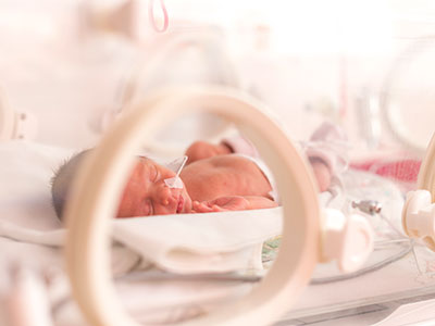
Tracking preemies’ blood flow to monitor brain maturation

Blood is the conduit through which our cells receive much of what they need to grow and thrive. The nutrients and oxygen that cells require are transported by this liquid messenger. Getting adequate blood flow is especially important during the rapid growth of gestation and early childhood – particularly for the brain, the weight of which roughly triples during the last 13 weeks of a typical pregnancy. Any disruption to blood flow during this time could dramatically affect the development of this critical organ.

Now, a new study by Children’s National Health System researchers finds that blood flow to key regions of very premature infants’ brains is altered, providing an early warning sign of disturbed brain maturation well before such injury is visible on conventional imaging. The prospective, observational study was published online Dec. 4, 2017 by The Journal of Pediatrics.

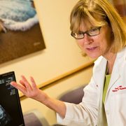
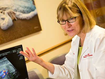
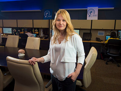
“During the third trimester of pregnancy, the fetal brain undergoes an unprecedented growth spurt. To power that growth, cerebral blood flow increases and delivers the extra oxygen and nutrients needed to nurture normal brain development,” says Catherine Limperopoulos, Ph.D., director of the Developing Brain Research Laboratory at Children’s National and senior author of the study. “In full-term pregnancies, these critical brain structures mature inside the protective womb where the fetus can hear the mother and her heartbeat, which stimulates additional brain maturation. For infants born preterm, however, this essential maturation process happens in settings often stripped of such stimuli.”

The challenge: How to capture what goes right or wrong in the developing brains of these very fragile newborns? The researchers relied on arterial spin labeling (ASL) magnetic resonance (MR) imaging, a noninvasive technique that labels the water portion of blood to map how blood flows through infants’ brains in order to describe which regions do or do not receive adequate blood supply. The imaging work can be done without a contrast agent since water from arterial blood itself illuminates the path traveled by cerebral blood.

“In our study, very preterm infants had greater absolute cortical cerebral blood flow compared with full-term infants. Within regions, however, the insula (a region critical to experiencing emotion), anterior cingulate cortex (a region involved in cognitive processes) and auditory cortex (a region involved in processing sound) for preterm infants received a significantly decreased volume of blood, compared with full-term infants. For preterm infants, parenchymal brain injury and the need for cardiac vasopressor support both were correlated with decreased regional CBF,” Limperopoulos adds.

The team studied 98 preterm infants who were born June 2012 to December 2015, were younger than 32 gestational weeks at birth and who weighed less than 1,500 grams. They matched those preemies by gestational age with 104 infants who had been carried to term. The brain MRIs were performed as the infants slept.

Blood flows where it is needed most with areas of the brain that are used more heavily commandeering more oxygen and nutrients. Thus, during brain development, CBF is a good indicator of functional brain maturation since brain areas that are the most metabolically active need more blood.

the cerebral blood flow (CBF) maps, corresponding anatomical image aligned to the CBF map, and the regions of interest examined

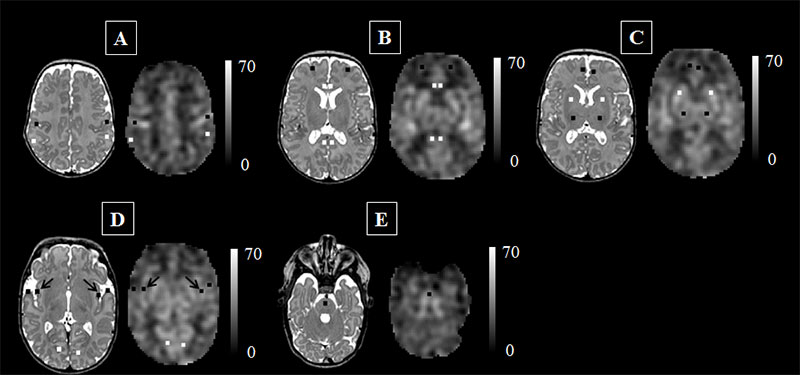
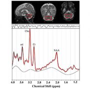
This figure represents the cerebral blood flow (CBF) maps, corresponding anatomical image aligned to the CBF map, and the regions of interest examined. The scale indicates the quantitative value of the CBF map and is expressed in mL/100g/min. The data are from a preterm infant scanned at term age without evidence of brain injury. The insula (see black arrows in panel ‘D’) may be particularly vulnerable to the added stresses of the preterm infant’s life outside the womb.
Credit: M. Bouyssi-Kobar, et al., The Journal of Pediatrics.

“The ongoing maturation of the newborn’s brain can be seen in the distribution pattern of cerebral blood flow, with the greatest volume of blood traveling to the brainstem and deep grey matter,” says Marine Bouyssi-Kobar, M.S., the study’s lead author. “Because of the sharp resolution provided by ASL-MR images, our study finds that in addition to the brainstem and deep grey matter, the insula and the areas of the brain responsible for sensory and motor functions are also among the most oxygenated regions. This underscores the critical importance of these brain regions in early brain development. In preterm infants, the insula may be particularly vulnerable to the added stresses of life outside the womb.”

Of note, compromised regional brain structures in adults are implicated in multiple neurodevelopmental disorders. “Altered development of the insula and anterior cingulate cortex in newborns may represent early warning signs of preterm infants at greater risk for long-term neurodevelopmental impairments,” Limperopoulos says.

Research reported in this post was supported by the Canadian Institutes of Health Research, MOP-81116; the SickKids Foundation, XG 06-069; and the National Institutes of Health under award number R01 HL116585-01.

Congratulations to Dorothy Bulas, M.D. – 2017 RSNA Outstanding Educator recipient

Dorothy Bulas

Dorothy Bulas, M.D., section head of ultrasound and fetal imaging at Children’s National Health System, was honored with the RSNA 2017 Outstanding Educator award at the Radiological Society of North America’s (RSNA) Annual Meeting, held November 26 – December 1 in Chicago, Illinois.

The winner of the award is selected annually by the RSNA Board of Directors based on the awardee’s significant contributions and long-term commitment – 15 years or more – to radiologic education.

“In addition to being a talented clinician and an accomplished researcher, Dr. Bulas is an extraordinary teacher who has made tireless contributions to the educational programs of RSNA,” said RSNA President Richard L. Ehman, M.D. “For more than three decades, she has been a passionate and effective advocate for improving pediatric radiology worldwide – especially in poorly served countries – by participating in educational outreach.”

Children’s National experts contribute to new Zika guidelines

Roberta DeBiasi and Sarah Mulkey

Roberta DeBiasi, M.D., M. S., and Sarah B. Mulkey, M.D., Ph.D., members of Children’s multidisciplinary Congenital Zika Virus Program, were among the experts invited to participate in a forum held in Atlanta at CDC headquarters in late August to formulate new Zika recommendations.

The Centers for Disease Control and Prevention (CDC) on Oct. 19, 2017 updated guidelines for evaluation of women, fetuses and infants exposed to the Zika virus during pregnancy. Although only women with symptoms will now be routinely tested, asymptomatic and symptomatic infants born to these women will still be tested for the Zika virus using blood and urine tests.

Infants who appear normal, whose mothers either had negative Zika results or who had not undergone testing, will not undergo Zika testing. These infants still will undergo a standard evaluation, including a detailed physical exam, hearing screen and routine developmental assessments. The revised Zika guidance includes input from practitioners on the front lines of the Zika epidemic, including Children’s National Health System clinicians.

“These changes in the recommendations for Zika testing should not be interpreted as Zika infection risks subsiding for pregnant women and their infants in the United States. It’s simply an acknowledgement of the limitations of current testing methods – which must occur within a narrow window after Zika exposure – and the poor predictive value of Zika testing right now,” says Roberta L. DeBiasi, M.D., M.S., chief of Children’s Division of Pediatric Infectious Diseases. Dr. DeBiasi and Sarah B. Mulkey, M.D., Ph.D., members of Children’s multidisciplinary Congenital Zika Virus Program, were among the experts invited to participate in the Zika forum held in Atlanta at CDC headquarters in late August to formulate the recommendations.

While all infants will receive a standard evaluation, expanded evaluations that include an ophthalmologic assessment, more detailed hearing evaluation and ultrasound of the newborn’s head will be reserved for infants born to mothers confirmed to be Zika positive or Zika probable, or for infants born with abnormalities potentially consistent with congenital Zika syndrome, regardless of maternal status.

The majority of U.S. infants who have been exposed to Zika in the womb appeared normal at birth, according to CDC registries. Now, the next wave of these normal-appearing babies will receive standard evaluations when they are born, including a newborn hearing screening. At each well-child visit, these infants will receive:

  • A comprehensive physical examination
  • An age-appropriate vision screening
  • Developmental monitoring and screening using validated tools

“This is a natural evolution in the diagnosis and screening strategy now that the peak of the first wave of Zika transmission appears to be over,” Dr. DeBiasi says. “While we continue to evaluate new possible cases of Zika infection among pregnant women in our practice, a sizable proportion of Children’s cases are Zika-exposed infants whose physical exam and neuroimaging appeared normal at birth. Through ongoing monitoring, we hope to learn more about these children’s long-term neurodevelopment outcomes.”

Fetal MRI plus ultrasound assess Zika-related brain changes

Sarah Mulkey

Magnetic resonance imaging and ultrasound provide complementary data needed to assess ongoing changes to the brains of fetuses exposed to Zika in utero, says Sarah B. Mulkey, M.D., Ph.D.

For Zika-affected pregnancies, fetal magnetic resonance imaging (MRI) used in addition to standard ultrasound (US) imaging can better assess potential brain abnormalities in utero, according to research presented by Children’s National Health System during IDWeek 2017. In cases of abnormal brain structure, fetal MRI can reveal more extensive areas of damage to the developing brain than is seen with US.

“MRI and US provide complementary data needed to assess ongoing changes to the brains of fetuses exposed to Zika in utero,” says Sarah B. Mulkey, M.D., Ph.D., a fetal/neonatal neurologist at Children’s National Health System and lead author of the research paper. “In addition, our study found that relying on ultrasound alone would have given one mother the false assurance that her fetus’ brain was developing normally while the sharper MRI clearly pointed to brain abnormalities.”

As of Sept. 13, the Centers for Disease Control and Prevention (CDC) reported that 1,901 U.S. women were exposed to Zika at some point during their pregnancies but their infants appeared normal at birth. Another 98 U.S. women, however, gave birth to infants with Zika-related birth defects.  And eight more women had pregnancy losses with Zika-related birth defects, according to CDC registries.

The longitudinal neuroimaging study led by Children’s National enrolled 48 pregnant women exposed to the Zika virus in the first or second trimester whose infection was confirmed by reverse transcription polymerase chain reaction, which detects Zika viral fragments shortly after exposure, and/or Immunoglobulin M testing, which reveals antibodies the body produces to neutralize the virus. Forty-six of the study volunteers live in Barranquilla, Colombia, where Zika infection is endemic. Two women live in the Washington region and were exposed to Zika during travel elsewhere.

All of the women underwent at least one diagnostic imaging session while pregnant, receiving an initial MRI or US at 25.1 weeks’ gestational age. Thirty-six women underwent a second MRI/US imaging pair at roughly 31 weeks’ gestation. Children’s National radiologists read every image.

Three of 48 pregnancies, or 6 percent, were marked by abnormal fetal MRIs:

  • One fetus had heterotopias (clumps of grey matter located at the wrong place) and abnormal cortical indent (a deformation at the outer layer of the cerebrum, a brain region involved in consciousness). The US taken at the same gestational age for this fetus showed its brain was developing normally.
  • Another fetus had parietal encephalocele (an uncommon skull defect) and Chiari malformation Type II (a life-threatening structural defect at the base of the skull and the cerebellum, the part of the brain that controls balance). The US for this fetus also detected these brain abnormalities.
  • The third fetus had a thin corpus callosum (bundle of nerves that connects the brain’s left and right hemispheres), an abnormally developed brain stem, temporal cysts, subependymal heterotopias and general cerebral/cerebellar atrophy. This fetal US showed significant ventriculomegaly (fluid-filled structures in the brain that are too large) and a fetal head circumference that decreased sharply from the 32nd to 36th gestational week, a hallmark of microcephaly.

After they were born, infants underwent a follow-up MRI without sedation and US. For nine infants, these ultrasounds revealed cysts in the choroid plexus (cells that produce cerebrospinal fluid) or germinal matrix (the source for neurons and glial cells that migrate during brain development). And one infant’s US after birth showed lenticulostriate vasculopathy (brain lesions).

“Because a number of factors can trigger brain abnormalities, further studies are needed to determine whether the cystic changes to these infants’ brains are attributable to Zika exposure in the womb or whether some other insult caused these troubling results,” Dr. Mulkey says.

What Children’s has learned about congenital Zika infection

Roberta DeBiasi

Roberta DeBiasi, M.D., M.S., outlined lessons learned during a pediatric virology workshop at IDWeek2017, one of three such Zika presentations led by Children’s National research-clinicians during this year’s meeting of pediatric infectious disease specialists.

The Congenital Zika Virus Program at Children’s National Health System provides a range of advanced testing and services for exposed and infected fetuses and newborns. Data that the program has gathered in evaluating and managing Zika-affected pregnancies and births may offer instructive insights to other centers developing similar programs.

The program evaluated 36 pregnant women and their fetuses from January 2016 through May 2017. Another 14 women and their infants were referred to the Zika program for postnatal consultations during that time.

“As the days grow shorter and temperatures drop, we continue to receive referrals to our Zika program, and this is a testament to the critical need it fulfills in the greater metropolitan D.C. region,” says Roberta L. DeBiasi, M.D., M.S., chief of the Division of Pediatric Infectious Diseases and co-leader of the program. “Our multidisciplinary team now has consulted on 90 dyads (mothers and their Zika-affected fetuses/infants). The lessons we learned about when and how these women were infected and how their offspring were affected by Zika may be instructive to institutions considering launching their own programs.”

Dr. DeBiasi outlined lessons learned during a pediatric virology workshop at IDWeek2017, one of three such Zika presentations led by Children’s National research-clinicians during this year’s meeting of pediatric infectious disease specialists.

“The Zika virus continues to circulate in dozens of nations, from Angola to the U.S. Virgin Islands. Clinicians considering a strategic approach to managing pregnancies complicated by Zika may consider enlisting an array of specialists to attend to infants’ complex care needs, including experts in fetal imaging, pediatric infectious disease, physical therapists, audiologists, ophthalmologists and radiologists skilled at reading serial magnetic resonance images as well as ultrasounds,” Dr. DeBiasi says. “At Children’s we have a devoted Zika hotline to triage patient and family concerns. We provide detailed instructions for referring institutions explaining protocols before and after childbirth, and we provide continuing education for health care professionals.”

Of the 36 pregnant women possibly exposed to Zika during pregnancy seen in the program’s first year, 32 lived in the United States and traveled to countries where Zika virus was circulating. Two women had partners who traveled to Zika hot zones. And two moved to the Washington region from places where Zika is endemic. Including the postnatal cases, 89 percent of patients had been bitten by Zika-tainted mosquitoes, while 48 percent of women could have been exposed to Zika via sex with an infected partner.

Twenty percent of the women were exposed before conception; 46 percent were exposed to Zika in the first trimester of pregnancy; 26 percent were exposed in the second trimester; and 8 percent were exposed in the final trimester. In only six of 50 cases (12 percent) did the Zika-infected individual experience symptoms.

Zika infection can be confirmed by detecting viral fragments but only if the test occurs shortly after infection. Twenty-four of the 50 women (nearly 50 percent) arrived for a Zika consultation outside that 12-week testing window. Eleven women (22 percent) had confirmed Zika infection and another 28 percent tested positive for the broader family of flavivirus infections that includes Zika. Another detection method picks up antibodies that the body produces to neutralize Zika virus. For seven women (14 percent), Zika infection was ruled out by either testing method.

“Tragically, four fetuses had severe Zika-related birth defects,” Dr. DeBiasi says. “Due to the gravity of those abnormalities, two pregnancies were not carried to term. The third pregnancy was carried to term, but the infant died immediately after birth. The fourth pregnancy was carried to term, but that infant survived less than one year.”

Brain impairment in newborns with CHD prior to surgery

Catherine Limperopoulous

Children’s National researchers led by Catherine Limperopoulos, Ph.D., demonstrate for the first time that the brains of high-risk infants show signs of functional impairment before they undergo corrective cardiac surgery.

Newborns with congenital heart disease (CHD) requiring open-heart surgery face a higher risk for neurodevelopmental disabilities, yet prior studies had not examined whether functional brain connectivity is altered in these infants before surgery.

Findings from a Children’s National Health System study of this question suggest the presence of brain dysfunction early in the lives of infants with CHD that may be associated with neurodevelopmental impairments years later.

Using a novel imaging technique, Children’s National researchers demonstrated for the first time that the brains of these high-risk infants already show signs of functional impairment even before they undergo corrective open heart surgery. Looking at the newborns’ entire brain topography, the team found intact global organization – efficient and effective small world networks – yet reduced functional connectivity between key brain regions.

“A robust neural network is critical for neurons to travel to their intended destinations and for the body to carry out nerve cells’ instructions. In this study, we found the density of connections among rich club nodes was diminished, and there was reduced connectivity between critical brain hubs,” says Catherine Limperopoulos, Ph.D., director of the Developing Brain Research Laboratory at Children’s National and senior author of the study published online Sept. 28, 2017 in NeuroImage: Clinical. “CHD disrupts how oxygenated blood flows throughout the body, including to the brain. Despite disturbed hemodynamics, infants with CHD still are able to efficiently transfer neural information among neighboring areas of the brain and across distant regions.”

The research team led by Josepheen De Asis-Cruz, M.D., Ph.D., compared whole brain functional connectivity in 82 healthy, full-term newborns and 30 newborns with CHD prior to corrective heart surgery. Conventional imaging had detected no brain injuries in either group. The team used resting state functional connectivity magnetic resonance imaging (rs-fcMRI), a imaging technique that characterizes fluctuating blood oxygen level dependent signals from different regions of the brain, to map the effect of CHD on newborns’ developing brains.

The newborns with CHD had lower birth weights and lower APGAR scores (a gauge of how well brand-new babies fare outside the womb) at one and five minutes after birth. Before the scan, the infants were fed, wrapped snugly in warm blankets, securely positioned using vacuum pillows, and their ears were protected with ear plugs and ear muffs.

While the infants with CHD had intact global network topology, a close examination of specific brain regions revealed functional disturbances in a subnetwork of nodes in newborns with cardiac disease. The subcortical regions were involved in most of those affected connections. The team also found weaker functional connectivity between right and left thalamus (the region that processes and transmits sensory information) and between the right thalamus and the left supplementary motor area (the section of the cerebral cortex that helps to control movement). The regions with reduced functional connectivity depicted by rs-fcMRI match up with regional brain anomalies described in imaging studies powered by conventional MRI and diffusion tensor imaging.

“Global network organization is preserved, despite CHD, and small world brain networks in newborns show a remarkable ability to withstand brain injury early in life,” Limperopoulos adds. “These intact, efficient small world networks bode well for targeting early therapy and rehabilitative interventions to lower the newborns’ risk of developing long-term neurological deficits that can contribute to problems with executive function, motor function, learning and social behavior.”

Children’s National Fetal Medicine Institute hosts 2nd annual International Symposium on the Fetal Brain

The Children’s National Health System Fetal Medicine Institute hosted the 2nd annual International Symposium on the Fetal Brain in August 2017 in Washington, D.C.

Speakers at this year’s symposium focused on the following four areas:

  • Brain Development in an Unsupportive In Utero Environment – Diagnosis and Consequences
  • Supporting Brain Development in the Ex Utero Fetus: How Far Are We From Optimal?
  • Genomic and Epigenomic Mechanisms Underlying Differences in Brain Development
  • The Emergence of Consciousness and Pain Sensation

Adré J. du Plessis, M.B.Ch.B., M.P.H., Director of the Fetal Medicine Institute and Division Chief of Fetal and Transitional Medicine hosted the conference. In his opening remarks Dr. du Plessis noted “Our goal has been to gather together a diverse group from across the spectrum of disciplines focused on the well-being of the fetal brain and to engage all disciplines together.”

Diana-Bianchi-at-ISFB

Diana Bianchi, MD gives her keynote presentation on non-invasive fetal testing at the second annual International Symposium on the Fetal Brain.

Invited, internationally renowned speakers presented on diverse topics, including Diana Bianchi, Director of the Eunice Kennedy Shriver National Institute of Child Health and Human Development who spoke on the “Non-Invasive Fetal Testing Beyond Karyotype: What’s in it for the Fetal Brain?”

A new component to the symposium was the clinically-focused breakfast breakout sessions, created based on feedback from attendees of the 2016 Symposium. Sessions covered varied topics such as “Fetal Ultrasound: the Cornerstone of Fetal Neurodiagnosis,” “The Essentials of Neurogenetic Testing,” “Developing a Transitional Fetal-Neonatal Program” and “Using MRI to Advance Fetal Neurodiagnosis.”

The conference started with an exciting discussion by Alistair Gunn, M.B.Ch.B., Head of the Department of Physiology at the University of Auckland. His presentation “Fetal Heart Rate: What It Does and Does Not Tell Us” explored the considerable body of evidence that essentially all decelerations are mediated by chemoreflex responses to repeated hypoxia and that the parasympathetic autonomic nervous system is the critical regulator of both fetal heart rate and heart rate variation in labor.

Following a voting process from the symposium’s external speakers, the inaugural Andrea Poretti Abstract Award was presented to Katherine Ottolini for her poster titled: Breastmilk Feeds Improve Brain Microstructural Development in Very Premature Infants.

For more information about the sessions and speakers at the 2017 Symposium, please visit our website.

Understanding the long-term consequences of prematurity

LCModel output from 32 GA baby

Children’s National Health System researchers processed H1-MRS data using LCModel software to calculate absolute metabolite concentrations for N-acetyl-aspartate (NAA), choline (Cho) and creatine (Cr). Preterm infants had significantly lower cerebellar NAA (p=<0.025) and higher Cho (p=<0.001) when compared with healthy term-equivalent infants. The area of the brain within the red box is the cerebellum, the region of interest for this study.

Premature birth, a condition that affects approximately 10 percent of births in the United States, often is accompanied by health problems ranging from difficulties breathing and eating to long-term neurocognitive delays and disabilities. However, the reasons for these problems have been unclear.

In a study published online Aug. 15, 2017 in Scientific Reports, a team of Children’s National Health System clinician-researchers reports that prematurity is associated with altered metabolite profiles in the infants’ cerebellum, the part of the brain that controls coordination and balance. Pre-term infants in the study had significantly lower levels of a chemical marker of nerve cell integrity and significantly higher concentrations of a chemical marker of cellular membrane turnover.

“These data suggest that interrupting the developing fetal brain’s usual growth plan during gestation – which can occur through early birth, infection or experiencing brain damage – might trigger a compensatory mechanism. The infant’s brain tries to make up for lost time or heal injured tissue by producing a certain type of cells more quickly than it normally would,” says Catherine Limperopoulos, Ph.D., director of the Developing Brain Research Laboratory at Children’s National and senior study author. “The more sensitive imaging technique that we used also revealed nerve cell damage from brain injuries extends beyond the site of injury, a finding that contrasts with what is found through conventional magnetic resonance imaging (MRI).”

It has long been clear that prematurity – birth before 37 weeks gestation – is accompanied by a number of immediate and long-term complications, from potential problems breathing and feeding at birth to impairments in hearing and sight that can last throughout an individual’s life.

Neurocognitive developmental delays often accompany pre-term birth, many of which can have long-lasting consequences. Studies have shown that children born prematurely are more likely to struggle in school, have documented learning disabilities and experience significant delays in developing gross and fine motor skills compared with children born at full-term.

Several studies have investigated the root cause of these issues in the cerebrum, the structure that takes up the majority of the brain and is responsible for functions including learning and memory, language and communication, sensory processing and movement. However, the cerebellum – a part of the brain that plays an important role in motor control – has not received as much research attention.

In the new study, Limperopoulos and colleagues used a specialized MRI technique that allowed them to parse out differences in which molecules are present in the cerebellum of full-term infants compared with premature infants. Their findings show a variety of differences that could offer clues to explain developmental differences between these two populations – and potentially identify ways to intervene to improve outcomes.

The researchers recruited 59 premature infants, born at 32 or fewer weeks’ gestation, and 61 healthy, full-term infants. Each baby received a special type of MRI known as proton magnetic resonance spectroscopy, or H1-MRS, that measures the concentrations of particular molecules in the brain. The full-term infants had these MRIs shortly after birth; the pre-term infants had them at 39 to 41 weeks gestational age, or around the time that they would have been born had the pregnancy continued to term.

Looking specifically at the cerebellum, the researchers found that the pre-term infants overall had significantly lower concentrations of N-acetyl-aspartate (NAA), a marker of the integrity of nerve cells. They also had significantly higher concentrations of choline, a marker of cell membrane integrity and membrane turnover.

Concentrations of creatine, a marker of stores of cellular energy, were about the same overall between the two groups. However, the researchers found that brain injuries, which affected 35 of the pre-term infants but none of the full-term infants, were associated with significantly lower concentrations of NAA, choline and creatine. Having a neonatal infection, which affected 21 of the pre-term infants but none of the full-term ones, was associated with lower NAA and creatine.

The findings could offer insight into exactly what’s happening in the brain when infants are born pre-term and when these vulnerable babies develop infections or their brains become injured – conditions that convey dramatically higher risks for babies born too early, Limperopoulos says. The differences between the full-term babies and the pre-term ones reflect disturbances these cells are experiencing at a biochemical level, she explains.

Limperopoulos and colleagues note that more research will be necessary to connect these findings to what is already known about developmental problems in pre-term infants. Eventually, she says, scientists might be able to use this knowledge to develop treatments that might be able to change the course of brain development in babies born too early, getting them on track with infants born at term.

“We know that the bodies of pre-term infants demonstrate a remarkable ability to catch up with peers who were born at full-term, in terms of weight and height. Our challenge is to ensure that preemies’ brains also have an opportunity to develop as normally as possible to ensure optimal long-term outcomes,” Limperopoulos says.

A closer look at the placenta to predict FGR

Catherine Limperopoulos

Using three-dimensional magnetic resonance imaging, a Children’s National research team that included Catherine Limperopoulos, Ph.D., characterized the shape, volume, morphometry and texture of placentas during pregnancy and, using a novel framework, predicted with high accuracy which pregnancies would be complicated by fetal growth restriction.

Early in development, cells from the fertilized egg form the placenta, a temporary organ that serves as an interface between the mother and her growing offspring. When things go right, as occurs in the vast majority of pregnancies, the placenta properly delivers nutrients from the mother’s diet and oxygen from the air she breathes to the developing fetus while siphoning away its waste products. This organ also plays important immune-modulating and endocrine roles.

However, in a number of pregnancies, the placenta does not do an adequate job. Unable to effectively serve the fetus, a variety of adverse conditions can develop, including preeclampsia, fetal growth restriction (FGR), preterm birth and even fetal death.

Despite the key role that the placenta plays in fetal health, researchers have few non-invasive ways to assess how well it works during pregnancy. In fact, placental disease might not be suspected until very late.

In a new study, a team of Children’s National Health System research scientists is beginning to provide insights into the poorly understood placenta.

Using three-dimensional (3D) magnetic resonance imaging (MRI), the research team characterized the shape, volume, morphometry and texture of placentas during pregnancy and, using a novel framework, predicted with high accuracy which pregnancies would be complicated by FGR.

“When the placenta fails to carry out its essential duties, both the health of the mother and fetus can suffer and, in extreme cases, the fetus can die. Because there are few non-invasive tools that reliably assess the health of the placenta during pregnancy, unfortunately, placental disease may not be discovered until too late – after impaired fetal growth already has occurred,” says Catherine Limperopoulos, Ph.D., co-director of research in the Division of Neonatology at Children’s National Health System and senior author of the study published online July 22 in Journal of Magnetic Resonance Imaging. “Identifying early biomarkers of placental disease that may impair fetal growth and well-being open up brand-new opportunities to intervene to protect vulnerable fetuses.”

The Children’s research team acquired 124 fetal scans from 80 pregnancies beginning at the 18th gestational week and continuing through the 39th gestational week. Forty-six women had normal pregnancies and healthy fetuses while 34 women’s pregnancies were complicated by FGR, defined by estimated fetal weight that fell below the 10th percentile for gestational age. The placenta was described by a combination of shape and textural features. Its shape was characterized by three distinct 3D features: Volume, thickness and elongation. Its texture was evaluated by three different sets of textural features computed on the entire placenta.

“The proposed machine learning-based framework distinguished healthy pregnancies from FGR pregnancies with 86 percent accuracy and 87 percent specificity. And it estimated the birth weight in both healthy and high-risk fetuses throughout the second half of gestation reasonably well,” says the paper’s lead author, Sonia Dahdouh, Ph.D., a research fellow in Children’s Developing Brain Research Laboratory.

“We are helping to pioneer a very new frontier in fetal medicine,” Limperopoulos adds. “Other studies have developed prediction tools based on fetal brain features in utero. To our knowledge, this would be the first proposed framework for semi-automated diagnosis of FGR and estimation of birth weight using structural MRI images of the placental architecture in vivo. This has the potential to address a sizable clinical gap since we lack methods that are both sufficiently sensitive and specific to reliably detect FGR in utero.”

The research team writes that its findings underscore the importance of future studies on a larger group of patients to expand knowledge about underlying placenta mechanisms responsible for disturbed fetal growth, as well as to more completely characterize other potential predictors of fetal/placental development in high-risk pregnancies, such as genetics, physiology and nutrition.

Lab led by Zhe Han, Ph.D., receives $1.75 million from NIH

Zhe Han, PhD

A new four-year NIH grant will enable Zhe Han, Ph.D., to carry out the latest stage in the detective work to determine how histone-modifying genes regulate heart development and the molecular mechanisms of congenital heart disease caused by these genetic mutations.

The National Institutes of Health (NIH) has awarded $1.75 million to a research lab led by Zhe Han, Ph.D., principal investigator and associate professor in the Center for Genetic Medicine Research, in order to build models of congenital heart disease (CHD) that are tailored to the unique genetic sequences of individual patients.

Han was the first researcher to create a Drosophila melanogaster model to efficiently study genes involved in CHD, the No.1 birth defect experienced by newborns, based on sequencing data from patients with the heart condition. While surgery can fix more than 90 percent of such heart defects, an ongoing challenge is how to contend with the remaining cases since mutations of a vast array of genes could trigger any individual CHD case.

In a landmark paper published in 2013 in the journal Nature, five different institutions sequenced the genomes of more than 300 patients with CHD and their families, identifying 200 mutated genes of interest.

“Even though mutations of these genes were identified from patients with CHD, these genes cannot be called ‘CHD genes’ since we had no in vivo evidence to demonstrate these genes are involved in heart development,” Han says. “A key question to be answered: How do we efficiently test a large number of candidate disease genes in an experimental model system?”

In early 2017, Han published a paper in Elife providing the answer to that lingering question. By silencing genes in a fly model of human CHD, the research team confirmed which genes play important roles in development. The largest group of genes that were validated in Han’s study were histone-modifying genes. (DNA winds around the histone protein, like thread wrapped around a spool, to become packed into a higher-level structure.)

The new four-year NIH grant will enable Han to carry out the next stage of the detective work to determine precisely how histone-modifying genes regulate heart development. In order to do so, his group will silence the function of histone-modifying genes one by one, to study their function in the fly heart development and to identify the key histone-modifying genes for heart development. And because patients with CHD can have more than one mutated gene, he will silence multiple genes simultaneously to determine how those genes work in partnership to cause heart development to go awry.

By the end of the four-year research project, Han hopes to be able to identify all of the histone-modified genes that play pivotal roles in development of the heart in order to use those genes to tailor make personalized fly models corresponding to individual patient’s genetic makeup.

Parents with mutations linked to CHD are likely to pass heart disease risk to the next generation. One day, those parents could have an opportunity to sequence their genes to learn the degree of CHD risk their offspring face.

“Funding this type of basic research enables us to understand which genes are important for heart development and how. With this knowledge, in the near future we could predict the chances of a baby being born with CHD, and cure it by using gene-editing approaches to prevent passing disease to the next generation,” Han says.

Spectral data shine light on placenta

preemie baby

A research project led by Subechhya Pradhan, Ph.D., aims to shed light on metabolism of the placenta, a poorly understood organ, and characterize early biomarkers of fetal congenital heart disease.

The placenta serves as an essential intermediary between a pregnant mother and her developing fetus, transporting in life-sustaining oxygen and nutrients, ferrying out waste and serving as interim lungs, kidneys and liver as those vital organs develop in utero.

While the placenta plays a vital role in supporting normal pregnancies, it remains largely a black box to science. A research project led by Subechhya Pradhan, Ph.D., and partially funded by a Clinical and Translational Science Institute Research Award aims to shed light on placenta metabolism and characterize possible early biomarkers of impaired placental function in fetal congenital heart disease (CHD), the most common type of birth defect.

“There is a huge information void,” says Pradhan, a research faculty member of the Developing Brain Research Laboratory at Children’s National Health System. “Right now, we do not have very much information about placenta metabolism in vivo. This would be one of the first steps to understand what is actually going on in the placenta at a biochemical level as pregnancies progress.”

The project Pradhan leads will look at the placentas of 30 women in the second and third trimesters of healthy, uncomplicated pregnancies and will compare them with placentas of 30 pregnant women whose fetuses have been diagnosed with CHD. As volunteers for a different study, the women are already undergoing magnetic resonance imaging, which takes detailed images of the placenta’s structure and architecture. The magnetic resonance spectroscopy scans that Pradhan will review show the unique chemical fingerprints of key metabolites: Choline, lipids and lactate.

Choline, a nutrient the body needs to preserve cellular structural integrity, is a marker of cell membrane turnover. Fetuses with CHD have higher concentrations of lactate in the brain, a telltale sign of a shortage of oxygen. Pradhan’s working hypothesis is that there may be differing lipid profiles and lactate levels in the placenta in pregnancies complicated by CHD.  The research team will extract those metabolite concentrations from the spectral scans to describe how they evolve in both groups of pregnant women.

“While babies born with CHD can undergo surgery as early as the first few days (or sometimes hours) of life to correct their hearts, unfortunately, we still see a high prevalence of neurodevelopmental impairments in infants with CHD. This suggests that neurological dysfunctional may have its origin in fetal life,” Pradhan says.

Having an earlier idea of which fetuses with CHD are most vulnerable has the potential to pinpoint which pregnancies need more oversight and earlier intervention.

Placenta spectral data traditionally have been difficult to acquire because the pregnant mother moves as does the fetus, she adds. During the three-minute scans, the research team will try to limit excess movement using a technique called respiratory gating, which tells the machine to synchronize image acquisition so it occurs in rhythm with the women’s breathing.

Understanding genetic synergy in cleft palate

Baby with Cleft Palate

Like mechanics fixing a faulty engine, Youssef A. Kousa, M.S., D.O., Ph.D., says researchers will not be able to remedy problems related to IRF6, a gene implicated in cleft palate, until they better understand how the gene works.

Like all of the individual elements of fetal development, palate growth is a marvel of nature. In part of this process, ledges of tissue on the sides of the face grow downwards on each side of the tongue, then upward, fusing at the midline at the top of the mouth. The vast majority of the time, this process goes correctly. However, some part of it goes awry for the 2,650 babies born in the United States each year with cleft palates and the thousands more born worldwide with the defect.

For nearly two decades, researchers have known that a gene known as IRF6 is involved in palate formation. Studies have shown that this gene contributes about 12 percent to 18 percent of the risk of cleft palate, more than any other gene identified thus far. IRF6 is active in epithelial tissues – those that line cavities and surfaces throughout the body – including the periderm, a tissue that lines the mouth cavity and plays an important role during development.

According to Youssef A. Kousa, M.S., D.O., Ph.D., a child neurology fellow at Children’s National Health System, the periderm acts like a nonstick layer, preventing the tongue or other structures from adhering to the growing palate and preventing it from sealing at the midline. While researchers have long suspected that IRF6 plays a strong role in promoting this nonstick quality, exactly how it exerts its influence has not been clear.

“Gaining a better understanding of this gene might help us to eventually address deficits or perturbations in the system that creates the palate,” Dr. Kousa says. “Like a mechanic fixing a faulty engine, we will not be able to remedy problems related to this gene until we know how the gene works.”

Youssef Kousa

“Gaining a better understanding of this gene might help us to eventually address deficits or perturbations in the system that creates the palate,” Dr. Kousa says. “Like a mechanic fixing a faulty engine, we will not be able to remedy problems related to this gene until we know how the gene works.”

In a study published July 19, 2017 by the Journal of Dental Research, Dr. Kousa and colleagues seek to decipher one piece of this puzzle by investigating how this key gene might interact with others that are active during fetal development. The researchers were particularly interested in genes that work together in a cascade of activity known as the tyrosine kinase receptor signaling pathway.

Because this pathway includes a large group of genes, Dr. Kousa and colleagues reasoned that they could answer whether IRF6 interacts with this pathway by looking at whether the gene interacts with the last member of the cascade, a gene called SPRY4. To do this, the researchers worked with experimental models that had mutations in IRF6, SPRY4 or both. If these two genes interact, the scientists hypothesized, carrying mutations in both genes at the same time should result in a dramatically different outcome compared with animals that carried mutations in just one gene.

Using selective breeding techniques, the researchers created animals that had mutations in either of these genes or in both. Their results suggest that IRF6 and SPRY4 indeed do interact: Significantly more of the oral surface was adhered to the tongue during fetal development in experimental models that had mutations in both genes compared with those that had just one single gene mutated. Examining the gene activity in the periderm cells of these affected animals, the researchers found that doubly mutated experimental models also had decreased activity in a third gene known as GRHL3, which also has been linked with cleft lip and palate.

Dr. Kousa says the research team plans to continue exploring this interaction to better understand the flow of events that lead from perturbations in these genes to formation of cleft palate. Some of the questions they would like to answer include exactly which gene or genes in the tyrosine kinase receptor signaling pathway specifically interact with IRF6 – since SPRY4 represents just the end of that pathway, others genes earlier in the pathway are probably the real culprits responsible for driving problems in palate formation. They also will need to verify if these interactions take place in humans in the same way they occur in preclinical models.

Eventually, Dr. Kousa adds, the findings could aid in personalized prenatal counseling, diagnosis and screening related to cleft palate, as well as preventing this condition during pregnancy. Someday, doctors might be able to advise couples who carry mutations in these genes about whether they are more likely to have a baby with a cleft palate or determine which select group of pregnancies need closer monitoring. Additionally, because research suggests that GRHL3 might interact with nutrients, including inositol, it might be possible to prevent some cases of cleft palate by taking additional supplements during pregnancy.

“The more we know about how these genes behave,” Dr. Kousa says, “the more we can potentially avoid fetal palate development going down the wrong path.”

Children’s National expert joins a national discussion on Zika and other emerging threats

Roberta DeBiasi

“Our goal is to provide the earliest and most accurate information to women affected by Zika exposure and infection during pregnancy, including capability for fetal MRI,” says Roberta DeBiasi, M.D., M.S., chief of the Division of Pediatric Infectious Diseases and co-director of the Congenital Zika Virus Program at Children’s National Health System.

An expert roundtable discussion, “Facing the Zika Crisis and Other Emerging Threats,” organized in collaboration with Purdue University, the Gallup Organization and the Bipartisan Policy Center, was recently held at the U.S. Capitol. Chief of the Division of Pediatric Infectious Diseases and Co-Director of the Congenital Zika Program at Children’s National Health System, Roberta DeBiasi, M.D.,M.S., weighed in on the discussion, sharing knowledge on the challenges to the United States health system and the continuous research and work that the Children’s National Congenital Zika program does.

Eighteen months ago, Children’s National received its first referral for a Washington, D.C. woman who had a Zika infected pregnancy in January 2016. This case prompted the development of the Congenital Zika program to serve as a consultation resource for affected women and infants, and to perform research to address the knowledge gaps concerning Zika infection during pregnancy: Young researchers tackle Zika’s unanswered questions.

“Our goal is to provide the earliest and most accurate information to women affected by Zika exposure and infection during pregnancy, including capability for fetal MRI,” says Dr. DeBiasi.

Since then, the Zika team has evaluated 65 mother-fetus/infant pairs. Researchers are actively learning the best methods for detection of infection and neurologic injury by continually conducting research and obtaining new and useful information that can be shared with others. The research mission of the Congenital Zika program is now focusing on several areas, which include the study of biomarkers to predict if the infant could be affected by the disease, the utility of a fetal MRI in conjunction with ultrasound, genetic risk factors in mothers and infants that might explain why some infants become infected and some do not, long term neurodevelopment of babies that are infected, and neuropathologic evaluations of brains from fetuses that have died from Zika.

The challenges and concerns that were presented for the United States health system include the willingness and ability to share information, the acceptance of the need for data sharing between institutions and determining if testing resources are adequate and appropriate.

Dr. DeBiasi says, “Institutions have become much better at looking at how to prepare for emerging infectious diseases on a broader level.” Proactively thinking, Dr. DeBiasi finds it useful for health systems to use their own task forces, such as the Ebola Response Task force at Children’s National, as a cohesive existing team that will be prepared for additional infectious disease threats that may arise.

Center for Neuroscience Research investigators join CIFASD

Kazue Hashimoto-Torii and Masaaki Torii, Collaborative Initiative on Fetal Alcohol Spectrum Disorders

Masaaki Torii, Ph.D., Kazue Hashimoto-Torii, Ph.D., and their research teams are joining Collaborative Initiative on Fetal Alcohol Spectrum Disorders, a consortium supported by the National Institutes of Health.

Kazue Hashimoto-Torii, Ph.D., Masaaki Torii, Ph.D., and the research teams they lead have joined a national research consortium for Fetal Alcohol Spectrum Disorders that is supported by the National Institutes of Health (NIH).

The Collaborative Initiative on Fetal Alcohol Spectrum Disorders (CIFASD) aims to leverage multidisciplinary approaches to develop effective interventions and treatments for Fetal Alcohol Spectrum Disorders.

“Both of our labs have been fortunate in receiving multiple R series research grants from the NIH. I am deeply honored that we now join this prestigious national consortium, which opens additional opportunities to collaborate with other labs with neurobehavioral, genetics and facial dysmorphology expertise as well as other specialized disciplines,” says Hashimoto-Torii, principal investigator in the Center for Neuroscience Research at Children’s National Health System.

Fetal Alcohol Spectrum Disorders are a constellation of conditions that result from exposure to alcohol in the womb that reflect the vastly different ways fetuses respond to that in utero insult. While early intervention is crucial, one challenge that continues to bedevil the field is trying to determine which pregnancies are most at risk.

“It is crucial to develop early and precise biomarkers for predicting children’s risk for cognitive and behavioral problems,” Hashimoto-Torii says. “Our labs will work on developing a novel approach for identifying such biomarkers.”

The Children’s researchers will examine epigenetic changes at the single cell level that may provide the earliest hint of cognitive and learning difficulties – long before children show any symptoms of such problems. Hashimoto-Torii’s lab will perform single-cell droplet digital polymerase chain reaction (PCR) based biomarker analysis of blood samples from experimental models and humans. Meanwhile, the lab run by Torii – also a principal investigator in the Center for Neuroscience Research – will collect blood samples from experimental models, perform comprehensive behavioral analysis, and evaluate potential correlations between behaviors seen in the experimental models and their drop-PCR results.

“Under the auspices of CIFASD, we ultimately hope to link these biomarkers from our lab with results that our colleagues are seeing in children in order to validate their ability to accurately predict outcomes from prenatal alcohol exposure,” she says.

Panel: Significant Zika risks linger for pregnant women and developing fetuses in US

Roberta DeBiasi

The threat from Zika “is not over. It is just beginning for the families who are affected by this,” says Roberta L. DeBiasi, M.D., M.S., chief of the Division of Pediatric Infectious Diseases and co-director of the Congenital Zika Virus Program at Children’s National Health System.

The Zika virus epidemic may have fallen off the radar for many media outlets, but significant risks continue to linger for pregnant women and developing fetuses, a panel of experts told staff working for U.S. Congressional leaders.

“The threat of this virus is real, and the threat continues,” Margaret Honein, Ph.D., M.P.H., of the Centers for Disease Control and Prevention’s (CDC) pregnancy and birth defects task force, said during the July 13 briefing held in the Russell Senate Office Building.

Dr. Honein told about 100 attendees that more than 200 Zika-affected babies have been born in the United States suffering from serious birth defects, such as rigid joints, inconsolable distress that causes them to cry continuously and difficulties swallowing. Some of these infants experience seizures that cause further brain damage.

Predicting what Zika will do next in the United States is very difficult, Dr. Honein said, adding that local outbreaks could occur “at any time.” A map she displayed showed Zika’s impact in shades of blue, with Zika infections documented in nearly every state and the highest number of infections – and deepest shade of blue­ – for California, Florida and Texas.

The threat from Zika “is not over. It is just beginning for the families who are affected by this,” agreed Roberta L. DeBiasi, M.D., M.S., chief of the Division of Pediatric Infectious Diseases and co-director of the Congenital Zika Virus Program at Children’s National Health System.

Since Children’s National launched its Zika program in May 2016, the multidisciplinary team has consulted on 65 mother-fetus/infant pairs, Dr. DeBiasi said. Because in utero Zika infection can result in a wide range of side effects, the Children’s team includes pediatric infectious diseases experts, fetal/neonatal neurologists to consult on seizures, audiologists to assess hearing, physical therapists and orthopaedists to contend with limb contractures, pulmonologists to relieve breathing problems and ophthalmologists to diagnose and treat vision disorders – among other specialists.

“You really need a program that has all of these areas of expertise available for a family,” Dr. DeBiasi told attendees. “It is not possible for a family to organize 27 different appointments if you have a child with these needs.”

Children’s Zika experts also collaborate with researchers in Colombia to gauge the ability of magnetic resonance imaging to produce earlier Zika diagnoses, to assess the role of viral load as biomarkers and to document Zika’s long-term impact on children’s neurodevelopment. The Colombia study has enrolled an additional 85 women/infant pairs.

In one presentation slide, Dr. DeBiasi showed sharp magnetic resonance imaging scans from their research study of a fetal brain at 18 and 22 weeks gestation that indicated clear abnormalities, including abnormal cortical folding. Ultrasound images taken at the exact same time points did not detect these abnormalities, she said.

Asked for advice by an attendee whose clinic treats women who regularly travel between California and Mexico, Dr. DeBiasi underscored the fact that Zika infection poses a risk to developing fetuses even if the pregnant woman has no symptoms of infection. “Whether or not they’re symptomatic, the risk is the same. It’s hard for people to understand that. That is No. 1,” she said.

Another challenge is for women who scrupulously follow the CDC’s guidance on lowering their infection risk while traveling. Upon return, those women may be unaware that they could still be exposed to Zika through unprotected sex with their partner who also has travelled, for as long as six months after travel.

Will the Zika epidemic re-emerge in 2017?

Anthony Fauci

Anthony S. Fauci, M.D., director of the National Institute of Allergy and Infectious Diseases at the National Institutes of Health, discussed the possibility of a reemergence of Zika virus at Children’s National Research and Education Week.

Temperatures are rising, swelling the population of Aedes mosquitoes that transmit the Zika virus and prompting an anxious question: Will the Zika epidemic re-emerge in 2017?

Anthony S. Fauci, M.D., director of the National Institute of Allergy and Infectious Diseases at the National Institutes of Health (NIH), sketched out contrasting scenarios. Last year in Puerto Rico, at least 13 percent of residents were infected with Zika, “a huge percentage of the population to get infected in any one outbreak,” Dr. Fauci says. But he quickly adds: “That means that 87 percent of the population” did not get infected. When the chikungunya virus swept through the Caribbean during an earlier outbreak, it did so in multiple waves. “We are bracing for a return of Zika, but we shall see what happens.” Dr. Fauci says.

When it comes to the continental United States, however, previous dengue and chikungunya outbreaks were limited to southern Florida and Texas towns straddling the Mexican border. Domestic Zika transmission last year behaved in much the same fashion.

“Do we think we’re going to get an outbreak [of Zika] that is disseminated throughout the country? The answer is no,” Dr. Fauci adds. “We’re not going to see a major Puerto Rico-type outbreak in the continental United States.”

Dr. Fauci’s remarks were delivered April 24 to a standing-room-only auditorium as part of Research and Education Week, an annual celebration of the cutting-edge research and innovation happening every day at Children’s National. He offered a sweeping, fact-filled summary of Zika’s march across the globe: The virus was first isolated from a primate placed in a treehouse within Uganda’s Zika forest to intentionally become infected; Zika lurked under the radar for the first few decades, causing non-descript febrile illness; it bounced from country to country, causing isolated outbreaks; then, it transformed into an infectious disease of international concern when congenital Zika infection was linked to severe neural consequences for babies born in Brazil.

zika virus

Zika virus lurked under the radar for several decades, causing non-descript febrile illness; it bounced from country to country, resulting in isolated outbreaks; then, it transformed into an infectious disease of international concern.

“I refer to Brazil and Zika as the perfect storm,” Dr. Fauci told attendees. “You have a country that is a large country with a lot of people, some pockets of poverty and economic depression –  such as in the northeastern states –  without good health care there, plenty of Aedes aegypti mosquitoes and, importantly, a totally immunologically naive population. They had never seen Zika before. The right mosquitoes. The right climate. The right people. The right immunological status. And then, you have the explosion in Brazil.”

In Brazil, 139 to 175 babies were born each year with microcephaly – a condition characterized by a smaller than normal skull – from 2010 to 2014. From 2015 through 2016, that sobering statistic soared to 5,549 microcephaly cases, 2,366 of them lab-confirmed as caused by Zika.

Microcephaly “was the showstopper that changed everything,” says Dr. Fauci. “All of a sudden, [Zika] went from a relatively trivial disease to a disease that had dire consequences if a mother was infected, particularly during the first trimester.”

As Zika infections soared, ultimately affecting more than 60 countries, the virus surprised researchers and clinicians a number of times, by:

  • Being spread via sex
  • Being transmitted via blood transfusion, a finding from Brazil that prompted the Food and Drug Administration to recommend testing for all U.S. donated blood and blood products
  • Decimating developing babies’ neural stem cells and causing a constellation of congenital abnormalities, including vision problems and contractions to surviving infants’ arms and legs
  • Causing Guillain-Barré syndrome
  • Triggering transient hearing loss
  • Causing myocarditis, heart failure and arrhythmias

When it comes to the U.S. national response, Dr. Fauci says one of the most crucial variables is how quickly a vaccine becomes available to respond to the emerging outbreak. For Zika, the research community was able to sequence the virus and launch a Phase I trial in about three months, “the quickest time frame from identification to trial in the history of all vaccinology,” he adds.

Zika is a single-stranded, enveloped RNA virus that is closely related to dengue, West Nile, Japanese encephalitis and Yellow fever viruses, which gives the NIH and others racing to produce a Zika vaccine a leg up. The Yellow fever vaccine, at 99 percent effectiveness, is one of the world’s most effective vaccines.

“I think we will wind up with an effective vaccine. I don’t want to be over confident,” Dr. Fauci  says. “The reason I say I believe that we will is because [Zika is] a flavivirus, and we have been able to develop effective flavivirus vaccines. Remember, Yellow fever is not too different from Zika.”

Damage may lurk in “normal” Zika-exposed brains

Sarah Mulkey Columbia Zika Study

An international study that includes Sarah B. Mulkey, M.D., Ph.D., aims to answer one of the most vexing questions about Zika: If babies’ brains appear “normal” at birth, have they survived Zika exposure in the womb with few neurological repercussions? Dr. Mulkey presented preliminary findings at PAS2017.

It has been well established by researchers, including scientists at Children’s National Health System, that the Zika virus is responsible for a slew of birth defects – such as microcephaly, other brain malformations and retinal damage – in babies of infected mothers. But how the virus causes these often devastating effects, and who exactly is affected, has not been explained fully.

Also unknown is whether exposed babies that appear normal at birth are truly unaffected by the virus or have hidden problems that might surface later. The majority of babies born to Zika-infected mothers in the United States appear to have no evidence of Zika-caused birth defects, but that’s no guarantee that the virus has not caused lingering damage.

Recently, Sarah B. Mulkey, M.D., Ph.D., made a trip to Colombia, where Children’s National researchers are collaborating on a clinical study. There, she tested Zika-affected babies’ motor skills as they sat, stood and lay facing upward and downward. The international study aims to answer one of the most vexing questions about Zika: If babies’ brains appear “normal” at birth, have they survived Zika exposure in the womb with few neurological repercussions?

“We don’t know the long-term neurological consequences of having Zika if your brain looks normal,” says Dr. Mulkey, a fetal-neonatal neurologist who is a member of Children’s Congenital Zika Virus Program. “That is what’s so scary, the uncertainty about long-term outcomes.”

According to the Centers for Disease Control and Prevention (CDC), one in 10 pregnancies across the United States with laboratory-confirmed Zika virus infection results in birth defects in the fetus or infant. For the lion’s share of Zika-affected pregnancies, then, babies’ long-term prospects remain a mystery.

“This is a huge number of children to be impacted and the impact, as we understand, has the potential to be pretty significant,” Dr. Mulkey adds.

Dr. Mulkey, the lead author, presented the research group’s preliminary findings during the 2017 annual meeting of the Pediatric Academic Societies (PAS). The presentation was one of several that focused on the Zika virus. Roberta L. DeBiasi, M.D., M.S., chief of the Division of Pediatric Infectious Diseases at Children’s National, organized two invited symposia devoted to the topic of Zika: Clinical perspectives and knowledge gaps; and the science of Zika, including experimental models of disease and vaccines. Dr. DeBiasi’s presentation included an overview of the 68 Zika-exposed or infected women and infants seen thus far by Children’s multidisciplinary Congenital Zika Virus Program.

“As the world’s largest pediatric research meeting, PAS2017 is an ideal setting for panelists to provide comprehensive epidemiologic and clinical updates about the emergence of Congenital Zika Syndrome and to review the pathogenesis of infection as it relates to the fetal brain,” Dr. DeBiasi says. “With temperatures already rising to levels that support spread of the Aedes mosquito, it is imperative for pediatricians around the world to share the latest research findings to identify the most effective interventions.”

As one example, Dr. Mulkey’s research sought to evaluate the utility of using magnetic resonance imaging (MRI) to evaluate fetal brain abnormalities in 48 babies whose mothers had confirmed Zika infection during pregnancy. Forty-six of the women/infant pairs enrolled in the prospective study are Colombian, and two are Washington, D.C. women who were exposed during travel to a Zika hot zone.

The women were infected with Zika during all three trimesters and experienced symptoms at a mean gestational age of 8.4 weeks. The first fetal MRIs were performed as early as 18 weeks’ gestation. Depending upon the gestational age when they were enrolled in the study, the participants had at least one fetal MRI as well as serial ultrasounds. Thirty-six fetuses had a second fetal MRI at about 31.1 gestational weeks. An experienced pediatric neuroradiologist evaluated the images.

Among the 48 study participants, 45 had “normal” fetal MRIs.

Three fetuses exposed to Zika in the first or second trimester had abnormal fetal MRIs:

  • One had heterotopia and an early, abnormal fold on the surface of the brain, indications that neurons did not migrate to their anticipated destination during brain development. This pregnancy was terminated at 23.9 gestational weeks.
  • One had parietal encephalocele, a rare birth defect that results in a sac-like protrusion of the brain through an opening in the skull. According to the CDC, this defect affects one in 12,200 births, or 340 babies, per year. It is not known if this rare finding is related to Zika infection.
  • One had a thin corpus callosum, dysplastic brainstem, heterotopias, significant ventriculomegaly and generalized cerebral/cerebellar atrophy.

“Fetal brain MRI detected early structural brain changes in fetuses exposed to the Zika virus in the first and second trimester,” Dr. Mulkey says. “The vast majority of fetuses exposed to Zika in our study had normal fetal MRI, however. Our ongoing study, underwritten by the Thrasher Research Fund, will evaluate their long-term neurodevelopment.”

Adré J. du Plessis, MB.Ch.B., M.P.H., director of the Fetal Medicine Institute and senior author of the paper, notes that this group “is a very important cohort to follow as long as Dr. Mulkey’s funding permits. We know that microcephaly is among the more devastating side effects caused by Zika exposure in utero. Unanswered questions remain about Zika’s impact on hearing, vision and cognition for a larger group of infants. Definitive answers only will come with long-term follow-up.”

Many of the Colombian families live in Sabanalarga, a relatively rural, impoverished area with frequent rain, leaving pockets of fresh water puddles that the mosquito that spreads Zika prefers, Dr. Mulkey adds. Families rode buses for hours for access to fetal MRI technology, which is not common in Colombia.

“The mothers are worried about their babies. They want to know if their babies are doing OK,” she says.

The brain’s fluid-filled spaces during growth

Catherine Limperopoulous

Catherine Limperopoulous, Ph.D., and her colleagues used volumetric MRIs to assess how the ventricles, cerebrospinal fluid and the rest of the fetal brain normally change over time.

The human brain is not one solid mass. Buried within its gray and white matter are a series of four interconnected chambers, called ventricles, which produce cerebrospinal fluid. These ventricles are readily apparent on the fetal ultrasounds that have become the standard of prenatal care in the United States and most developed countries around the world. Abnormalities in the ventricles’ size or shape – or both – can give doctors an early warning that fetal brain development might be going awry.

But what is abnormal? It is not always clear, says Catherine Limperopoulos, Ph.D., director of the Developing Brain Research Laboratory at Children’s National Health System. Limperopoulos explains that despite having many variations in fetal ventricles, some infants have completely normal neurodevelopmental outcomes later. On the other hand, some extremely subtle variations in shape and size can signal problems.

On top of these complications are the tools clinicians typically use to assess the ventricles. Limperopoulos explains that most early indications of ventricle abnormalities come from ultrasounds, but the finer resolution of magnetic resonance imaging (MRI) can provide a more accurate assessment of fetal brain development. Still, both standard MRI and ultrasound provide only two-dimensional pictures, making it difficult to quantify slight differences in the volume of structures.

To help solve these problems, Limperopoulos and her colleagues recently published a paper in Developmental Neuroscience that takes a different tack. The team performed volumetric MRIs – a technique that provides a precise three-dimensional measure of structural volumes – on the brains of healthy fetuses to assess how the ventricles, cerebrospinal fluid and the rest of the brain normally change over time. Limperopoulos’ team recently performed a similar study to assess normal volumetric development in the brain’s solid tissues.

Previous studies published on comparable topics typically used information gathered from subjects who initially had clinical concerns but eventually were dismissed from these studies for not having worrisome diagnoses in the end. This might not truly reflect a typical population of pregnant women, Limperopoulos says.

Working with 166 pregnant women with healthy pregnancies spanning from 18 to 40 weeks gestation, the researchers performed volumetric MRIs on their singleton fetuses that covered every week of this second half of pregnancy. This technique allowed them to precisely calculate the volumes of structures within the fetal brain and get an idea of how these volumes changed over time within the group.

Their results show that over the second and third trimester:

  • The lateral ventricles, the largest ventricles found in the cerebrum with one for each brain hemisphere, grew about two-fold;
  • The third ventricle, found in the forebrain, grew about 23-fold;
  • The fourth ventricle, found in the hindbrain, grew about eight-fold;
  • And the extra-axial cerebrospinal fluid, found under the lining of the brain, increased about 11-fold.

The total brain volume increased 64-fold over this time, with the parenchyma – the solid brain tissue that encompasses gray and white matter – growing significantly faster than the cerebrospinal fluid-filled spaces.

Limperopoulos points out that the ability to measure the growth of the brain’s fluid-filled spaces relative to the surrounding brain tissue can provide critical information to clinicians caring for developing fetuses. In most cases, knowing what is normal allows doctors to reassure pregnant women that their fetus’ growth is on track. Abnormalities in these ratios can provide some of the first signals to alert doctors to blockages in cerebrospinal fluid flow, abnormal development, or the loss of brain tissue to damage or disease. Although the neurodevelopmental outcomes from each of these conditions can vary significantly, traditional ultrasounds or MRIs might not be able to distinguish these possibilities from each other. Being able to differentiate why cerebrospinal fluid spaces have abnormal shapes or sizes might allow doctors to better counsel parents, predict neurological outcomes, or potentially intervene before or after birth to mitigate brain damage.

“By developing a better understanding of what’s normal,” Limperopoulos says, “we can eventually identify reliable biomarkers of risk and guide interventions to minimize risks for vulnerable fetuses.”

A brain’s protector may also be its enemy

Kazue Hashimoto Torii

By looking back to the earliest moments of embryonic brain development, Kazue Hashimoto-Torii, Ph.D. and her collaborators sought to explain the molecular and cellular bases for complex congenital brain disorders that can result from exposure to harmful agents.

When the brain is exposed to an environmental stressor all is not immediately lost. Brain cells have mechanisms that protect them against the ravages of alcohol and other toxic substances. One of these is a protein the cells make, known as Heat Shock Factor 1 (Hsf1), which helps to shield them from damage. The fetal brain also can make Hsf1, which protects its particularly vulnerable cells from environmental stressors that pregnant mothers are exposed to during gestation.

However, a new study suggests that this system is not perfect. Research led by Children’s National Health System scientists suggests that when too much Hsf1 is produced, it actually can impair the brain during development. While this finding was made in a preclinical model, it raises questions about neural risks for human infants if their mothers drink alcohol in the first or second trimester of pregnancy.

When fetuses are chronically exposed to harmful agents such as alcohol, ethanol or methyl mercury in utero, the experience can negatively affect fetal brain development in unpredictable ways. Some fetal brains show little or no damage, while others suffer severe damage. By looking at the earliest moments of embryonic brain development, an international research team that includes five Children’s National authors sought to explain the molecular and cellular bases for complex congenital brain disorders that can result from exposure to such harmful agents.

“From a public health perspective, there is ongoing debate about whether there is any level of drinking by pregnant women that is ‘safe,’ ” says Kazue Hashimoto-Torii, Ph.D., principal investigator in the Center for Neuroscience Research at Children’s National and senior author of the paper published May 2 in Nature Communications. “We gave ethanol to pregnant preclinical models and found their offspring’s neural cells experienced widely differing responses to this environmental stress. It remains unclear which precise threshold of stress exposure represents the tipping point, transforming what should be a neuroprotective response into a damaging response. Even at lower levels of alcohol exposure, however, the risk for fetal neural cells is not zero,” Hashimoto-Torii adds.

The cerebral cortex – the thin outer layer of the cerebrum and cerebellum that enables the brain to process information – is particularly vulnerable to disturbances in the womb, the study authors write. To fend off insult, neural cells employ a number of self-preservation strategies, including launching the protective Hsf1-Heat shock protein (Hsp) signaling pathway that is used by a wide range of organisms, from single-cell microbes to humans. Developing fetuses activate Hsf1-Hsp signaling upon exposure to environmental stressors, some to no avail.

To help unravel the neurological mystery, the researchers used a method that allows a single molecule to fluoresce during stress exposure. They tapped specific environmental stressors, such as ethanol, hydrogen peroxide and methyl mercury – each of which are known to produce oxidative stress at defined concentrations. And, using an experimental model, they examined the Hsf1 activation pattern in the developing cerebral cortex by creating a marker, an encoding gene tagged with a type of fluorescent protein that makes it glow bright red.

“Our results suggest that heterogeneous events of abnormal brain development may occur probabilistically – which explains patterns of cortical malformations that vary with each individual, even when these individuals are exposed to similar levels of environmental stressors,” Hashimoto-Torii adds.

Among the more striking findings, neural cells with excessively high levels of Hsf1-Hsp activation due to ethanol exposure experience disruptions to normal development, with delayed migration by immature cortical neurons. For the fetal brain to develop normally, neurons need to migrate to precise places in the brain at just the right time to enable robust neural connections. When neurons fail to arrive at their destinations or get there too late, there can be gaps in the neural network, compromising efficient and effective communication across the brain’s various regions.

“Even a short period of Hsf1 overactivation during prenatal development causes critical neuronal migration deficiency. The severity of deficiency depends on the duration of Hsf1 overactivation,” Hashimoto-Torii says. “Expression patterns vary, however, across various tissues. Stochastic response within individual cells may be largely responsible for variability seen within tissue and organs.”

The research team found one bright spot: Cortical neurons that stalled due to lack of the microtubule-associated molecule Dcx were able to regain their ability to migrate properly when the gene was replenished after birth. A reduction in Hsf1 activity after birth, however, did not show the same ability to trigger the “reset” button on neural development.

“The finding suggests that genes other than microtubule-associated genes may play pivotal roles in ensuring that migrating neurons reach their assigned destinations in the brain at the right time – despite the added challenge of excessive Hsf1 activation,” according to Hashimoto-Torii.

Sarah Mulkey receives NIH career development grant

Sarah Mulkey

Sarah B. Mulkey, M.D., Ph.D., a fetal-neonatal neurologist in the Division of Fetal and Translational Medicine at Children’s National Health System, has received a KL2 award from the Clinical and Translational Science Institute at Children’s National, which is funded through the National Institutes of Health. This grant, totaling $135,000 over two years, will allow Dr. Mulkey to reserve dedicated research time — apart from her clinical duties — to pursue a research project studying the autonomic nervous system in newborns.

Dr. Mulkey’s project will focus on developing a better understanding of this part of the nervous system — responsible for unconscious control of basic bodily functions, such as heart rate and breathing — in healthy, full-term babies, and how this system integrates with other brain regions responsible for mood and stress responses. Dr. Mulkey and colleagues then will compare these findings to those from babies whose autonomic nervous systems might have abnormal development, such as infants born pre-term or those with congenital heart defects or intrauterine growth restriction. The findings could help researchers develop new interventions to optimize autonomic nervous system development in vulnerable patients and improve long-term neurologic and psychological health in children.

“This award is an incredible opportunity for a young investigator since it provides protected time both for research and career development,” Dr. Mulkey says. “We need more clinicians in pediatric research to improve medical care and outcomes for children. This award makes it possible for me to devote significant time to research in order to contribute to new knowledge about babies throughout my career.”

To that end, NIH’s National Center for Advancing Translational Sciences has created a new LinkedIn page to highlight the innovative work of KL2 scholars.