Natella Yurievna Rakhmanina named to regional HIV planning commission
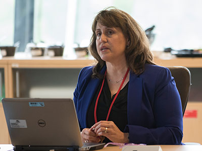
Natella Yurievna Rakhmanina, M.D., Ph.D., FAAP, AAHIVS, director of Ryan White HIV Services at Children’s National Health System, was appointed a commissioner to the Washington, D.C., Regional Planning Commission on Health and HIV.
Dr. Rakhmanina will be among the District of Columbia board and commission appointees honored during a swearing-in ceremony on Sept. 17, 2018, at the Walter Washington Convention Center.
Looking back over the last decade, she says the District has made impressive progress in lowering the prevalence rate of human immunodeficiency virus (HIV), which in 2002 had 1,686 per 100,000 District residents diagnosed with AIDS.
“It was really high. I was stunned coming to clinic and seeing a large number of kids and adolescents in care and many suffering significant complications, as our treatment options were limited at the time,” she says.
Since that time, DC Health has made “incredible investments” and adopted innovative approaches, such as name-based reporting of HIV and a Red Carpet program, to ensure newly diagnosed people are quickly linked with care. As a proud partner of DC Health’s HIV/AIDS, Hepatitis, STD and TB Administration, Children’s National launched a campaign in 2009 to universally test adolescents for HIV in two pediatric emergency departments (ED), she says.
“All teenagers aged 13 and older who arrive for any medical diagnosis are offered an oral HIV test. Children’s National ED-based HIV screening program alone has tested 30,000 children at both of our emergency departments,” she says. “We’re still not at our goal. However, the prevalence of HIV had dropped to 1.9 percent in the latest department of health analysis. We are doing better. We have much fewer people dying from AIDS. We are diagnosing earlier.”
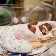
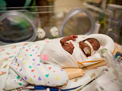
What’s more, trends in mother-to-child transmission, a major route of transmission for pediatric HIV, also have improved in D.C.
“In 2006, our maternal HIV transmission rates were among the highest in the nation. But, in 2013, 2014 and 2015 there were zero cases. We have seen some setbacks recently, however. In 2016, there were three perinatally acquired cases and four in 2017, but these cases came out of the larger Metropolitan D.C. area,” she explains. “Every perinatally transmitted case for us is a red star. We work very closely with the regional departments of health. We really want to get back to zero cases of maternal transmission in the region.”
The regional planning commission meets several times per year to decide how to distribute federal funding in Washington and the Metropolitan D.C. area to support HIV prevention, diagnosis, treatment and care.
“My voice on the council is to make sure I speak up for services for mothers, children and adolescents,” Dr. Rakhmanina says. “The biggest challenge of HIV care remains treating children. There’s a good selection of medicines for adults, but not all are suited for kids. Young children in particular can’t be given one pill once a day. Really young children can’t swallow a pill. Using a liquid formulation, which kids prefer, may mean opening three different bottles twice daily and swallowing a liquid that often doesn’t taste good.”
Adolescents diagnosed with HIV also find medication adherence challenging, she says.
“At that age, they face a lot of challenges to self-acceptance and disease management, in part, because it’s not a physical disability. A young person with HIV may not feel anything,” she says. “They struggle with staying on daily medications. Many of them tell us they don’t want to think about HIV and face stigma.”
Another ongoing challenge is ensuring moms living with HIV remain on medicines after they’ve given birth.
“They’re tremendously committed to continuing treatment while pregnant: Treatment means their babies are born free of HIV,” she says. “That is a great success. Once the baby is born, many times the women bring their babies to be tested, but the woman’s own health becomes less of a priority. We see a drop in adherence once they have the baby.”
By serving on the commission, Dr. Rakhmanina aims to push to extend Children’s commitment to excellence beyond its walls.