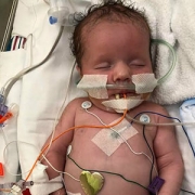
An-Massaro

Keeping an eye on autonomic function for infants with HIE

An-Massaro

“By including heart rate variability measurements and other markers of autonomic function in our current predictive armamentarium,” says An Massaro, M.D., “we may be able to offer new hope for infants with HIE.”

In about two to three in every 1,000 full-term births, babies develop a neurological condition called hypoxic ischemic encephalopathy (HIE) when their brains receive insufficient oxygen. HIE can be a devastating condition, leading to severe developmental or cognitive delays or motor impairments that become more evident as the child grows older. Despite improvements in care – including therapeutic hypothermia, a whole-body cooling method administered shortly after birth that can slow brain damage – about half of children with this condition die from neurological complications by age 2.

Finding ways to identify children with the most severe HIE could help researchers focus their efforts and provide even more intense neuroprotective care, explains An Massaro, M.D., a neonatologist at Children’s National Health System. But thus far, it’s been unclear which symptoms reflect the extent of HIE-induced brain damage.

That’s why Dr. Massaro and colleagues embarked on a study published in the May 2018 issue of Journal of Pediatrics. The team sought to determine whether dysfunction of the autonomic nervous system (ANS) – the auto-pilot part of the nervous system responsible for unconscious bodily functions, such as breathing and digestion – reflected in routine care events can be used as a marker for brain injury severity.

The researchers collected data from 25 infants who were treated for HIE with therapeutic hypothermia at Children’s National. Thanks to multi-modal monitoring, these babies’ medical records hold a treasure trove of information, explains Rathinaswamy B. Govindan, Ph.D., a staff scientist in Children’s Advanced Physiological Signals Processing Lab.

In addition to including continuous heart rate tracings and blood pressure readings that are standard for many infants in the neonatal intensive care unit (NICU), they also recorded cerebral near infrared spectroscopy, a monitor that measures brain tissue oxygen levels. The investigators performed detailed analyses to evaluate how these monitor readings change in response to a variety of routine care events, such as diaper changes, heel sticks, endotracheal tube manipulations and pupil examinations.

The researchers stratified these infants based on how dysfunctional their ANS behaved by using heart rate variability as a marker: The fewer natural fluctuations in heart rate, the more damaged their ANS was thought to be. And they also used non-invasive brain magnetic resonance imaging (MRI) to determine brain damage. They then compared this information with the babies’ physiological responses during each care event.

Their findings show that infants with impaired ANS, based on depressed heart rate variability before the care event, had significantly different responses to these care events compared with babies with intact ANS.

  • For stimulating interventions, such as diaper changes and heel sticks, both heart rate and blood pressure increased in babies with intact ANS but decreased in babies with impaired ones.
  • Shining a light in their pupils led to an expected decreased heart rate with stable blood pressure in ANS-intact infants, but in ANS-impaired infants, there was no responsive change in heart rate and, additionally, a decrease in blood pressure was observed.
  • Responses were similar between the two groups during breathing tube manipulations, except for a slight increase in heart rate a few minutes later in the ANS-impaired group.

These results, Govindan explains, suggest that a real-time, continuous way to assess ANS function may offer insights into the expected physiological response for a given infant during routine NICU care.

“This is exactly the type of additional information that intensivists need to pinpoint infants who may benefit from additional neuroprotective support,” he says. “Right now, it is standard practice to monitor brain activity continuously using electroencephalogram and to check the status of the brain using MRI to assess the response to therapeutic cooling. Neither of these assessments can be readily used by neonatologists at the bedside in real-time to make clinical decisions.”

Assessing ANS function in real-time can help guide neuroprotective care in high-risk newborns by providing insight into the evolving nature of brain damage in these infants, Dr. Massaro adds.

Beyond simply serving as a biomarker into brain injury, poor ANS function also could contribute to the development of secondary injury in newborns with HIE by stymieing the normal changes in heart rate and blood pressure that help oxygenate and heal injured brains. The researchers found that the cumulative duration of autonomic impairment was significantly correlated with the severity of brain injury visible by MRI in this group of infants.

“By including heart rate variability measurements and other markers of autonomic function in our current predictive armamentarium,” says Dr. Massaro, “we may be able to offer new hope for infants with HIE.”

In addition to Dr. Massaro, the Senior Author, study co-authors include Lead Author, Heather Campbell, M.D.; Rathinaswamy B. Govindan, Ph.D., Children’s Advanced Physiological Signals Processing Lab; Srinivas Kota, Ph.D.; Tareq Al-Shargabi, M.S.; Marina Metzler, B.S.; Nickie Andescavage, M.D., Children’s neonatalogist; Taeun Chang, M.D., Children’s neonatal and fetal neurologist; L. Gilbert Vezina, M.D., attending in Children’s Division of Diagnostic Imaging and Radiology; and Adré J. du Plessis, M.B.Ch.B., M.P.H., chief of Children’s Division of Fetal and Transitional Medicine.

This research was supported by the Clinical and Translational Science Institute at Children’s National under awards UL1TR000075 and 1KL2RR031987-01 and the Intellectual and Developmental Disabilities Research Consortium within the National Institutes of Health under award P30HD040677.

Anna Penn

Protecting the fetal brain from harm

Anna Penn

Ongoing placental dysfunction and allopregnanolone loss, not the increase that was expected due to stress, may alter cortical development in complicated pregnancies and put babies at risk, says Anna Penn, M.D., Ph.D.

Researchers long have known that allopregnanolone (ALLO), a derivative of the hormone progesterone, is produced in adults’ brains during times of acute stress and modulates how easily the brain’s neurons fire. ALLO also is produced in the placenta during fetal development, one of more than 200 different hormones that each uniquely contribute to fostering a smooth pregnancy and maintaining a fetus’ overall health. Although ALLO is thought to protect the developing brain in pregnancies complicated by conditions that might harm it, such as high blood pressure, how its levels evolve during pregnancy and in newborns shortly after birth has remained unknown.

Now, a new study presented during the Pediatric Academic Societies (PAS) 2018 annual meeting suggests that the placenta ramps up ALLO production over the second trimester, peaking just as fetuses approach full term.

To investigate this phenomenon, Anna Penn, M.D., Ph.D., a neonatologist/neuroscientist at Children’s National Health System, and colleagues created a designer experimental model to study how premature loss of ALLO alters orderly brain development. Knowing more about the interplay between ALLO and normal development of the cortex, the outer layer of the cerebrum, is a first step that could lead to strategies to rescue this vital brain region.

“The cortex is basically the brain’s command-and-control center for higher functions. In our experimental model, it develops from the middle of gestation through to the end of gestation. If ALLO levels are disrupted just as these cells are being born, neurons migrating to the cortex are altered and the developing neural network is compromised,” says Dr. Penn, senior author of the research presented at PAS 2018. “We’re concerned this same phenomenon occurs in human infants whose preterm birth disrupts their supply of this essential hormone.”

To better understand the human placental hormone pattern, the research team analyzed cord blood or serum samples collected within the first 36 hours of life for 61 preterm newborns born between 24 to 36 gestational weeks. They compared those preemie samples with samples drawn from 61 newborns carried to term who were matched by race, gender, size for gestational age, delivery method and maternal demographics.

They used liquid-chromatography-tandem mass spectrometry, a technique that can precisely analyze trace levels of compounds, to compare levels of 27 different steroids, including ALLO and its precursors as well as better-known adrenal gland hormones, such as cortisol and 17-Hydroxyprogesterone.

“Pregnancies complicated by hypertension tended to correlate with lower ALLO levels, though this finding did not reach statistical significance. This suggests that ongoing placental dysfunction and ALLO loss, not the increase that we expected to be caused by stress, may alter cortical development in these pregnancies and put babies at risk,” Dr. Penn adds. “In addition, having the largest neonatal sample set to date in which multiple steroid hormones have been measured can provide insight into the shifting hormone patterns that occur around 36 weeks gestation, just prior to term. Hopefully, restoring the normal hormonal milieu for preemies or other at-risk newborns will improve neurological outcomes in the future.”

In addition to Dr. Penn, study co-authors include Caitlin Drumm, MedStar Georgetown University Hospital; Sameer Desale, MedStar Health Research Institute; and Kathi Huddleston, Benjamin Solomon and John Niederhuber, Inova Translational Medicine Institute.

Preemie Baby

Brain food for preemies

Preemie Baby

Babies born prematurely – before 37 weeks of pregnancy – often have a lot of catching up to do. Not just in size. Preterm infants typically lag behind their term peers in a variety of areas as they grow up, including motor development, behavior and school performance.

New research suggests one way to combat this problem. The study, led by Children’s researchers and presented during the Pediatric Academic Societies 2018 annual meeting, suggests that the volume of carbohydrates, proteins, lipids and calories consumed by very vulnerable premature infants significantly contributes to increased brain volume and white matter development, even though additional research is needed to determine specific nutritional approaches that best support these infants’ developing brains.

During the final weeks of pregnancy, the fetal brain undergoes an unprecedented growth spurt, dramatically increasing in volume as well as structural complexity as the fetus approaches full term.

One in 10 infants born in the U.S. in 2016 was born before 37 weeks of gestation, according to the Centers for Disease Control and Prevention. Within this group, very low birthweight preemies are at significant risk for growth failure and neurocognitive impairment. Nutritional support in the neonatal intensive care unit (NICU) helps to encourage optimal brain development among preterm infants. However, their brain growth rates still lag behind those seen in full-term newborns.

“Few studies have investigated the impact of early macronutrient and caloric intake on microstructural brain development in vulnerable preterm infants,” says Katherine Ottolini, lead author of the Children’s-led study. “Advanced quantitative magnetic resonance imaging (MRI) techniques may help to fill that data gap in order to better direct targeted interventions to newborns who are most in need.”

The research team at Children’s National Health System enrolled 69 infants who were born younger than 32 gestational weeks and weighed less than 1,500 grams. The infants’ mean birth weight was 970 grams and their mean gestational age at birth was 27.6 weeks.

The newborns underwent MRI at their term-equivalent age, 40 weeks gestation. Parametric maps were generated for fractional anisotropy in regions of the cerebrum and cerebellum for diffusion tensor imaging analyses, which measures brain connectivity and white matter tract integrity. The research team also tracked nutritional data: Grams per kilogram of carbohydrates, proteins, lipids and overall caloric intake.

“We found a significantly negative association between fractional anisotropy and cumulative macronutrient/caloric intake,” says Catherine Limperopoulos, Ph.D., director of Children’s Developing Brain Research Laboratory and senior author of the research. “Curiously, we also find significantly negative association between macronutrient/caloric intake and regional brain volume in the cortical and deep gray matter, cerebellum and brainstem.”

Because the nutritional support does contribute to cerebral volumes and white matter microstructural development in very vulnerable newborns, Limperopoulos says the significant negative associations seen in this study may reflect the longer period of time these infants relied on nutritional support in the NICU.

In addition to Ottolini and Limperopoulos, study co-authors include Nickie Andescavage, M.D., Attending, Children’s Neonatal-Perinatal Medicine; and Kushal Kapse.

newborn in incubator

How EPO saves babies’ brains

newborn in incubator

Researchers have discovered that treating premature infants with erythropoietin can help protect and repair their vulnerable brains.

The drug erythropoietin (EPO) has a long history. First used more than three decades ago to treat anemia, it’s now a mainstay for treating several types of this blood-depleting disorder, including anemia caused by chronic kidney disease, myelodysplasia and cancer chemotherapy.

More recently, researchers discovered a new use for this old drug: Treating premature infants to protect and repair their vulnerable brains. However, how EPO accomplishes this feat has remained unknown. New genetic analyses presented at the Pediatric Academic Societies 2018 annual meeting that was conducted by a multi-institutional team that includes researchers from Children’s National show that this drug may work its neuroprotective magic by modifying genes essential for regulating growth and development of nervous tissue as well as genes that respond to inflammation and hypoxia.

“During the last trimester of pregnancy, the fetal brain undergoes tremendous growth. When infants are born weeks before their due dates, these newborns’ developing brains are vulnerable to many potential insults as they are supported in the neonatal intensive care unit during this critical time,” says An Massaro, M.D., an attending neonatologist at Children’s National Health System and lead author of the research. “EPO, a cytokine that protects and repairs neurons, is a very promising therapeutic approach to support the developing brains of extremely low gestational age neonates.”

The research team investigated whether micro-preemies treated with EPO had distinct DNA methylation profiles and related changes in expression of genes that regulate how the body responds to such environmental stressors as inflammation, hypoxia and oxidative stress.  They also investigated changes in genes involved in glial differentiation and myelination, production of an insulating layer essential for a properly functioning nervous system. The genetic analyses are an offshoot of a large, randomized clinical trial of EPO to treat preterm infants born between 24 and 27 gestational weeks.

The DNA of 18 newborns enrolled in the clinical trial was isolated from specimens drawn within 24 hours of birth and at day 14 of life. Eleven newborns were treated with EPO; a seven-infant control group received placebo.

DNA methylation and whole transcriptome analyses identified 240 candidate differentially methylated regions and more than 50 associated genes that were expressed differentially in infants treated with EPO compared with the control group. Gene ontology testing further narrowed the list to five candidate genes that are essential for normal neurodevelopment and for repairing brain injury:

“These findings suggest that EPO’s neuroprotective effect may be mediated by epigenetic regulation of genes involved in the development of the nervous system and that play pivotal roles in how the body responds to inflammation and hypoxia,” Dr. Massaro says.

In addition to Dr. Massaro, study co-authors include Theo K. Bammler, James W. MacDonald, biostatistician, Bryan Comstock, senior research scientist, and Sandra “Sunny” Juul, M.D., Ph.D., study principal investigator, all of University of Washington.

Research and Education Week awardees embody the diverse power of innovation

cnmc-research-education-week

“Diversity powers innovation” was brought to life at Children’s National April 16 to 20, 2018, during the eighth annual Research and Education Week. Children’s faculty were honored as President’s Award winners and for exhibiting outstanding mentorship, while more than 360 scientific poster presentations were displayed throughout the Main Atrium.

Two clinical researchers received Mentorship Awards for excellence in fostering the development of junior faculty. Lauren Kenworthy, Ph.D received the award for Translational Science and Murray M. Pollack, M.D., M.B.A., was recognized in the Clinical Science category as part of Children’s National Health System’s Research and Education Week 2018.

Dr. Kenworthy has devoted her career to improving the lives of people on the autism spectrum and was cited by former mentees as an inspirational and tireless counselor. Her mentorship led to promising new lines of research investigating methods for engaging culturally diverse families in autism studies, as well as the impact of dual language exposure on cognition in autism.

Meanwhile, Dr. Pollack was honored for his enduring focus on motivating early-career professionals to investigate outcomes in pediatric critical care, emergency medicine and neonatology. Dr. Pollack is one of the founders of the Collaborative Pediatric Critical Care Research Network. He developed PRISM 1 and 2, which has revolutionized pediatric intensive care by providing a methodology to predict mortality and outcome using standardly collected clinical data. Mentees credit Dr. Pollack with helping them develop critical thinking skills and encouraging them to address creativity and focus in their research agenda.

In addition to the Mentorship and President’s Awards, 34 other Children’s National faculty, residents, interns and research staff were among the winners of Poster Presentation awards. The event is a celebration of the commitment to improving pediatric health in the form of education, research, scholarship and innovation that occurs every day at Children’s National.

Children’s Research Institute (CRI) served as host for the week’s events to showcase the breadth of research and education programs occurring within the entire health system, along with the rich demographic and cultural origins of the teams that make up Children’s National. The lineup of events included scientific poster presentations, as well as a full slate of guest lectures, educational workshops and panel discussions.

“It’s critical that we provide pathways for young people of all backgrounds to pursue careers in science and medicine,” says Vittorio Gallo, Ph.D., Children’s chief research officer and CRI’s scientific director. “In an accelerated global research and health care environment, internationalization of innovation requires an understanding of cultural diversity and inclusion of different mindsets and broader spectrums of perspectives and expertise from a wide range of networks,” Gallo adds.

“Here at Children’s National we want our current and future clinician-researchers to reflect the patients we serve, which is why our emphasis this year was on harnessing diversity and inclusion as tools to power innovation,” says Mark L. Batshaw, M.D., physician-in-chief and chief academic officer of Children’s National.

“Research and Education Week 2018 presented a perfect opportunity to celebrate the work of our diverse research, education and care teams, who have come together to find innovative solutions by working with local, national and international partners. This event highlights the ingenuity and inspiration that our researchers contribute to our mission of healing children,” Dr. Batshaw concludes.

Awards for the best posters were distributed according to the following categories:

  • Basic and translational science
  • Quality and performance improvement
  • Clinical research
  • Community-based research and
  • Education, training and program development.

Each winner illustrated promising advances in the development of new therapies, diagnostics and medical devices.

Diversity powers innovation: Denice Cora-Bramble, M.D., MBA
Diversity powers innovation: Vittorio Gallo, Ph.D.
Diversity powers innovation: Mark L. Batshaw, M.D.

child measuring belly with tape measure

Children’s obesity research team presents compelling new findings

child measuring belly with tape measure

Faculty from Children’s National Health System’s Department of Psychology & Behavioral Health set out to learn if any demographic, psychiatric, or cognitive factors play a role in determining if an adolescent should be eligible for bariatric surgery, and what their weight loss outcomes might be. Presenting at the Society for Pediatric Psychology Annual Conference earlier this month, a group of researchers, fellows and clinicians, including surgeons from Children’s National showcased their findings. One of the posters developed by Meredith Rose, LGSW, ML, who works as an interventionist on a Children’s National clinical research team, received special recognition in the Obesity Special Interest Group category.

One presentation reported on a total of 222 pediatric patients with severe obesity, which is defined as 120 percent of the 95th percentile for Body Mass Index. Mean age of the participants was 16 years of age, 71 percent were female and 80 percent where Hispanic or non-White. As part of their preparation for surgery, all patients were required to complete a pre-bariatric surgery psychological evaluation, including a clinical interview and Schedule for Affective Disorders and Schizophrenia (KSADS-PL) screening. The studies by the Children’s teams were based on a medical record review of the pre-screening information. Adolescents being evaluated for surgery had high rates of mental health diagnoses, particularly anxiety and depression, but also included Attention Deficit Hyperactivity Disorder, eating disorders, and intellectual disability.

Another Children’s presentation at the conference looked at weight loss outcomes for adolescents based on IQ and intellectual disability. Overall, neither Full Scale IQ from the Wechsler Abbreviated Intelligence Scale – 2nd edition, nor the presence of an intellectual disability predicted weight loss following surgery.

“The sum of our research found that kids do really well with surgery,” said Eleanor Mackey, PhD, assistant professor of psychology and behavioral health. “Adolescents, regardless of the presence of intellectual disability areas are likely to lose a significant amount of weight following surgery,” added Dr. Mackey.

“This is a particularly important fact to note because many programs and insurers restrict weight loss surgery to ‘perfect’ candidates, while these data points demonstrate that our institution does not offer or deny surgery on the basis of any cognitive characteristics,” says Evan P. Nadler, M.D., associate professor of surgery and pediatrics. “Without giving these kids a chance with surgery, we know they face a lifetime of obesity, as no other intervention has shown to work long-term in this patient population. Our research should empower psychologists and physicians to feel more confident recommending bariatric surgery for children who have exhausted all other weight loss options.”

The research team concluded that examining how individual factors, such as intellectual disability, psychiatric diagnoses, and demographic factors are associated with the surgery process is essential to ensuring adequate and empirically supported guidelines for referral for, and provision of bariatric surgery in adolescents. Next steps by the team will include looking into additional indicators of health improvement, like glucose tolerance, quality of life, or other lab values, to continue evaluating the benefits of surgery for this population.

Sudeepta Basu

GABA concentration in pre-term brain increases with gestational age

Sudeepta Basu

“A more complete understanding of the diagnostic and prognostic importance of GABA and glutamate in the preterm brain will help us to direct treatment strategies for the most vulnerable preterm infants at risk of brain injury,” says Sudeepta K. Basu, M.D.

The major neurotransmitters gamma-aminobutyric acid (GABA) and glutamate are pivotal to fetal and newborn brain development and influence evolution of brain injury and repair following preterm birth. Magnetic resonance spectroscopy (MRS) enables in vivo measurement of brain metabolites. However, GABA and glutamate are found in the developing brain in low concentrations, and their weak signal can be swamped by the stronger signal of more dominant metabolites.

A Children’s research team reports findings from a pilot study utilizing an innovative technique of MRS to reliably measure in vivo GABA in the developing preterm brain. The groundbreaking research done by the team that includes Principal Investigator Sudeepta K. Basu, M.D., neonatology attending at Children’s National Health System, is very unique and original since there are no existing data of in vivo GABA concentrations in the developing cerebellum. Under the mentorship of Catherine Limperopoulos, Ph.D., director of Children’s Developing Brain Research Laboratory, the team of multi-disciplinary specialists is pursuing cutting-edge technologies in advanced MRI neuroimaging to explore brain development and injury in preterm infants.

The research, presented at the Eastern Society for Pediatric Research (ESPR) annual meeting by Dr. Basu, was honored with the “2018 Meritorious Poster Award.” The research titled “Distinct temporal trends of GABA and glutamate in the cerebellum and frontal cortex of preterm infants” reports, for the first time, positive temporal trends in the specific regions of the developing brain intricately involved in cognitive and motor functions. This work lays the foundation for developing novel ways to diagnose, monitor and investigative brain protective therapies for vulnerable prematurely born infants.

The Children’s team performed non-sedated MRS in 44 preterm infants whose mean gestational age at birth was 26.5 weeks, placing voxels at the middle of the cerebellum and the right frontal cortex. GABA and GIx (glutamate combined with glutamine) were positively correlated with post-menstrual age in the frontal cortex, but not the cerebellum.  At the ESPR meeting, the team also presented for the first time that caffeine, a neuroprotective agent in preemies, leads to increased in vivo GABA concentration in the developing frontal cortex.

“Open questions include whether these findings reflect varying paces of maturation and vulnerability to injury among specific regions of the brain. Also, the relationship between clinical factors and medication exposure and changes in the concentration of these neurotransmitters may guide brain protective therapies in future,” Dr. Basu says. “A more complete understanding of the diagnostic and prognostic importance of GABA and glutamate in the preterm brain will help us to direct treatment strategies for the most vulnerable preterm infants at risk of brain injury.”

Children’s senior fellows from Division of Neonatology made four platform presentations during the ESPR conference:

  • “Caffeine increases GABA/Cr ratio in frontal cortex of preterm infants on spectroscopy.” Aditi Gupta; Sudeepta K. Basu, M.D.; Mariam Said, M.D.; Subechhya Pradhan, Linda White; Kushal Kapse; Jonathan Murnick, M.D., Ph.D.; Taeun Chang, M.D.; and Catherine Limperopoulos, Ph.D.
  • “Impact of early nutrition on microstructural brain development in VLBW Infants.” Katherine M. Ottolini, Nickie Andescavage, M.D.; Kushal Kapse; and Catherine Limperopoulos, Ph.D.
  • “Direct measurement of neonatal cardiac output utilizing the CO status monitor.” Simranjeet S. Sran, Mariam Said, M.D.; and Khodayar Rais-Bahrami, M.D.
  • “Cerebro-cerebellar diaschisis in preterm infants following unilateral cerebral parenchymal injury.” Huma Mirza, Yao Wu, Kushal Kapse, Jonathan Murnick, M.D., Ph.D.; Taeun Chang, M.D.; and Catherine Limperopoulos, Ph.D.
Javad Nazarian

Private foundation and researchers partner to cure childhood cancers

Javad Nazarian

Researchers nationally and internally stand the best chance of fulfilling Gabriella Miller’s dream of curing childhood cancers by effectively working together, says Javad Nazarian, Ph.D.

“Thank you for helping me reach my goal.” The handwritten note was penned by Gabriella Miller, a patient treated at Children’s National Health System who ultimately succumbed to an aggressive form of pediatric brain cancer.

Gabriella, then 9 years old, dreamed of curing childhood cancer, including diffuse intrinsic pontine glioma (DIPG), the aggressive pediatric brain tumor that took her life.

Attendees will gather April 14, 2018, for an annual gala held by the Smashing Walnuts Foundation – a group Gabriella started – to celebrate their progress on achieving her goal and to chart future strategic approaches.

“While this foundation was the brainchild of a single person, researchers nationally and internally stand the best chance of fulfilling her dream by working together more effectively,” says Javad Nazarian, Ph.D., M.S.C., the gala’s main speaker. Nazarian is scientific director of Children’s Brain Tumor Institute and is scientific co-chair of the Children’s Brain Tumor Tissue Consortium.

To that end, Children’s National was named a member of a public-private research collective awarded up to $14.8 million by the National Institutes of Health (NIH) to launch a data resource center that cancer sleuths around the world can tap into to accelerate discovery of novel treatments for childhood tumors.

This April, the NIH announced that researchers it funded had completed PanCancer Atlas, a detailed genomic analysis on a data set of molecular and clinical information from more than 10,000 tumors representing 33 types of cancer, including DIPG.

And this January, the NIH announced that it would accept applications from researchers performing whole-genome sequencing studies at one of its Gabriella Miller Kids First research program sequencing facilities. The centers will produce genome, exome and transcriptome sequencing.

Expanding access to these growing troves of data requires a close eye on nuts-and-bolts issues, such as securing sufficient physical data storage space to house the data, Nazarian adds. It’s essential for research teams around the world to have streamlined access to data sets they can analyze as well as contribute to.

“In addition to facilitating researchers’ access to this compiled data, we want to ensure that patients and families feel they are partners in this enterprise by also offering opportunities for them to share meaningful clinical data,” Nazarian says.

Nazarian has been instrumental in expanding the comprehensive biorepository at Children’s National, growing it from just a dozen samples six years ago to thousands of specimens donated by patients with all types of pediatric brain tumors, including DIPG.

“We are so grateful to our patients and families. They share our passion for finding cures and validating innovative treatments for pediatric cancers that defy current treatment. They provide funding through their foundations. Families touched by tragedy offer samples to help the next family avoid reliving their experience,” Nazarian says. “It is in their names – and in Gabriella’s name – that we continue to push ourselves to ‘crack the cure’ for childhood brain cancer.”

Sean Donahue

Pediatric ophthalmology celebrates 75th anniversary in Washington, D.C.

Sean Donahue

Angeline M. Parks Visiting Professor Sean P. Donahue, M.D., Ph.D., (front left) enjoys a light moment during the celebration of the 75th anniversary while Anthony Sandler, M.D., Children’s surgeon in chief, senior vice president of the Joseph E. Robert Jr. Center for Surgical Care and director of the Sheikh Zayed Institute, speaks to the group.

After 75 years dedicated to the eyes of children, the world’s pediatric ophthalmologists gathered in Washington, D.C., the specialty’s birthplace, to share the latest research and innovation in the field. The group gathered for a joint meeting of the International Strabismological Association (ISA) and the American Association for Pediatric Ophthalmology and Strabismus (AAPOS), which was held March 18-22, 2018.

“This year marks the 75th anniversary of our specialty, which was founded right here, at Children’s National, in Washington, D.C., when Dr. Frank Costenbader restricted his practice exclusively to children and began to train residents in the nuance of treating children’s eyes,” says Mohamad S. Jaafar, M.D., chief of the Division of Ophthalmology at Children’s National Health Center. “It is a tremendous honor to welcome my colleagues back to the birthplace of pediatric ophthalmology on this grand occasion.”

In advance of the larger meeting, Children’s Division of Ophthalmology welcomed some of the international attendees to Children’s National for a special gathering on Saturday, March 17, 2018.

The event at Children’s featured a special lecture by this year’s Angeline M. Parks Visiting Professor, Sean P. Donahue, M.D., Ph.D. Dr. Donahue is the Sam and Darthea Coleman Chair in Pediatric Ophthalmology and Chief of Pediatric Ophthalmology at the Children’s Hospital at Vanderbilt. This Annual Visiting Professorship was established by the members of the Costenbader Society (The Children’s National Pediatric Ophthalmology Alumni Society) in memory of Angeline M. Parks, the wife of pediatric ophthalmologist Marshall M. Parks, M.D., to carry on her legacy of establishing a warm and supportive environment between physician and spouse, which benefits the physicians and their young patients.

Three former division chiefs of Ophthalmology at Children’s National, Drs. Costenbader, Parks and Friendly, have national lectureships established in their names to reflect their contributions to the field. Dr. Frank Costenbader, the society’s namesake, established the sub-specialty of pediatric ophthalmology. Dr. Parks founded the Children’s Eye Foundation and the AAPOS, and David S. Friendly, M.D., codified pediatric ophthalmology fellowship training across the United States.

Honor Awards for Children’s pediatric ophthalmologists at ISA-AAPOS

During the ISA-AAPOS meeting, two current Children’s National pediatric ophthalmologists were recognized with Honor Awards for their long-term dedication to pediatric ophthalmology, their patients, and their engagement in the AAPOS to advance the field.

William Madigan, M.D., vice chief of Ophthalmology at Children’s, a professor of surgery at the Uniformed Services University of the Health Sciences, and a clinical professor of Ophthalmology and Pediatrics at the George Washington University School of Medicine and Health Sciences. He was recognized by AAPOS for his long-time service, including:

  • Chair of the organization’s audit committee and the Costenbader Lecture selection committee.
  • Membership on the fellowship directors’ committee that developed nationwide requirements for pediatric ophthalmology fellowships and established the certification process to insure high quality and uniform education in the specialty.
  • Invited lectures in Shanghai, China; Geneva, Switzerland; and Sao Paolo, Brazil, among others.
  • Many posters and presentations about clinical and research topics of importance for members of the AAPOS and other distinguished professional societies.

Marijean Miller, M.D., director of Neonatal Ophthalmology, division research director at Children’s National and clinical professor of Ophthalmology and Pediatrics at the George Washington University School of Medicine and Health Sciences, was recognized by AAPOS for her cumulative contributions to the society, including:

  • Multiple memberships on vital committees, including AAPOS’s training and accreditation committee and audit committee.
  • Presentation of original research via posters and oral presentations on topics including best practices in neonatal clinical care, innovative tools and applications and advocacy for patients and their families.

“We are so grateful to have a team that continues the tradition of excellence in pediatric ophthalmology here at Children’s National,” Dr. Jaafar says. “Drs. Madigan and Miller exemplify the dedication of our division to caring for the children we serve, and to advancing our field. Congratulations to both!”

Sarah Mulkey

MRI finds novel brain defects in Zika-exposed newborns

Sarah Mulkey

“Imaging is constantly helping us make new discoveries with this virus, and in these two cases we found things that had not been previously described,” says Sarah Mulkey, M.D., Ph.D.

Magnetic resonance imaging (MRI) has identified two brain abnormalities never before reported in newborns with prenatal exposure to the Zika virus. Children’s National Health System researchers reported these findings from a study of more than 70 fetuses or newborns with Zika exposure in utero. The study was published in the January 2018 edition of Pediatric Neurology.

The two novel defects – cranial nerve enhancement and cerebral infarction – may join the growing list of neurological findings associated with congenital Zika infection.

“Imaging is constantly helping us make new discoveries with this virus, and in these two cases we found things that had not been previously described,” says Sarah Mulkey, M.D., Ph.D., the study’s lead author and a fetal-neonatal neurologist at Children’s National. Dr. Mulkey works in Children’s Congenital Zika Virus Program, one of the nation’s first comprehensive, dedicated Zika programs.

The research team recommends that postnatal brain MRI be considered in addition to ultrasound for newborns exposed to Zika in utero. “Brain MRI can be performed in the newborn often without sedation and provides an opportunity to look for brain abnormalities we might not catch otherwise – or might not detect until much later,” says Dr. Mulkey.

Birth defects are seen in 6 to 11 percent of pregnancies affected by Zika, and some of the neurological complications in infants are not apparent until well after birth.

Of the two infants in which the new abnormalities were observed, both had normal head size at birth. Neither had smaller-than-normal head size (microcephaly), one of the more severe effects associated with congenital Zika syndrome.

One infant had a normal neurological evaluation at 2 days of age. However, a brain MRI conducted the following day, using gadolinium contrast due to concern of infection, showed enhancement of multiple cranial nerves. “Nerve root enhancement is very rare in a newborn and had not been described with Zika before,” Dr. Mulkey says. “Yet, there was no neurological deficit that we could identify by physical exam.”

The research team acknowledges that the clinical significance of this finding is not yet known.

In the second patient, brain MRI conducted without contrast at 16 days of age revealed a small area consistent with chronic infarction (ischemic stroke) that likely occurred during the third trimester.

“We followed the mother throughout her pregnancy, and both MRI and ultrasound imaging were normal at 28 weeks gestation,” Dr. Mulkey says. “A postnatal ultrasound was also normal, but the postnatal MRI showed a stroke that had occurred at least one month prior to the MRI and after the last fetal study.”

She adds: “This is the first published report of fetal stroke associated with Zika infection, and it may add to our knowledge of what can occur with congenital Zika infection.”

Unlike most congenital infections, Zika virus does not appear to cause viral-induced placental inflammation, which can lead to fetal stroke. So, the authors say they cannot be sure that congenital Zika contributed to the infarct in this case. However, they write, “Given the relatively low incidence of perinatal ischemic infarct and the lack of other maternal- or birth-related risk factors for this patient, Zika infection is considered a possible etiology.”

In both patients, neonatal brain MRI identified subclinical findings that had not previously been described as part of congenital Zika syndrome. As the body of evidence about the Zika virus has grown, the spectrum of associated brain abnormalities has expanded to include considerably more findings than isolated microcephaly.

Data gathered in 2017 from the Centers for Disease Control and Prevention’s Zika pregnancy and infant registry indicates that 25 percent of eligible U.S. infants receive recommended postnatal imaging. Dr. Mulkey said this represents many possible missed opportunities for earlier identification of brain abnormalities.

“Brain MRI should be considered in all newborns exposed to Zika virus in utero, even in the presence of normal birth head circumference, normal cranial ultrasound and normal fetal imaging,” she says. “In both of these patients, the changes we observed were not evident on cranial ultrasound or on fetal MRI and fetal ultrasound.”

In addition to Dr. Mulkey, Children’s co-authors include L. Gilbert Vezina, M.D., Neuroradiology Program director; Dorothy I. Bulas, M.D., chief of Diagnostic Imaging and Radiology; Zarir Khademian, M.D., radiologist; Anna Blask, M.D., radiologist; Youssef A. Kousa, M.S., D.O., Ph.D., child neurology fellow; Lindsay Pesacreta, FNP; Adré  J. du Plessis, M.B.Ch.B., M.P.H., Fetal Medicine Institute director; and Roberta L. DeBiasi, M.D., M.S., senior author and Pediatric Infectious Disease division chief; and Caitlin Cristante, B.S.

Financial support for this research was provided by the Thrasher Research Fund.

Joseph Scafidi

Developing brains are impacted, but can recover, from molecularly targeted cancer drugs

Joseph Scafidi

“The plasticity of the developing brain does make it susceptible to treatments that alter its pathways,” says Joseph Scafidi, D. O., M.S. “Thankfully, that same plasticity means we have an opportunity to mitigate the damage from necessary and lifesaving treatments by providing the right support after the treatment is over.”

One of the latest developments in oncology treatments is the advancement of molecularly targeted therapeutic agents. These drugs can be used to specifically target and impact the signaling pathways that encourage tumor growth, and are also becoming a common go to for ophthalmologists to treat retinopathy of prematurity in neonates.

But in the developing brain of a child or adolescent, these pathways are also crucial to the growth and development of the brain and central nervous system.

“These drugs have been tested in vitro, or in tumor cells, or even in adult studies for efficacy, but there was no data on what happens when these pathways are inhibited during periods when their activation is also playing a key role in the development of cognitive and behavioral skills, as is the case in a growing child,” says Joseph Scafidi, D. O., M.S., a neuroscientist and pediatric neurologist who specializes in neonatology at Children’s National Health System.

As it turns out, when the drugs successfully inhibit tumor growth by suppressing receptors, they can also significantly impact the function of immature brains, specifically changing cognitive and behavioral functions that are associated with white matter and hippocampal development.

The results appeared in Cancer Research, and are the first to demonstrate the vulnerability of the developing brain when this class of drugs is administered. The pre-clinical study looked at the unique impacts of drugs including gefitinib (Iressa), sunitib malate (Sutent) and rapamycin (Sirolimus) that target specific pathways responsible for the rapid growth and development that occurs throughout childhood.

The agents alter signaling pathways in the developing brain, including decreasing the number of oligodendrocytes, which alters white matter growth. Additionally, the agents also impact the function of specific cells within the hippocampus related to learning and memory. When younger preclinical subjects were treated, impacts of exposure were more significant. Tests on the youngest pre-clinical subjects showed significantly diminished capacity to complete cognitive and behavioral tasks and somewhat older, e.g. adolescent, subjects showed somewhat fewer deficits. Adult subjects saw little or no deficit.

“The impacts on cognitive and behavioral function for the developing brain, though significant, are still less detrimental than the widespread impacts of chemotherapy on that young brain,” Dr. Scafidi notes. “Pediatric oncologists, neuro-oncologists and ophthalmologists should be aware of the potential impacts of using these molecularly targeted drugs in children, but should still consider them as a treatment option when necessary.”

The effects are reversible

Researchers also found measurable improvements in these impaired cognitive and behavioral functions when rehabilitation strategies such as environmental stimulation, cognitive therapy and physical activity were applied after drug exposure.

“The plasticity of the developing brain does make it susceptible to treatments that alter its pathways,” says Dr. Scafidi. “Thankfully, that same plasticity means we have an opportunity to mitigate the damage from necessary and lifesaving treatments by providing the right support after the treatment is over.”

Many major pediatric oncology centers, including the Center for Cancer and Blood Disorders at Children’s National, already incorporate rehabilitation strategies such as cognitive therapy and increased physical activity to help pediatric patients return to normal life following treatment. The results from this study suggest that these activities after treatment for pediatric brain tumors may play a vital role in improving recovery of brain cognitive and behavioral function in the pediatric population.

This research was funded by grants to Dr. Scafidi from the National Brain Tumor Society, Childhood Brain Tumor Foundation and the National Institutes of Health.

banner year

2017: A banner year for innovation at Children’s National

banner year

In 2017, clinicians and research faculty working at Children’s National Health System published more than 850 research articles about a wide array of topics. A multidisciplinary Children’s Research Institute review group selected the top 10 articles for the calendar year considering, among other factors, work published in high-impact academic journals.

“This year’s honorees showcase how our multidisciplinary institutes serve as vehicles to bring together Children’s specialists in cross-cutting research and clinical collaborations,” says Mark L. Batshaw, M.D., Physician-in-Chief and Chief Academic Officer at Children’s National. “We’re honored that the National Institutes of Health and other funders have provided millions in awards that help to ensure that these important research projects continue.”

The published papers explain research that includes using imaging to describe the topography of the developing brains of infants with congenital heart disease, how high levels of iron may contribute to neural tube defects and using an incisionless surgery method to successfully treat osteoid osteoma. The top 10 Children’s papers:

Read the complete list.

Dr. Batshaw’s announcement comes on the eve of Research and Education Week 2018 at Children’s National, a weeklong event that begins April 16, 2018. This year’s theme, “Diversity powers innovation,” underscores the cross-cutting nature of Children’s research that aims to transform pediatric care.

Pedbot video game

New robotic therapies for cerebral palsy

Little girl on hippobot

The hippobot is a mechanical horseback riding simulator that provides hippotherapy for children.

Cerebral palsy is the most common type of movement disorder in children, affecting 1 in 500 babies born each year. For these infants, learning to sit up, stand and walk can be a big challenge which often requires years of physical therapy to stretch and strengthen their muscles. A team led by Kevin Cleary, Ph.D., technical director of the Bioengineering Initiative at Children’s National Health System, and Sally Evans, M.D., director of Pediatric Rehabilitation Medicine at Children’s National, has created two new types of robotic therapy that they hope will make physical therapy more enjoyable and accessible for children.

Hippobot equine therapy simulator

One of the most effective types of therapy for children with cerebral palsy is hippotherapy, which uses horseback riding to rehabilitate children with neurological and musculoskeletal disabilities. The movement of horses helps riders with cerebral palsy improve endurance, balance and core strength, which in turns helps them gain the ability to sit without support. If a child with cerebral palsy does not master independent sitting early in life, he or she may never gain the ability to stand or walk. Unfortunately, many children never have the chance to experience hippotherapy due to geographical constraints and cost issues.

To increase patient access to hippotherapy, the bioengineering team (Reza Monfaredi, Ph.D.; Hadi Fooladi Talari, M.S.; Pooneh Roshani Tabrizi, Ph.D.; and Tyler Salvador, B.S.) developed the hippobot — a mechanical horseback riding simulator that provides hippotherapy for children ages 4 to 10 in the office setting. To create the hippobot, the researchers mounted a carousel horse on a six-degree of freedom commercial motion platform (the platform moves in the x, y and z directions and rotates about roll, pitch and yaw axes). They then programmed the platform to simulate a horse walking, trotting and cantering.

“Several experienced horse riders have tried the motion platform and commented that it gives a realistic feel,” says Dr. Cleary.

The team then incorporated optical tracking of the hippobot rider’s spine and pelvis to monitor their posture and created a virtual reality video display that simulates a horse moving down a pier. As other animals come towards the horse, the rider must lean right or left to avoid them.  The trackers on their back show which way they are leaning and feed that information into the gaming system.

“We wanted to see how the patient’s spine reacts as the horse moves through different patterns, and if the patients get better at maintaining their posture over several sessions,” says Dr. Cleary.

To date the system has been used with several children with cerebral palsy under an IRB-approved study. All of the participants enjoyed riding the horse and came back for multiple sessions.

The hippobot system was developed in close collaboration with the Physical Medicine and Rehabilitation Division at Children’s National, including Olga Morozova, M.D., Justin Burton, M.D., and Justine Belschner, P.T.

Pedbot ankle rehabilitation system

Pedbot video game

Patients use pedbot as an input device to pilot an airplane through a series of hoops. The level of the difficulty of the game can be easily adjusted based on the patient’s capability and physical condition.

More than half of children with cerebral palsy also have gait impairment as a result of excessive plantar flexion and foot inversion/eversion, or equinovarus/equinovalgus at their ankle and foot. To help these patients, Dr. Cleary’s team developed the pedbot — a small robot platform that enables better strengthening, motor control and range of motion in the ankle joint.

“Children with cerebral palsy have difficulty walking in part because they have trouble controlling their feet,” explains Dr. Evans. “Use of pedbot as part of therapy can help to give them increased control of their feet.”

Most ankle rehabilitation robots are limited in their movements, and have only one or two degrees of freedom, focusing on ankle dorsiflexion/plantarflexion and sometimes inversion/eversion. Pedbot is unique in that it has three degrees of freedom with a remote center of motion in the ankle joint area that allows it to move in ways other devices can’t.

The pedbot platform can move in three translational directions (x, y and z) and also rotate about three axes (the x, y and z axes). As an analogy, this is similar to the movement of a flight simulator. The system also includes motors and encoders at each axis and can be used in passive and active modes.

In both modes, the patient sits on a therapy chair with their foot strapped to the robotic device. In the passive mode, the therapist assists the patient in training motions along each axis. The robot can then repeat the motion under therapist supervision while incrementally increasing the range of motion as desired by the therapist.

For the active mode or “gaming” mode, the team developed a video game based on an airplane motif. Patients use pedbot as an input device to pilot an airplane through a series of hoops. The level of the difficulty of the game can be easily adjusted based on the patient’s capability and physical condition.

To date, four patients have participated in an IRB-approved clinical trial for the pedbot. All of the patients enjoyed the game and they were willing to continue to participate as suggested by a physiotherapist.

The pedbot team, in addition to the engineers mentioned above, includes Catherine Coley, P.T.; Stacey Kovelman, P.T.; and Sara Alyamani, B.A. In future work, they plan to expand the system to include electromyography measurements with Paola Pergami, M.D.,Ph.D. They also are planning to develop a low cost, 3D printed version for the home market so children can do Pedbot therapy every day.

Nathan Smith

Sounding the alarm on fluorescent calcium dyes

“This study serves as a warning to other neuroscientists,” says Nathan A. Smith, Ph.D., a new principal investigator in the Center for Neuroscience Research at Children’s National Health System and first author of the study.

Scientists using chemical based fluorescent dyes to study the calcium dynamics of the cells within the central nervous system suspected that something external was disrupting normal cell function in their studies.

Neuroscientists at the University of Rochester Medical Center and Children’s National Health System have confirmed their suspicions by capturing data that shows, for the first time, how these fluorescent calcium dyes are causing cell damage when loaded over an extended period of time.  Their findings appeared in Science Signaling.

“This study serves as a warning to other neuroscientists,” says Nathan A. Smith, Ph.D., a new principal investigator in the Center for Neuroscience Research at Children’s National Health System and first author of the study. “As many of my colleagues have noted, we’ve known that something was going on, but now, we have evidence that the longer these dyes stay in cells, or the longer time it takes to load them into cells, the more problems we see with normal cell function.”

The study comparatively analyzed the effects of chemical and genetically encoded calcium ion indicators on cellular functions, which had not been previously performed. The outcomes showed that all of the fluorescent calcium dyes, including Fluo-4, Rhod-2, and FURA-2, had negative consequences for a number of cellular functions. For example, the dyes inhibited the sodium potassium pump (Na,K-ATPase), a membrane protein essential for many cellular membrane functions including the exchange between intracellular sodium ions (Na+) and extracellular potassium ions (K+). Inhibiting the Na,K-ATPase process results in the buildup of K+ ions in the synapses, leading to errant neural firing that has been linked to a number of disorders, including epilepsy. Additional observed impacts of exposure to the dyes included reduced cell viability, decreased glucose uptake, increased lactate release and cell swelling.

“Now, our field needs to take a step back and reevaluate findings that may have been influenced by these chemical trackers, to make sure that our observations were driven by our intended manipulations and not this additional factor,” Dr. Smith continues.

Non-chemical alternatives

Tracking calcium ions and their dynamics within the central nervous system through fluorescent calcium indicators is the primary method of measuring glial activity and glial interactions with other cell types. Unlike neurons, glial cells are electrically non-excitable; therefore electrical-based recording methods used to measure neuronal activity are ineffective.

In recent years, neuroscientists have developed additional methods to track calcium ions: genetically encoded calcium indicators (GECIs), which have grown in use since 2008. These GECIs have evolved over the years to have higher signal to noise ratios, target specific cell types and sub-compartments, and remain stable over time.*

Dr. Smith’s lab uses GECIs exclusively to monitor the calcium dynamics of glial brain cells called astrocytes as well as their interactions with other cells such as neurons. His work now seeks to understand how those interactions influence neural networks and how they operate differently in neurodevelopmental disorders, including attention-deficit hyperactivity disorder (ADHD), epilepsy and others.

“Our field has continued using these traditional calcium indicator dyes in labs because they are familiar and affordable,” Dr. Smith notes. “Our findings are a clear call to action that it’s time to revisit some of those approaches in favor of new technology.”

newborn in incubator

Tracking oxygen saturation with vital signs to identify vulnerable preemies


Khodayar-Rais-Bahrami

PDF Version

What’s known

Critically ill infants in neonatal intensive care units (NICU) require constant monitoring of their vital signs. Invasive methods, such as using umbilical arterial catheters to check blood pressure, are the gold standard but pose significant health risks. Low-risk noninvasive monitoring, such as continuous cardiorespiratory monitors, can measure heart rate, respiratory rate and blood oxygenation. A noninvasive technique called near-infrared spectroscopy (NIRS) can gauge how well tissues, including the brain, are oxygenated. While NIRS long has been used to monitor oxygenation in conditions in which blood flow is altered, such as bleeding in the brain, how NIRS values relate to other vital sign measures in NICU babies was unknown.

What’s new

A research team led by Khodayar Rais-Bahrami, M.D., a neonatologist at Children’s National Health System, investigated this question in 27 babies admitted to Children’s NICU. The researchers separated these subjects into two groups: Low birth weight (LBW, less than 1.5 kg or 3.3 pounds) and moderate birth weight (MBW, more than 1.5 kg). Then, they looked for correlations between information extracted from NIRS, such as tissue oxygenation (specific tissue oxygen saturation, StO2) and the balance between oxygen supply and consumption (fractional tissue oxygen extraction, FTOE), and various vital signs. They found that StO2 increased with blood pressure for LBW babies but decreased with blood pressure for MBW babies. Brain and body FTOE in LBW babies decreased with blood pressure. In babies with abnormal brain scans, brain StO2 increased with blood pressure and brain FTOE decreased with blood pressure. Together, the researchers suggest, these measures could give a more complete picture of critically ill babies’ health.

Questions for future research

Q: Can NIRS data be used as a surrogate for other forms of monitoring?

Q: How could NIRS data help health care professionals intervene to improve the health of critically ill infants in the NICU?

Source: Significant correlation between regional tissue oxygen saturation and vital signs of critically ill infants.” B. Massa-Buck, V. Amendola, R. McCloskey and K. Rais-Bahrami. Published by Frontiers in Pediatrics Dec. 21, 2017.

Pregnant-Mom

MRI opens new understanding of fetal growth restriction

Pregnant-Mom

Quantitative MRI can identify placental dysfunction complicated by fetal growth restriction earlier, creating the possibility for earlier intervention to minimize harm to the developing fetus.

A team of researchers has found that quantitative magnetic resonance imaging (MRI) can identify pregnancies where placental dysfunction results in fetal growth restriction (FGR), creating the possibility for earlier FGR detection and intervention to augment placental function and thus minimize harm to the fetal brain.

The study, published online in the Journal of Perinatology, reports for the first time that in vivo placental volume is tied to global and regional fetal brain volumes.

Placental insufficiency is a known risk factor for impaired fetal growth and neurodevelopment. It may cause the fetus to receive inadequate oxygen and nutrients, making it difficult to grow and thrive. The earlier placental insufficiency occurs in a pregnancy, the more serious it can be. But detecting a failing placenta before the fetus is harmed has been difficult.

One additional challenge is that a fetus may be small because the placenta is not providing adequate nourishment. Or the fetus simply may be genetically predisposed to be smaller. Being able to tell the difference early can have a lifelong impact on a baby. Infants affected by FGR can experience behavioral problems, learning difficulties, memory and attention deficits, and psychiatric issues as the child grows into adolescence and adulthood.

“Our study proved that MRI can more accurately determine which pregnancies are at greater risk for impaired fetal health or compromised placenta function,” says Nickie Andescavage, M.D., the study’s lead author and a specialist in neonatology and neonatal neurology and neonatal critical care at Children’s National Health System. “The earlier we can identify these pregnancies, the more thoughtful we can be in managing care.”

Dr. Andescavage’s research focus has been how fetal growth affects labor, delivery and postnatal complications.

Nickie-Andescavage-Niforatos

“Our study proved that MRI can more accurately determine which pregnancies are at greater risk for impaired fetal health or compromised placenta function,” says Nickie Andescavage, M.D., the study’s lead author.

“We don’t have a good understanding of why FGR happens, but we do know it’s hard to identify during pregnancy because often there are no signs,” says Dr. Andescavage. “Even when detected, it’s hard to follow. But if we’re aware of it, we can better address important questions, like when to deliver an at-risk fetus.”

In the study, the team measured placental and fetal brain growth in healthy, uncomplicated pregnancies and in pregnancies complicated by FGR. A total of 114 women participated, undergoing ultrasound, Doppler ultrasound and MRI imaging to measure placental volume and fetal brain volume.

An ultrasound test is often what detects FGR, but the measurements generated by ultrasound can be non-specific. In addition, reproducibility issues with 3D sonography limit its use as a standalone tool for placental assessment. Once FGR is detected via ultrasound, this study showed that complementary MRI provides more accurate structural measures of the fetal brain, as well as more detail and insight into placental growth and function.

“Our team has studied FGR for a few years, using imaging to see that’s happening with the fetus in real time,” says Dr. Andescavage. “The relationship of placental volume and fetal brain development had not been previously studied in utero.”

In pregnancies complicated by FGR, MRI showed markedly decreased placental and brain volumes. The team observed significantly smaller placental, total brain, cerebral and cerebellar volumes in these cases than in the healthy controls. The relationship between increasing placental volume and increasing total brain volume was similar in FGR and in normal pregnancies. However, the study authors write “the overall volumes were smaller and thus shifted downward in pregnancies with FGR.”

In addition, FGR-complicated pregnancies that also showed abnormalities in Doppler ultrasound imaging had even smaller placental, cerebral and cerebellar volumes than pregnancies complicated by FGR that did not have aberrations in Doppler imaging.

Since this study showed that quantitative fetal MRI can accurately detect decreased placental and brain volumes when FGR is present, Dr. Andescavage believes this imaging technique may give doctors important new insights into the timing and possibly the mechanisms of brain injury in FGR.  “Different pathways can lead to FGR. With this assessment strategy, we could potentially elucidate those,” she adds.

Using quantitative MRI to identify early deviations from normal growth may create opportunities for future interventions to protect the developing fetal brain. New treatments on the horizon promise to address placental health. MRI could be used to investigate these potential therapies in utero. When those therapies become available, it could allow doctors to monitor treatment effects in utero.

Study co-authors include Adré J. du Plessis, M.B.Ch.B., M.P.H., Director of Children’s Fetal Medicine Institute; Marina Metzler; Dorothy Bulas, M.D., FACR, FAIUM, FSRU, Chief of Children’s Division of Diagnostic Imaging and Radiology; L. Gilbert Vezina, M.D., Director of Children’s Neuroradiology Program; Marni Jacobs; Catherine Limperopoulos, Ph.D., Director of Children’s Developing Brain Research Laboratory and study senior author; Sabah N. Iqbal, MedStar Washington Hospital Center; and Ahmet Alexander Baschat, Johns Hopkins Center for Fetal Therapy.

Research reported in this post was supported by the Canadian Institutes of Health Research, MOP-81116; the National Institutes of Health under award numbers UL1TR000075 and KL2TR000076; and the Clinical and Translational Science Institute at Children’s National.

Neonatal baby

Multidisciplinary experts help CDC’s Zika research

“We are very excited about this next phase in our Zika research,” says Roberta L. DeBiasi, M.D., M.S. “It is a natural extension of our earlier participation as subject matter experts assisting as the CDC developed and published guidelines to inform the care of Zika-exposed and Zika-infected infants across the nation and U.S. territories.”

The Centers for Disease Control and Prevention (CDC) is funding three multidisciplinary experts from the Congenital Zika Virus Program at Children’s National Health System to collaborate on two of the CDC’s longitudinal Zika research projects in Colombia, South America.

“Zika en embarazadas y niños en Colombia” (ZEN) is a research study jointly designed by Colombia’s Instituto Nacional de Salud (INS) and the CDC to evaluate the association between Zika virus infection and adverse maternal, fetal and infant health outcomes. The study is following a large cohort of Colombian women from the first trimester of pregnancy, their male partners and their infants.

Under the six-month contract, Roberta L. DeBiasi, M.D., M.S., Sarah B. Mulkey, M.D., Ph.D., and Cara Biddle, M.D., M.P.H., will serve as consultants for the ZEN study providing expertise in pediatric infectious diseases, neurology, neurodevelopment and coordination of the complex care needs of Zika-affected infants.

The federal funding will underwrite the consultants’ work effort, as well as travel to the CDC’s headquarters in Atlanta and to research sites in Colombia. To that end, Drs. DeBiasi, Mulkey and Biddle participated in a December 2017 kickoff meeting, joining ZEN team leaders based in the U.S. at the CDC, as well as the INS in Colombia, with whom they will conduct research and collaborate academically.

Cara-Biddle-and-Sarah-Mulkey

Cara Biddle, M.D., M.P.H., and Sarah B. Mulkey, M.D., Ph.D., also will serve as consultants for the ZEN study.

“We are very excited about this next phase in our Zika research,” says Dr. DeBiasi, chief of the Division of Pediatric Infectious Diseases and co-director of the Children’s Zika program. “It is a natural extension of our earlier participation as subject matter experts assisting as the CDC developed and published guidelines to inform the care of Zika-exposed and Zika-infected infants across the nation and U.S. territories.”

Children’s National is leading its own longitudinal studies in Colombia that explore such questions as whether Zika-exposed infants whose neuroimaging appears normal when they are born experience any longer-term neurological issues and the role of genetics in neurologic injury following congenital Zika virus exposure and infection.

Carlos Ferreira Lopez

Researchers discover new gene variant for inherited amino acid-elevating disease

Carlos Ferreira Lopez

PDF Version

What’s known

Hypermethioninemia is a rare condition that causes elevated levels of methionine, an essential amino acid in humans. This condition stems from genetic variations inherited from one or both parents. Some forms of hypermethioninemia are recessive, meaning that two copies of defective genes are necessary to cause this disease. Other forms are dominant, meaning that only one copy can cause hypermethioninemia. Recessive forms of the disease tend to have more serious consequences, causing elevated methionine levels throughout life and leading to changes in the brain’s white matter visible on magnetic resonance imaging that can cause neurological problems. The dominant forms are generally thought to be largely benign and require minimal follow-up.

What’s new

A research team led by Carlos Ferreira Lopez, M.D., a medical geneticist at Children’s National Health System, discovered a new gene variant that had not been associated with hypermethioinemia previously when an infant who had tested positive for elevated methionine on newborn blood-spot screening came in for a follow-up evaluation. While the majority of dominant hypermethioninemia are caused by a genetic mutation known as MAT1A p.Arg264His, the child didn’t have this or any of the common recessive hypermethioninemia mutations. Genetic testing showed that she carried a different mutation to the MAT1A gene known as p.Ala259Val, of which she carried only a single copy. The child fit the typical profile of having the dominant form of the disease, with methionine levels gradually declining over time. Testing of her mother showed that she carried the same gene variant, with few consequences other than a hepatitis-like illness as a child. Because liver disease can accompany dominant hypermethioninemia, the infant’s doctors will continue periodic follow-up to ensure she remains healthy.

Questions for future research

Q: Besides the potential for harmful liver effects, does dominant hypermethioninemia have other negative consequences?

Q: How common is this gene variant, and are certain people at more risk for carrying it?

Source: Confirmation that MAT1A p.Ala259Val mutation causes autosomal dominant hypermethioninemia. Muriello, M.J., S. Viall, T. Bottiglieri, K. Cusmano-Ozog and C. R. Ferreira. Published by Molecular Genetics and Metabolism Reports December 2017.

William Gaillard

Putting childhood epilepsy in the spotlight at American Epilepsy Society Meeting

William Gaillard

“We aim to build the evidence base for treatments that are effective specifically for children with epilepsy,” says William D. Gaillard, M.D., chief of Child Neurology, Epilepsy and Neurophysiology, and director of the Comprehensive Pediatric Epilepsy Program.

While epilepsy affects people of all ages, the unique way it manifests in infants, children and adolescents can be attributed in part to the complexities of the growing and developing brain. Researchers from the Children’s National Comprehensive Pediatric Epilepsy Program brought their expertise on the challenges of understanding and treating epilepsy in children to the recent American Epilepsy Society Annual Meeting, the largest professional gathering on epilepsy in the world.

“We aim to build the evidence base for treatments that are effective specifically for children with epilepsy,” says William D. Gaillard, M.D., chief of Child Neurology, Epilepsy and Neurophysiology, and director of the Comprehensive Pediatric Epilepsy Program. “We have learned much from studies in adult populations but technologies like functional MRI allow us to get in-depth understanding, often in non-invasive ways, of precisely how epilepsy is impacting a child.”

Dr. Gaillard was also recently elected to serve as the Second Vice President of the American Epilepsy Society. “The AES is the largest multidisciplinary professional and scientific society dedicated to the understanding, treatment and eradication of epilepsy and associated disorders, and I am honored to serve as the new Second Vice President,” he said.

The team’s presentations and poster sessions focused on several key areas in pediatric epilepsy:

Better ways to see, measure and quantify activity and changes in the brain for children with epilepsy before, during and after surgery

  • Novel applications of fMRI for children with epilepsy
    • Evaluation of an fMRI tool that tracks verbal and visual memory in children with epilepsy – one of the first to capture memory functions in this population of children using noninvasive fMRI;
    • Early study of the use of “resting-state” fMRI to map language skills before epilepsy surgery – an important first step toward noninvasively evaluating children who are too young or neurologically impaired to follow tasks in traditional MRI studies;
  • A study of whether intraoperative MRI, i.e. imaging during neurosurgery, allows for more complete removal of abnormal brain tissue associated with focal cortical dysplasia in children, which is a common cause of intractable epilepsy;
  • A preliminary case review of existing data to see if arterial spin labeling MRI, which measures blood flow to the brain, has potential to identify blood flow changes in specific locations of the brain where seizures occur;
  • An analysis of language laterality – the dominant side of the brain controlling language –  questioning the true reasons that the brains of children with epilepsy have differences in the hemisphere that predominantly controls language;
  • A review of some common assessments of language and working memory that are used pre- and post-operatively to gauge the impacts of pediatric epilepsy surgery. The study found that using multiple assessments, and studying results individually rather than as a group average, resulted in a more complete picture of the outcomes of surgery on these areas of brain function;
  • A preliminary study examining whether continuous EEG monitoring of neonates with hypoxic ischemic encephalopathy, or lack of oxygen to the brain, can be a reliable predictor of neurodevelopmental outcomes while the infant is undergoing therapeutic hypothermia.

“In order to expand our understanding of causes, impacts and outcomes, the range of research is broad given the complexity of epilepsy,” says Madison M. Berl, Ph.D. “This is the only way we can contribute to the goal of providing our colleagues and the families they serve with better resources to make informed decisions about how best to assess and treat pediatric epilepsy.”

The molecular, genetic and biological factors that contribute to onset and severity of pediatric epilepsy

  • A retrospective study of young patients with malformations in cortical development that are important causes of childhood epilepsy;
  • Investigation of a simple saliva test to effectively identify the presence of two common viral infections, human herpesvirus-6B and Epstein-Barr virus, that may be contributors to onset of epilepsy in otherwise normally functioning brains;
  • A preliminary review of the possible relationship between febrile infection-related epilepsy syndrome and the co-occurrence of another neuro-inflammatory condition – hemophagocytic lymphohistiocytosis.

Madison Berl, Ph.D., director of research in the Division of Pediatric Neuropsychology, and a pediatric neuropsychologist in the Comprehensive Pediatric Epilepsy Program, adds, “In order to expand our understanding of causes, impacts and outcomes, the range of research is broad given the complexity of epilepsy. This is the only way we can contribute to the goal of providing our colleagues and the families they serve with better resources to make informed decisions about how best to assess and treat pediatric epilepsy.”

effects of cardiopulmonary bypass surgery on the white matter of piglets.

The effects of cardiopulmonary bypass on white matter development

 cardiopulmonary bypass

Nobuyuki Ishibashi, M.D., and a team of researchers looked the effects of cardiopulmonary bypass surgery on the white matter of an animal model.

Mortality rates for infants born with congenital heart disease (CHD) have dramatically decreased over the past two decades, with more and more children reaching adulthood. However, many survivors are at risk for neurodevelopmental abnormalities  associated with cardiopulmonary bypass surgery (CPB), including long-term injuries to the brain’s white matter and neural connectivity impairments that can lead to neurological dysfunction.

“Clinical studies have found a connection between abnormal neurological outcomes and surgery, but we don’t know what’s happening at the cellular level,” explains Nobuyuki Ishibashi, M.D., Director of the Cardiac Surgery Research Laboratory at Children’s National. To help shed light on this matter, Ishibashi and a team of researchers looked at the effects of CPB on the white matter of an animal model.

The research team randomly assigned models to receive one of three CPB-induced insults: a sham surgery (control group); full-flow bypass for 60 minutes; and 25°C circulatory arrest for 60 minutes. The team then used fractional anisotropy — a technique that measures the directionality of axon mylenation — to determine white matter organization in the models’ brains. They also used immunohistology techniques to assess the integrity of white matter oligodendrocytes, astrocytes and microglia.

The results, published in the Journal of the American Heart Association, show that white matter experiences region-specific vulnerability to insults associated with CPB, with fibers within the frontal cortex appearing the most susceptible. The team also found that fractional anisotropy changes after CPB were insult dependent and that regions most resilient to CPB-induced fractional anisotropy reduction were those that maintained mature oligodendrocytes.

From these findings, Ishibashi and his co-authors conclude that reducing alterations of oligodendrocyte development in the frontal cortex can be both a metric and a goal to improve neurodevelopmental impairment in the congenital heart disease population. “Because we are seeing cellular damage in these regions, we can target them for future therapies,” explains Ishibashi.

The study also demonstrates the dynamic relationship between fractional anisotropy and cellular events after pediatric cardiac surgery, and indicates that the technique is a clinically relevant biomarker in white matter injury after cardiac surgery.