“My best advice to future clinician-scientists is to stay curious and open-minded; I doubt I could have predicted my current research interest or described the path between the study of early oligodendrocyte maturation to in vivo placental development, but each experience along the way – both academic and clinical – has led me to where I am today,” Nickie Andescavage, M.D., writes.
Too often, medical institutions erect an artificial boundary between caring for the developing fetus inside the womb and caring for the newborn whose critical brain development continues outside the womb.
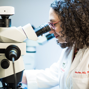
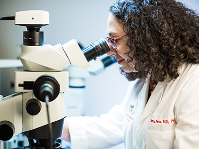
“To improve neonatal outcomes, we must transform our current clinical paradigms to begin treatment in the intrauterine period and continue care through the perinatal transition through strong collaborations with obstetricians and fetal-medicine specialists,” writes Nickie Andescavage, M.D., an attending in Neonatal-Perinatal Medicine at Children’s National.
Dr. Andescavage’s commentary was published online March 25, 2019, in Pediatrics Research and accompanies recently published Children’s research about differences in placental development in the setting of placental insufficiency. Her commentary is part of a new effort by Nature Publishing Group to spotlight research contributions from early career investigators.
The placenta, an organ shared by a pregnant woman and the developing fetus, plays a critical but underappreciated role in the infant’s overall health. Under the mentorship of Catherine Limperopoulos, Ph.D., director of MRI Research of the Developing Brain, and Adré J. du Plessis, M.B.Ch.B., MPH, chief of the Division of Fetal and Transitional Medicine, Dr. Andescavage works with interdisciplinary research teams at Children’s National to help expand that evidence base.
While attending Cornell University as an undergraduate, Dr. Andescavage had an early interest in neuroscience and neurobehavior. As she continued her education by attending medical school at Columbia University, she corroborated an early instinct to work in pediatrics.
It wasn’t until the New Jersey native began pediatric residency at Children’s National that those complementary interests coalesced into a focus on brain autoregulation and autonomic function in full-term and preterm infants and imaging the brains of both groups. In normal, healthy babies the autonomic nervous system regulates heart rate, blood pressure, digestion, breathing and other involuntary activities. When these essential controls go awry, babies can struggle to survive and thrive.
“My best advice to future clinician-scientists is to stay curious and open-minded; I doubt I could have predicted my current research interest or described the path between the study of early oligodendrocyte maturation to in vivo placental development, but each experience along the way – both academic and clinical – has led me to where I am today,” Dr. Andescavage writes in the commentary.
https://innovationdistrict.childrensnational.org/wp-content/uploads/2019/04/Nickie-Andescavage.png300400Innovation Districthttps://innovationdistrict.childrensnational.org/wp-content/uploads/2025/09/InnovationDistrict_CN_WebHeader-1396px-1030x151.pngInnovation District2019-04-16 14:30:532026-04-13 09:40:33To understand the preterm brain, start with the fetal brain
Following the noted success of CAR-T cells in treating leukemia, Eugene Hwang, M.D., and a team of physicians at Children’s National are studying the efficacy of using these white blood cell “armies” to fight central nervous system (CNS) tumors.
Following the noted success of CAR-T cells in treating leukemia, physicians at Children’s National are studying the efficacy of using these white blood cell “armies” to fight central nervous system (CNS) tumors. Employing a strategy of “supertraining” the cells to target and attack three tumor targets as opposed to just one, Eugene Hwang, M.D., and the team at Children’s are optimistic about using this immunotherapy technique on a patient population that hasn’t previously seen much promise for treatment or cure. The therapy is built on the backbone of T cell technology championed by Catherine Bollard, M.B.Ch.B., M.D., director of the Center for Cancer and Immunology Research, which is only available at Children’s National. Hwang sees this trial as an exciting start to using T cells to recognize resistant brain cancer. “We have never before been able to pick out markers on brain cancer and use the immune system to help us attack the cancer cells. This strategy promises to help us find treatments that are better at killing cancer and lessening side effects,” he says.
This Phase 1 dose-escalation is designed to determine the safety and feasibility of rapidly generated tumor multiantigen associated specific cytotoxic T lymphocytes (TAA-T) in patients with newly diagnosed diffuse intrinsic pontine gliomas (DIPGs) or recurrent, progressive or refractory non-brainstem CNS malignancies. Pediatric and adult patients who have high-risk CNS tumors with known positivity for one or more Tumor Associated Antigens (TAA) (WT1, PRAME and/or surviving) will be enrolled in one of two groups: Group A includes patients with newly diagnosed DIPGs who will undergo irradiation as part of their upfront therapy and Group B includes patients with recurrent, progressive or refractory CNS tumors including medulloblastoma, non-brainstem high-grade glioma, and ependymoma, among others. TAA-T will be generated from a patient’s peripheral blood mononuclear cells (PBMCs) or by apheresis. This protocol is designed as a phase 1 dose-escalation study. Group A patients: TAA-T will be infused any time >2 weeks after completion of radiotherapy. Group B patients: TAA-T will be infused any time >2 after completing the most recent course of conventional (non-investigational) therapy for their disease AND after appropriate washout periods as detailed in eligibility criteria.
The Children’s National Center for Cancer and Blood Disorders is committed to providing the best care for pediatric patients. Our experts play an active role in innovative clinical trials to advance pediatric cancer care. We offer access to novel trials and therapies, some of which are only available here at Children’s National. With research interests covering nearly aspect of pediatric cancer care, our work is making great advancements in childhood cancer.
https://innovationdistrict.childrensnational.org/wp-content/uploads/2019/04/Eugene-Hwang.png300400Innovation Districthttps://innovationdistrict.childrensnational.org/wp-content/uploads/2025/09/InnovationDistrict_CN_WebHeader-1396px-1030x151.pngInnovation District2019-04-01 10:36:292019-04-01 10:46:17Clinical Trial Spotlight: Creating a super army to target CNS tumors
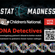
Children’s National Health System has been selected to compete in STAT Madness for the second consecutive year. Our entry for the bracket-style competition is “Sensitive liquid biopsy platform to detect tumor-released mutated DNA using patient blood and CSF,” a new technique that will allow kids to get better treatment for an aggressive type of pediatric brain tumor.
In 2018, Children’s first-ever STAT Madness entry advanced through five brackets in the national competition and, in the championship round, finished second. That innovation, which enables more timely diagnoses of rare diseases and common genetic disorders, helping to improve kids’ health outcomes around the world, also was among four “Editor’s Pick” finalists, entries that spanned a diverse range of scientific disciplines.
“Children’s National researchers collaboratively work across divisions and departments to ensure that innovations discovered in our laboratories reach clinicians in order to improve patient care,” says Mark Batshaw, M.D., Children’s Executive Vice President, Chief Academic Officer and Physician-in-Chief. “It’s gratifying that Children’s multidisciplinary approach to improving the lives of children with brain tumors has been included in this year’s STAT Madness competition.”
Pediatric brain cancers are the leading cause of cancer-related death in children younger than 14. Children with tumors in their midline brain structures have the worst outcomes, and kids diagnosed with diffuse midline gliomas, including diffuse intrinsic pontine glioma, have a median survival of just 12 months.
“We heard from our clinician colleagues that many kids were coming in and their magnetic resonance imaging (MRI) suggested a particular type of tumor. But it was always problematic to identify the tumor’s molecular subtype,” says Javad Nazarian, Ph.D., MSC, a principal investigator in Children’s Center for Genetic Medicine Research. “Our colleagues wanted a more accurate measure than MRI to find the molecular subtype. That raised the question of whether we could actually look at their blood to determine the tumor subtype.”
Children’s liquid biopsy, which remains at the research phase, starts with a simple blood draw using the same type of needle as is used when people donate blood. When patients with brain tumors provide blood for other laboratory testing, a portion of it is used for the DNA detective work. Just as a criminal leaves behind fingerprints, tumors shed telltale clues in the blood. The Children’s team searches for the histone 3.3K27M (H3K27M), a mutation associated with worse clinical outcomes.
“With liquid biopsy, we were able to detect a few copies of tumor DNA that were hiding behind a million copies of healthy DNA,” Nazarian says. “The blood draw and liquid biopsy complement the MRI. The MRI gives the brain tumor’s ZIP code. Liquid biopsy gives you the demographics within that ZIP code.”
Working with collaborators around the nation, Children’s National continues to refine the technology to improve its accuracy. The multi-institutional team published findings online Oct. 15, 2018, in Clinical Cancer Research.
Even though this research technique is in its infancy, the rapid, cheap and sensitive technology already is being used by people around the globe.
“People around the world are sending blood to us, looking for this particular mutation, H3K27M, ” says Lindsay B. Kilburn, M.D., a Children’s neuro–oncologist, principal investigator at Children’s National for the Pacific Pediatric Neuro-Oncology Consortium, and study co-author. “In many countries or centers, children do not have access to teams experienced in taking a biopsy of tumors in the brainstem, they can perform a simple blood draw and have that blood processed and analyzed by us. In only a few days, we can provide important molecular information on the tumor subtype previously only available to patients that had undergone a tumor biopsy.”
“With that DNA finding, physicians can make more educated therapeutic decisions, including prescribing medications that could not have been given previously,” Nazarian adds.
The STAT Madness round of 64 brackets opened March 4, 2019, and the championship round voting concludes April 5 at 5 p.m. (EST).
In addition to Nazarian and Dr. Kilburn, study co-authors include Eshini Panditharatna, Madhuri Kambhampati, Heather Gordish-Dressman, Ph.D., Suresh N. Magge, M.D., John S. Myseros, M.D., Eugene I. Hwang, M.D. and Roger J. Packer, M.D., all of Children’s National; Mariam S. Aboian, Nalin Gupta, Soonmee Cha, Michael Prados and Co-Senior Author Sabine Mueller, all of University of California, San Francisco; Cassie Kline, UCSF Benioff Children’s Hospital; John R. Crawford, UC San Diego; Katherine E. Warren, National Cancer Institute; Winnie S. Liang and Michael E. Berens, Translational Genomics Research Institute; and Adam C. Resnick, Children’s Hospital of Philadelphia.
Financial support for the research described in the report was provided by the V Foundation for Cancer Research, Goldwin Foundation, Pediatric Brain Tumor Foundation, Smashing Walnuts Foundation, The Gabriella Miller Kids First Data Resource Center, Zickler Family Foundation, Clinical and Translational Science Institute at Children’s National under award 5UL1TR001876-03, Piedmont Community Foundation, Musella Foundation for Brain Tumor Research, Matthew Larson Foundation, The Lilabean Foundation for Pediatric Brain Cancer Research, The Childhood Brain Tumor Foundation, the National Institutes of Health and American Society of Neuroradiology.
https://innovationdistrict.childrensnational.org/wp-content/uploads/2019/03/Stat-Madness-2019.png300400Innovation Districthttps://innovationdistrict.childrensnational.org/wp-content/uploads/2025/09/InnovationDistrict_CN_WebHeader-1396px-1030x151.pngInnovation District2019-03-06 13:43:012024-06-05 12:23:09Vote for Children’s National in STAT Madness
A new research study suggests that adolescents who get a good night’s sleep after a sports-related concussion might be linked to a shorter recovery time.
Research presented at the American Academy of Pediatrics Conference in Orlando, Fla., concluded that young athletes who slept well after a concussion were more likely to recover within two weeks, while those that didn’t receive a good night’s rest increased their likelihood to endure symptoms for 30 days or more.
The design and method was observational, where sleep factors and recovery are examined in association with each other. While the design does not allow a strong causal relationship to be established, it does not report control of other possible mediating variables, its sample size and strength of the findings are strongly suggestive, and provide a rationale for further study of sleep as a critical factor in recovery.
According to Gerard Gioia, Ph.D., chief of the Division of Pediatric Neuropsychology at Children’s National Health System, clinicians should ensure that sleep is properly assessed post-concussion and appropriate sleep hygiene strategies should be provided to the patient and family.
The average age of the 356 participants in the study was 14. Researchers conducting the study had the participants complete a questionnaire called the Pittsburgh Sleep Quality Index. Based on the answers reported, the teens were grouped into two categories: 261 good sleepers and 95 poor sleepers.
“The study highlights the importance of sleep, a critical factor in the recovery from a concussion,” says Dr. Gioia, “These findings are highly consistent with our own clinical experience in treating children and adolescents with concussions in that poor sleep are a significant limiting factor in recovery.”
During the follow-up visits three months later, both groups of patients had improved, however the good sleepers continued to have significantly better symptoms and sleep scores.
https://innovationdistrict.childrensnational.org/wp-content/uploads/2019/02/Teenage-boy-sleeping-masonry.jpg300400Innovation Districthttps://innovationdistrict.childrensnational.org/wp-content/uploads/2025/09/InnovationDistrict_CN_WebHeader-1396px-1030x151.pngInnovation District2019-02-22 10:41:022019-02-22 10:41:02Longer concussion recovery in children connected to poor sleep
Children’s research-clinicians created a novel preclinical model that mimics the persistent interneuron loss seen in preterm human infants, identifying interneuron subtypes that could become future therapeutic targets to prevent or lessen neurodevelopmental risks.
Research-clinicians at Children’s National Health System have created a novel preclinical model that mimics the persistent interneuron loss seen in preterm human infants, identifying interneuron subtypes that could become future therapeutic targets to prevent or lessen neurodevelopmental risks, the team reports Jan. 31, 2019, in eNeuro. The open access journal for Society for Neuroscience recognized the team’s paper as its “featured” article.
In the prefrontal cortex (PFC) of infants born preterm, there are decreased somatostatin and calbindin interneurons seen in upper cortical layers in infants who survived for a few months after preterm birth. This neuronal damage was mimicked in an experimental model of preterm brain injury in the PFC, but only when the newborn experimental models had first experienced a combination of prenatal maternal immune activation and postnatal chronic sublethal hypoxia. Neither neuronal insult on its own produced the pattern of interneuron loss in the upper cortical layers observed in humans, the research team finds.
“These combined insults lead to long-term neurobehavioral deficits that mimic what we see in human infants who are born extremely preterm,” says Anna Penn, M.D., Ph.D., a neonatologist in the Division of Neonatology and the Fetal Medicine Institute and a developmental neuroscientist at Children’s National Health System, and senior study author. “Future success in preventing neuronal damage in newborns relies on having accurate experimental models of preterm brain injury and well-defined outcome measures that can be examined in young infants and experimental models of the same developmental stage.”
According to the Centers for Disease Control and Prevention 1 in 10 infants is born preterm, before the 37th week of pregnancy. Many of these preterm births result from infection or inflammation in utero. After delivery, many infants experience other health challenges, like respiratory failure. These multi-hits can exacerbate brain damage.
Prematurity is associated with significantly increased risk of neurobehavioral pathologies, including autism spectrum disorder and schizophrenia. In both psychiatric disorders, the prefrontal cortex inhibitory circuit is disrupted due to alterations of gamma-aminobutyric acid (GABA) interneurons in a brain region involved in working memory and social cognition.
Cortical interneurons are created and migrate late in pregnancy and early infancy. That timing leaves them particularly vulnerable to insults, such as preterm birth.
In order to investigate the effects of perinatal insults on GABAergic interneuron development, the Children’s research team, led by Helene Lacaille, Ph.D., in Dr. Penn’s laboratory, subjected the new preterm encephalopathy experimental model to a battery of neurobehavioral tests, including working memory, cognitive flexibility and social cognition.
“This translational study, which examined the prefrontal cortex in age-matched term and preterm babies supports our hypothesis that specific cellular alterations seen in preterm encephalopathy can be linked with a heightened risk of children experiencing neuropsychiatric disorders later in life,” Dr. Penn adds. “Specific interneuron subtypes may provide specific therapeutic targets for medicines that hold the promise of preventing or lessening these neurodevelopmental risks.”
In addition to Dr. Penn and Lead Author Lacaille, Children’s co-authors include Claire-Marie Vacher; Dana Bakalar, Jiaqi J. O’Reilly and Jacquelyn Salzbank, all of Children’s Center for Neuroscience Research.
Financial support for research described in this post was provided by the National Institutes of Health under award R01HD092593, District of Columbia Intellectual Developmental Disabilities Research Center under award U54HD090257, Cerebral Palsy Alliance Research Foundation, Children’s National Board of Visitors, Children’s Research Institute and Fetal Medicine Institute.
https://innovationdistrict.childrensnational.org/wp-content/uploads/2019/02/Anna-Penn-using-a-microscope.png300400Innovation Districthttps://innovationdistrict.childrensnational.org/wp-content/uploads/2025/09/InnovationDistrict_CN_WebHeader-1396px-1030x151.pngInnovation District2019-02-19 10:55:162024-06-05 11:57:13New model mimics persistent interneuron loss seen in prematurity
Study authors Aaron Sathyanesan, Ph.D., Joseph Abbah, B.Pharm., Ph.D., Srikanya Kundu, Ph.D. and Vittorio Gallo, Ph.D.
Chronic sublethal hypoxia is associated with locomotor miscoordination and long-term cerebellar learning deficits in a clinically relevant model of neonatal brain injury, according to a study led by Children’s National Health System researchers published by Nature Communications. Using high-tech optical and physiological methods that allow researchers to turn neurons on and off and an advanced behavioral tool, the research team found that Purkinje cells fire significantly less often after injury due to perinatal hypoxia.
The research team leveraged a fully automated, computerized apparatus – an Erasmus Ladder – to test experimental models’ adaptive cerebellar locomotor learning skills, tracking their missteps as well as how long it took the models to learn the course.
The research project, led by Aaron Sathyanesan, Ph.D., a Children’s postdoctoral research fellow, was honored with a F1000 prime “very good rating.” The Children’s research team used both quantitative behavior tests and electrophysiological assays, “a valuable and objective platform for functional assessment of targeted therapeutics in neurological disorders,” according to the recommendation on a digital forum in which the world’s leading scientists and clinicians highlight the best articles published in the field.
Calling the Erasmus Ladder an “elegant” behavioral system, Richard Lu, Ph.D., and Kalen Berry write that the Children’s National Health System research team “revealed locomotor behavior and cerebellar learning deficits, and further utilized multielectrode recording/optogenetics approaches to define critical pathophysiological features, such as defects in Purkinje cell firing after neonatal brain injury.”
Lu, Beatrice C. Lampkin Endowed Chair in Cancer Epigenetics, and Berry, an associate faculty member in the Cancer and Blood Diseases Institute, both at Cincinnati Children’s, note that the Children’s results “suggest that GABA signaling may represent a potential therapeutic target for hypoxia-related neonatal brain injury that, if provided at the correct time during development post-injury, could offer lifelong improvements.”
In addition to Sathyanesan, Children’s co-authors include Co-Lead Author, Srikanya Kundu, Ph.D., and Joseph Abbah, both of Children’s Center for Neuroscience Research, and Vittorio Gallo, Ph.D., Children’s Chief Research Officer and the study’s senior author.
Research covered in this story was supported by the Intellectual and Developmental Disability Research Center under award number U54HD090257.
https://innovationdistrict.childrensnational.org/wp-content/uploads/2018/08/Vittorio-Gallo-and-coauthors-atmospheric_2018.png300400Innovation Districthttps://innovationdistrict.childrensnational.org/wp-content/uploads/2025/09/InnovationDistrict_CN_WebHeader-1396px-1030x151.pngInnovation District2019-01-29 09:34:202024-05-16 14:52:37Children’s perinatal hypoxia research lauded
Research by an international team that includes Children’s National faculty, published online Jan. 25, 2019 in Human Molecular Genetics, suggests that genetic mutations that cause cleft lip and palate also may contribute to neural tube defects, such as spina bifida.
Oral clefts are some of the most common birth defects worldwide, affecting about one in every 700 births. In the U.S., more than 4,000 babies are born each year with cleft lip, with or without cleft palate.
This defect isn’t simply a cosmetic manner: Oral clefts can severely affect feeding, speech and hearing, and they cause about 3,300 deaths annually worldwide.
To better understand these conditions, researchers have isolated a number of genetic mutations that appear to play contributing roles. These include those in a gene known as Interferon Regulatory Factor 6. New research by an international team that includes Children’s National faculty, published online Jan. 25, 2019 in Human Molecular Genetics, suggests that these mutations also may contribute to neural tube defects such as spina bifida.
In the first weeks of fetal development, the neural plate curves, creating a neural tube that, once fused shut, becomes the fetal brain and fetal spinal cord. Neural tube defects, which can range from mild to severe, are characterized by incomplete development of the brain, spinal cord or meninges. These defects can potentially result in paralysis or even fetal or neonatal demise. According to the National Institutes of Health, spina bifida, which affects the spinal cord, is the most common neural tube defect in the U.S., affecting up to 2,000 infants each year.
“Despite its high frequency, spina bifida remains among the least understood structural birth defects,” says Brian C. Schutte, an associate professor of Microbiology and Molecular Genetics, Pediatrics and Human Development at Michigan State University and the study’s senior author. “There is strong evidence that genetic factors are a leading cause of such structural birth defects, but in most cases, the cause is unknown. Our team’s study is the first published research to demonstrate that DNA variants in the gene IRF6 can cause spina bifida,” Schutte says.
What’s more, the research team identified a mechanism to explain how altering IRF6 leads to neural tube defects. This mechanism links IRF6 function to two other genes – known as transcription Factor AP2A (TFAP2A) and Grainyhead Like 3 (GRHL3) – that are also known to be required for the development of the neural tube, lip and palate.
“We’re all on the hunt for the reasons when, how and why birth defects happen,” adds Youssef A. Kousa, MS, D.O., Ph.D., a clinical fellow in the Division of Child Neurology at Children’s National Health System and the study’s lead author. “Our main goal is prevention. This paper is a significant development because our team has identified a group of genes that can potentially contribute to very common types of birth defects: craniofacial as well as neural tube defects.”
The scientific odyssey is a wonderful example of serendipity. Kousa, then working in Schutte’s lab, was studying the effects of a new mutant experimental model strain on development of the palate. But one day, he walked into Schutte’s office holding a deformed preclinical embryo and said: “Brian, look at this!”
“Weird things happen in biology,” Schutte replied and counseled him to return if it happened again. Less than two weeks later, Kousa was back with several more of the deformed preclinical embryos, saying: “OK, Brian. It happened again.”
Within hours Kousa had unearthed recently published research that included an image of a similarly affected preclinical embryo. The pair then sketched out possible intersecting genetic pathways, as they brainstormed the myriad ways to end up with that specific phenotype. Initially, they tested their hypotheses in experimental models and eventually corroborated findings through human genetic studies.
The human studies could only be performed by collaborations. Schutte shared their initial observations with human genetics researchers scattered across the country. Those labs then generously agreed to test whether DNA variants in IRF6 were associated with neural tube defects in samples from patients that they had collected over decades of research.
The team found that Tfap2a, Irf6 and Grhl3 are components of a gene regulatory network required for neurulation, a folding process that results in the neural tube bending and then fusing to become the basis of the embryo’s nervous system, from brain to spinal cord.
“Since this network is also required for formation of the lip, palate, limbs and epidermis, which develop at different times and places during embryogenesis, we suggest that the Tfap2a–Irf6–Grhl3 network is a fundamental pathway for multiple morphogenetic processes,” the researchers write.
Interferon Regulatory Factor 6 functions best when there is neither too much expression nor too little. Overexpression of Irf6 suppresses Transcription Factor Activation Protein 2A and Grainyhead Like 3, causing exencephaly, a neural tube defect characterized by the brain being located outside of the skull. Counterintuitively, experimental models that had too little Irf6 also ended up with reduced levels of Tfap2a and Grhl3 that led to a structural birth defect, but at the opposite end of the neural tube.
To test whether the experimental model findings held true in humans, they sequenced samples from people who had spina bifida and anencephaly – the rare birth defect that Kousa spotted in the experimental models – and found IRF6 function was conserved in people. Because of the genetic complexity of these birth defects, and the challenges inherent in collecting samples from cases of severe birth defects, many research teams were invited to participate in the study.
As testament to their collegiality, researchers from Stanford University, University of Texas at Austin, University of Iowa, University of Texas at Houston and Duke University agreed to share precious samples from the California Birth Defects Monitoring Program, from the Hereditary Basis of Neural Tube Defects study and from their own institutional sample collections.
“As we get better at personalized medicine, we could use this information to one day help to counsel families about their own risk and protective factors,” Kousa adds. “If we can identify the genetic pathway, we might also be able to modify it to prevent a birth defect. For example, prenatal supplementation with folic acid has led to a decrease in babies born with neural tube defects, but not all neural tube defects are sensitive to folic acid. This knowledge will help us develop individual-based interventions.”
Financial support for the research covered in this post was provided by the National Institutes of Health under grants DE13513, F31DE022696, DE025060, P01HD067244 and GM072859; startup funding from Michigan State University and the UT-Health School of Dentistry in Houston; and the Centers for Disease Control and Prevention under award number 5U01DD001033.
In addition to Kousa and Schutte, study co-authors include Huiping Zhu, Yunping Lei and Richard H. Finnell, University of Texas at Austin; Walid D. Fakhouri, University of Texas Health Science Center at Houston; Akira Kinoshita, Nagasaki University; Raeuf R. Roushangar, Nicole K. Patel, Tamer Mansour, Arianna L. Smith, and Dhruv B. Sharma, Michigan State University; A.J. Agopian and Laura E. Mitchell, University of Texas School of Public Health; Wei Yang and Gary M. Shaw, Stanford University School of Medicine; Elizabeth J. Leslie, Emory University; Xiao Li, Tamara D. Busch, Alexander G. Bassuk and Brad A. Amendt, University of Iowa; Edward B. Li and Eric C. Liao, Massachusetts General Hospital; Trevor J. Williams, University of Colorado Denver at Anschutz Medical Campus; Yang Chai, University of Southern California; and Simon Gregory and Allison Ashley-Koch, Duke University Medical Center.
https://innovationdistrict.childrensnational.org/wp-content/uploads/2019/01/little-girl-with-spina-bifida.jpg300400Innovation Districthttps://innovationdistrict.childrensnational.org/wp-content/uploads/2025/09/InnovationDistrict_CN_WebHeader-1396px-1030x151.pngInnovation District2019-01-25 12:58:112024-12-30 12:46:57Oral clefts may stem from a shared genetic cause as neural tube defects
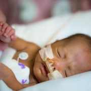
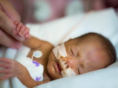
According to Children’s research presented during the Institute for Healthcare Improvement 2018 Scientific Symposium, standardizing feeding practices – including the timing for fortifying breast milk and formula with essential elements like zinc and protein – improves growth trends for the tiniest preterm infants.
About 1 in 10 infants is born before 37 weeks gestation. These premature babies have a variety of increased health risks, including deadly infections and poor lung function.
Emerging research suggests that getting their length and weight back on track could help. According to Children’s research presented during the Institute for Healthcare Improvement 2018 Scientific Symposium, standardizing feeding practices – including the timing for fortifying breast milk and formula with essential elements like zinc and protein – improves growth trends for the tiniest preterm infants.
The quality-improvement project at Children’s National Health System targeted very low birth weight infants, who weigh less than 3.3 pounds (1,500 grams) at birth. These fragile infants are born well before their internal organs, lungs, brain or their digestive systems have fully developed and are at high risk for ongoing nutritional challenges, health conditions like necrotizing enterocolitis (NEC) and overall poor development.
The research team measured progress by tracking the micro-preemies’ mean delta weight Z-score for weight gain, which measures nutritional status.
“In this cohort, mean delta weight Z-scores improved by 43 percent, rising from -1.8 to the goal of -1.0, when we employed an array of interventions. We saw the greatest improvement, 64 percent, among preterm infants who had been born between 26 to 28 weeks gestation,” says Michelande Ridoré, MS, Children’s NICU quality-improvement program lead who presented the group’s preliminary findings. “It’s very encouraging to see improved growth trends just six months after introducing these targeted interventions and to maintain these improvements for 16 months.”
Within Children’s neonatal intensive care unit (NICU), micro-preemies live in an environment that mimics the womb, with dimmed lighting and warmed incubators covered by blankets to muffle extraneous noise. The multidisciplinary team relied on a number of interventions to improve micro-preemies’ long-term nutritional outcomes, including:
Reducing variations in how individual NICU health care providers approach feeding practices
Fortifying breast milk (and formula when breast milk was not available), which helps these extra lean newborns add muscle and strengthen bones
Early initiation of nutrition that passes through the intestine (enteral feeds)
Re-educating all members of the infants’ care teams about the importance of standardized feeding and
Providing a decision aid about feeding intolerance.
Dietitians were included in the daily rounds, during which the multidisciplinary team discusses each infant’s care plan at their room, and used traffic light colors to describe how micro-preemies were progressing with their nutritional goals. It’s common for these newborns to lose weight in the first few days of life.
Infants in the “green” zone had regained their birth weight by day 14 of life and possible interventions included adjusting how many calories and protein they consumed daily to reflect their new weight.
Infants in the “yellow” zone between day 15 to 18 of life remained lighter than what they weighed at birth and were trending toward lower delta Z-scores. In addition to assessing the infant’s risk factors, the team could increase calories consumed per day and add fortification, among other possible interventions.
Infants in the “red” zone remained below their birth weight after day 19 of life and recorded depressed delta Z-scores. These infants saw the most intensive interventions, which could include conversations with the neonatologist and R.N. to discuss strategies to reverse the infant’s failure to grow.
Future research will explore how the nutritional interventions impact newborns with NEC, a condition characterized by death of tissue in the intestine. These infants face significant challenges gaining length and weight.
Institute for Healthcare Improvement 2018 Scientific Symposium presentation
“Improved growth of very low birthweight infants in the neonatal intensive care unit.”
Caitlin Forsythe, MS, BSN, RNC-NIC, NICU clinical program coordinator, Neonatology, and lead author; Michelande Ridoré, MS, NICU quality-improvement program lead; Victoria Catalano Snelgrove, RDN, LD, CNSC, CLC, pediatric clinical dietitian; Rebecca Vander Veer, RD, LD, CNSC, CLC, pediatric clinical dietitian; Erin Fauer, RDN, LD, CNSC, CLC, pediatric clinical dietitian; Judith Campbell, RNC, IBCLC, NICU lactation consultant; Eresha Bluth, MHA, project administrator; Anna Penn, M.D., Ph.D., neonatologist; Lamia Soghier, M.D., MEd, Medical Unit Director, Neonatal Intensive Care Unit; and Mary Revenis, M.D., NICU medical lead on nutrition and senior author; all of Children’s National Health System.
https://innovationdistrict.childrensnational.org/wp-content/uploads/2016/10/PreemieImage-e1494013414625.jpg300400Innovation Districthttps://innovationdistrict.childrensnational.org/wp-content/uploads/2025/09/InnovationDistrict_CN_WebHeader-1396px-1030x151.pngInnovation District2019-01-23 11:33:392024-02-02 14:39:31Getting micro-preemie growth trends on track
Pedbot’s home version adapts the same airplane-themed video game to a smaller therapeutic platform that is more affordable to build.
The novel ankle rehabilitation robot built at Children’s National to help children with cerebral palsy build ankle strength and control through video gaming is taking a big step forward. Engineers have created a smaller, more affordable version of the robotic platform using 3D printed parts, to explore the effectiveness of a home-based therapy program.
“We’re seeing preliminary success in our trial for in clinic use of the Pedbot. Now we’re hoping to see if making the technology accessible at home means that 1) Kids use it more often and 2) More frequent, regular use over time leads to better range of motion,” says Kevin Cleary, Ph.D., the Sheikh Zayed Institute for Pediatric Surgical Innovation’s bioengineering technical director and engineering lead for Pedbot.
Pedbot’s video game, designed by software engineer Hadi Fooladi, M.S., allows kids to pilot an airplane through a series of hoops at varying speeds as determined by the therapist and programmer. The game isn’t the only thing that’s unique about this therapeutic robot, however.
Just like the clinic version, the home model moves in three translational directions (x, y and z) and rotates about three axes (the x, y and z axes), similar to the movement of a flight simulator. The result is a robot that helps the patient exercise across a greater range of motion and build muscle strength in a way that more closely mimics real-life ankle function.
Pedbot Home potentially eliminates an additional major therapeutic barrier – the clinic appointment.
“The great thing about Pedbot is you’re constantly working to reach a moving target, and the therapist can vary the movement type as much or as little as needed for each patient,” says Catherine Coley, DPT, a physical therapist at Children’s National who is a member of the Pedbot development team. “We think the home version might make it easier for the child to succeed with a long term therapy program by removing the need for repeat clinic visits.”
“What if a child could come home from school and do their therapy at home after dinner? Would doing it every day for 20 minutes benefit the child more than just coming to see us once or twice a week for an hour? Can we make it easier for our patients to cooperate and follow through with therapy homework? These are some of the questions that we hope we can answer during our trial for the home version,” says Sally Evans, M.D., division chief of Pediatric Rehabilitation Medicine at Children’s National and clinical lead for the project.
The cross-functional Pedbot team includes engineers Reza Monfaredi Ph.D. and Tyler Salvador, B.S., as well as additional physical therapists, Stacey Kovelman, P.T. and Justine Belchner, P.T., and Sara Alyamani, B.A. Future expansions will include the addition of electromyography measurements in collaboration with Paola Pergami, M.D., Ph.D. and incorporation of other patient populations with Beth Wells, M.D.
Pedbot Home is currently being piloted in the home setting, with the goal of enrolling additional families to participate in a trial within the next year. The work is supported by a $500,000 federal grant from the Department of Health and Human Services’ National Institute on Disability, Independent Living, and Rehabilitation Research.
https://innovationdistrict.childrensnational.org/wp-content/uploads/2018/02/Pedbot-video-game.jpg300400Innovation Districthttps://innovationdistrict.childrensnational.org/wp-content/uploads/2025/09/InnovationDistrict_CN_WebHeader-1396px-1030x151.pngInnovation District2019-01-23 10:47:472019-04-12 09:52:34Pedbot’s next step – Home-based therapy
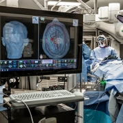
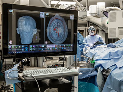
The virtual reality surgical system projects images into the operating room, allowing neurosurgeons to revisit the surgical plan in real time.
Neurosurgeons at Children’s National Health System are getting a new three-dimensional (3D) perspective on their cases thanks to an FDA-approved breakthrough virtual reality surgical system.
Children’s National is the first pediatric health system in metropolitan Washington, D.C., to use this state-of-the art system, created by Surgical Theater. It seamlessly integrates patient-specific surgical planning and navigation, professional education and rehearsal.
The technology acquisition was made possible through a generous gift from Sidney & Phyllis Bresler, in honor of their children Alex, Jonathan and Amanda and grandson Theo Charles Bresler, and in loving memory of Joshua Stouck.
“Virtual reality modeling enables us to further explore, analyze and find the best approach for each unique surgical procedure,” said Children’s National President and CEO Kurt Newman, M.D. “This generous gift from Sidney & Phyllis Bresler should translate into better outcomes for many of the more than 17,500 patients who receive surgery at our hospital each year, and will benefit generations to come. We are deeply grateful for the Breslers’ commitment to pediatric innovation.”
The 3D, 360-degree view gives surgeons a cutting-edge digital tool to plan procedures in depth using an accurate capture of the patient’s unique anatomy, and also allows the surgeon to illustrate the surgical path in greater detail than ever before for patients and their families.
“Technology such as Surgical Theater’s represents a quantum leap for neurosurgeons, both in and out of the operating room,” said Robert Keating, M.D., chief of Neurosurgery at Children’s National, in a press release from the company. “It allows us to marry state-of-the-art 3D simulation to the real world; for the patient and family as well as doctors in training, and ultimately offers a new tool for the neurosurgical armamentarium in approaching complex lesions in the brain, such as AVM’s, tumors, epilepsy and functional cases.”
https://innovationdistrict.childrensnational.org/wp-content/uploads/2019/01/surgical-theater.jpg300400Innovation Districthttps://innovationdistrict.childrensnational.org/wp-content/uploads/2025/09/InnovationDistrict_CN_WebHeader-1396px-1030x151.pngInnovation District2019-01-22 11:03:582023-07-03 10:43:30Virtual reality allows surgical planning from every angle
On Jan. 4, 2019, Nobuyuki Ishibashi, M.D., the director of the Cardiac Surgery Research Laboratory and an investigator with the Center for Neuroscience Research at Children’s National Health System, published a review in Trends in Neurosciences about the mechanisms of cortical dysmaturation, or disturbances in cortical development, that can occur in children born with congenital heart disease (CHD). By understanding the early-life impact and relationship between cardiac abnormalities and cortical neuronal development, Dr. Ishibashi and the study authors hope to influence strategies for neonatal neuroprotection, mitigating the risk for developmental delays among CHD patients.
Dr. Ishibashi answers questions about this review and CHD-neurodevelopmental research:
Tell us more about your research. Why did you choose to study these interactions in this patient population?
My research focuses on studying how CHD and neonatal cardiac surgery affect the rapidly-developing brain. Many children with CHD, particularly the most complex anomalies, suffer from important behavioral anomalies and neurodevelopmental delays after cardiac surgery. As a surgeon scientist, I want to optimize treatment strategy and develop a new standard of care that will reduce neurodevelopmental impairment in our patients.
How does this study fit into your larger body of work? What are a few take-home messages from this paper?
Our team and other laboratories have recently identified a persistent perinatal neurogenesis that targets the frontal cortex – the brain area responsible for higher-order cognitive functions. The main message from this article is that further understanding of the cellular and molecular mechanisms underlying cortical development and dysmaturation will likely help to identify novel strategies to treat and improve outcomes in our patients suffering from intellectual and behavioral disabilities.
What do you want pediatricians and researchers to know about this study? Why is it important right now?
Although the hospital mortality risk is greatly reduced, children with complex CHD frequently display subsequent neurological disabilities affecting intellectual function, memory, executive function, speech and language, gross and fine motor skills and visuospatial functions. In addition to the impact of the neurological morbidity on the patients themselves, the toll on families and society is immense. Therefore it is crucial to determine the causes of altered brain maturation in CHD.
How do you envision this research influencing future studies and pediatric health outcomes? As a researcher, how will you proceed?
In this article we placed special emphasis on the need for well-designed preclinical studies to define disturbances in cortical neurogenesis due to perinatal brain injury. I believe that further study of the impact of hypoxemia on brain development is of broad relevance — not just for children with congenital heart disease, but for other populations where intellectual and behavioral dysfunctions are a source of chronic morbidity, such as survivors of premature birth.
What discoveries do you envision being at the forefront of this field?
One of the important questions is: During which developmental period, prenatal or postnatal, is the brain most sensitive to developmental and behavioral disabilities associated with hypoxemia? Future experimental models will help us study key effects of congenital cortical development anomalies on brain development in children with CHD.
What impact could this research make? What’s the most striking finding and how do you think it will influence the field?
Although cortical neurogenesis at fetal and adult stages has been widely studied, the development of the human frontal cortex during the perinatal period has only recently received greater attention as a result of new identification of ongoing postnatal neurogenesis in the region responsible for important intellectual and behavioral functions. Children’s National is very excited with the discoveries because it has opened new opportunities that may lead to regeneration and repair of the dysmature cortex. If researchers identify ways to restore endogenous neurogenic abilities after birth, the risk of neurodevelopment disabilities and limitations could be greatly reduced.
Is there anything else you would like to add that we didn’t ask you about? What excites you about this research?
In this article we highlight an urgent need to create a truly translational area of research in CHD-induced brain injury through further exploration and integration of preclinical models. I’m very excited about the highly productive partnerships we developed within the Center for Neuroscience Research at Children’s National, led by an internationally-renowned developmental neuroscientist, Vittorio Gallo, Ph.D., who is a co-senior author of this article. Because of our collaboration, my team has successfully utilized sophisticated and cutting-edge neuroscience techniques to study brain development in children born with CHD. To determine the causes of altered brain maturation in congenital heart disease and ultimately improve neurological function, we believe that a strong unity between cardiovascular and neuroscience research must be established.
Additional study authors include Camille Leonetti, Ph.D., a postdoctoral research fellow with the Center for Neuroscience Research and Children’s National Heart Institute, and Stephen Back, M.D., Ph.D., a professor of pediatrics at Oregon Health and Science University.
The research was supported by multiple grants and awards from the National Institutes of Health, inclusive of the National Heart Lung and Blood Institute (RO1HL139712), the National Institute of Neurological Disorders and Stroke (1RO1NS054044, R37NS045737, R37NS109478), the National Institute on Aging (1RO1AG031892-01) and the National Institute of Child Health and Human Development (U54HD090257).
Additional support for this review was awarded by the American Heart Association (17GRNT33370058) and the District of Columbia Intellectual and Developmental Disabilities Research Center, which is supported through the Eunice Kennedy Shriver National Institute of Child Health and Human Development program grant 1U54HD090257.
https://innovationdistrict.childrensnational.org/wp-content/uploads/2017/06/Nobuyuki-Ishibashi.jpg300400Innovation Districthttps://innovationdistrict.childrensnational.org/wp-content/uploads/2025/09/InnovationDistrict_CN_WebHeader-1396px-1030x151.pngInnovation District2019-01-15 11:22:462023-09-18 11:53:11Cortical dysmaturation in congenital heart disease
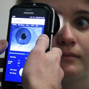
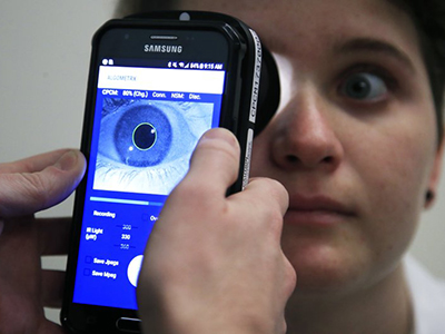
Clinical Research Assistant Kevin Jackson uses AlgometRx Platform Technology on Sarah Taylor’s eyes to measure her degree of pain. Children’s National is testing an experimental device that aims to measure pain according to how pupils react to certain stimuli. (AP Photo/Manuel Balce Ceneta)
Pediatric anesthesiologist Julia C. Finkel, M.D., of Children’s National Health System, gazed into the eyes of a newborn patient determined to find a better way to measure the effectiveness of pain treatment on one so tiny and unable to verbalize. Then she realized the answer was staring back at her.
Armed with the knowledge that pain and analgesic drugs produce an involuntary response from the pupil, Dr. Finkel developed AlgometRx, a first-of-its-kind handheld device that measures a patient’s pupillary response and, using proprietary algorithms, provides a diagnostic measurement of pain intensity, pain type and, after treatment is administered, monitors efficacy. Her initial goal was to improve the care of premature infants. She now has a device that can be used with children of any age and adults.
“Pain is very complex and it is currently the only vital sign that is not objectively measured,” says Dr. Finkel, who has more than 25 years of experience as a pain specialist. “The systematic problem we are facing today is that healthcare providers prescribe pain medicine based on subjective self-reporting, which can often be inaccurate, rather than based on an objective measure of pain type and intensity.” To illustrate her point, Dr. Finkel continues, “A clinician would never prescribe blood pressure medicine without first taking a patient’s blood pressure.”
The current standard of care for measuring pain is the 0-to-10 pain scale, which is based on subjective, observational and self-reporting techniques. Patients indicate their level of pain, with zero being no pain and ten being highest or most severe pain. This subjective system increases the likelihood of inaccuracy, with the problem being most acute with pediatric and non-verbal patients. Moreover, Dr. Finkel points out that subjective pain scores cannot be standardized, heightening the potential for misdiagnosis, over-treatment or under-treatment.
Dr. Finkel, who serves as director of Research and Development for Pain Medicine at the Sheikh Zayed Institute for Pediatric Surgical Innovation at Children’s National, says that a key step in addressing the opioid crisis is providing physicians with objective, real-time data on a patient’s pain level and type, to safely prescribe the right drug and dosage or an alternate treatment.,
She notes that opioids are prescribed for patients who report high pain scores and are sometimes prescribed in cases where they are not appropriate. Dr. Finkel points to the example of sciatica, a neuropathic pain sensation felt in the lower back, legs and buttocks. Sciatica pain is carried by touch fibers that do not have opioid receptors, which makes opioids an inappropriate choice for treating that type of pain.
A pain biomarker could rapidly advance both clinical practice and pain research, Dr. Finkel adds. For clinicians, the power to identify the type and magnitude of a patient’s nociception (detection of pain stimuli) would provide a much-needed scientific foundation for approaching pain treatment. Nociception could be monitored through the course of treatment so that dosing is targeted and personalized to ensure patients receive adequate pain relief while reducing side effects.
“A validated measure to show whether or not an opioid is indicated for a given patient could ease the health care system’s transition from overreliance on opioids to a more comprehensive and less harmful approach to pain management,” says Dr. Finkel.
She also notes that objective pain measurement can provide much needed help in validating complementary approaches to pain management, such as acupuncture, physical therapy, virtual reality and other non-pharmacological interventions.
Dr. Finkel’s technology, called AlgometRx, has been selected by the U.S. Food and Drug Administration (FDA) to participate in its “Innovation Challenge: Devices to Prevent and Treat Opioid Use Disorder.” She is also the recipient of Small Business Innovation Research (SBIR) grant from the National Institute on Drug Abuse.
https://innovationdistrict.childrensnational.org/wp-content/uploads/2019/01/AlgometRX.jpg300400Innovation Districthttps://innovationdistrict.childrensnational.org/wp-content/uploads/2025/09/InnovationDistrict_CN_WebHeader-1396px-1030x151.pngInnovation District2019-01-10 07:35:322024-09-06 15:28:51Breakthrough device objectively measures pain type, intensity and drug effects
The second most-read article in 2018 in the Journal of Clinical Sleep Medicine, published by the American Academy of Sleep Medicine (AASM), was about using actigraphy to evaluate sleep disorders and circadian rhythm sleep-wake disorders.
FDA-approved actigraphy devices are typically kept on the wrist or ankle and track movement activity, which researchers can use as part of a larger toolset to analyze how much activity occurs right before and during sleep.
The conditions for evaluating pediatric health conditions are as follows:
The AASM suggests that clinicians use actigraphy in the assessment of pediatric patients with insomnia disorder. (Conditional)
The AASM suggests that clinicians use actigraphy in the assessment of pediatric patients with circadian rhythm sleep-wake disorder. (Conditional)
The AASM suggests that clinicians use actigraphy to monitor total sleep time prior to testing with the Multiple Sleep Latency Test in adult and pediatric patients with suspected central disorders of hypersomnolence. (Conditional)
The AASM recommends that clinicians not use actigraphy in place of electromyography for the diagnosis of periodic limb movement disorder in adult and pediatric patients. (Strong)
In an interview with Neurology Today, Daniel Lewin, Ph.D., a sleep medicine specialist, pediatric psychologist and associate director of the sleep medicine program at Children’s National Health System, offered advice, alongside other sleep medicine experts, about the new guidelines:
“It’s a very powerful tool, but it does require some knowledge of basic sleep mechanisms and of how the tool can be used and what variables can be extracted from the tool,” Dr. Lewin said in the interview with Susan Kreimer.
Anne Goldstein, M.D., M.S., assistant professor of neurology at the University of Michigan Sleep Disorders Center, tells Kreimer that “Actigraphy records only movement and that non-moving is often misinterpreted as sleep.”
Dr. Lewin has used actigraphy in sleep research studies but notes the use of these devices come with extensive training. Other researchers expressed similar sentiments with Neurology Today, noting the value of the sleep assessment tool to capture preliminary sleep behavior assessments, similar to a self-reported sleep log, while noting their limitations, such as capturing sleep patterns over extended periods of time, instead of in 14-day increments.
“When you’re living a typical active human life, sleep can wax and wane, depending on travel patterns, work responsibilities and stress levels,” Nathaniel F. Watson, M.D., professor of neurology at the University of Washington School of Medicine in Seattle and director of the UW Medicine Sleep Clinic, tells Kreimer. “This variability in sleep highlights the need for additional technologies capable of assessing sleep over longer periods of time.”
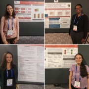
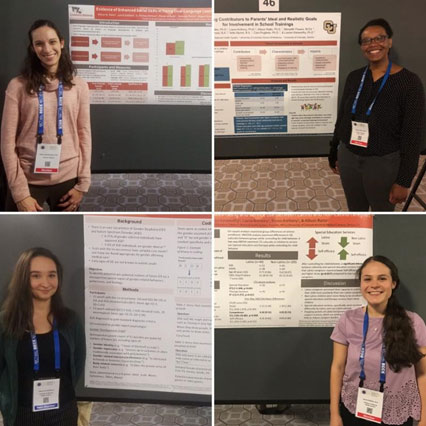
CASD Faculty Member and Clinical Psychologist, Dr. Allison Ratto (top left); Postdoctoral Fellow, Dr. Marissa Miller, (top right); and Research Assistants, Eleonora Sadikova (bottom left) and Laura Saldana (bottom right) presented posters at ABCT.
Technology’s potential to improve care delivery and reduce human suffering were the key focus of discussion at the recent Annual Convention of the Association for Behavioral and Cognitive Therapies (ABCT), held in Washington, D.C.
Within ABCT’s Autism Spectrum and Developmental Disabilities Special Interest Group (ASDD SIG), presentations showcased tools that leverage technology to better meet the needs of both autistic people and the clinicians who care for them. Researchers from the Center for Autism Spectrum Disorders (CASD) at Children’s National took center stage at the ASD focused group to share information about novel developments underway that harness technology for children and families.
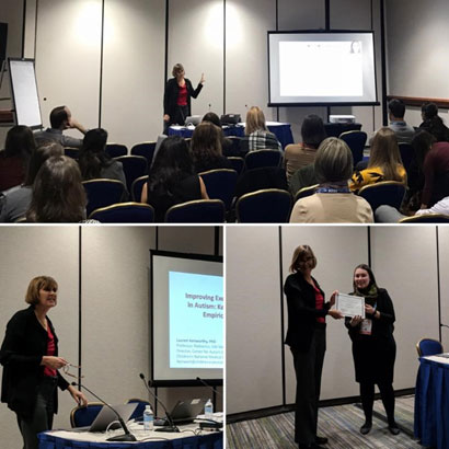
Lauren Kenworthy, Ph.D., director of CASD, served as the keynote speaker for the ASDD SIG Meeting. She also chaired a panel, “Leveraging Technology to Improve Autism Acceptance and Treatment” and presented, ” Online Parent Training Modules to Improve Executive Function in Autistic Children” about the e-Unstuck and On Target Parent Training Study, which adapts CASD’s successful classroom-based Unstuck and On Target toolkit for children ages 5 to 10 to an online platform so more families can benefit from the program’s skills and strategies.
Dr. Kenworthy was honored with the 2018 Transformative Contribution Award from the ABCT Autism Spectrum and Developmental Disabilities Special Interest Group for her lifetime of contributions to better understanding and better interventions for young people with ASD.
“It was a special honor to receive this recognition from ABCT this year, when the annual meeting is here in our home city,” says Dr. Kenworthy. “The Center for Autism Spectrum Disorders is focused on developing technology solutions that deliver therapies to everyone who needs them, no matter where they live, and technology is one powerful and promising way we can bridge care gaps both in the Washington, D.C. region and really, around the world.”
Dr. Lauren Kenworthy presenting during the panel she chaired (top); presenting to the ASDD SIG (bottom left); and receiving the ASDD SIG Transformative Award from ASDD SIG Awards Committee Chair, Dr. Tyler Hassenfeldt (bottom right).
In addition to Dr. Kenworthy, several other CASD researchers presented research during panels and poster presentations, including:
Panel Presentation: Efficacy of a Parent-Mediated Sexual Education Curriculum for Youth With ASD”– Cara Pugliese, Ph.D.
Poster presentations:
“Evidence of Enhanced Social Skills in Young Dual-Language Learners on the Autism Spectrum”- Allison Ratto, Ph.D. (first author)
“Exploring Contributors to Parents’ Ideal and Realistic Goals for Involvement in School Training”-Marissa Miller, Ph.D. (first author)
“Examining Caregiver Well-Being and Service Use between Latino and Non-Latino Caregivers”-Laura Saldana (first author)
“Pre-Pubertal Signs of Future Gender Dysphoria in Youth with ASD”-Eleonora Sadikova (first author)
The Association for Behavioral and Cognitive Therapies Annual Convention has been held for more than half a century. The gathering includes 3,500-plus mental health professionals and students who specialize in cognitive and behavioral therapies.
https://innovationdistrict.childrensnational.org/wp-content/uploads/2018/12/CASD-posters.jpg426426Innovation Districthttps://innovationdistrict.childrensnational.org/wp-content/uploads/2025/09/InnovationDistrict_CN_WebHeader-1396px-1030x151.pngInnovation District2018-12-21 12:04:572024-09-06 15:02:34Bridging gaps in autism care through technology
“A combination of prenatal MRI and US was able to detect Zika-related brain abnormalities during pregnancy, giving families timely information to prepare for the potential complex care needs of these infants,” says Sarah B. Mulkey, M.D., Ph.D.
Worldwide, thousands of babies have been born to mothers who were infected during pregnancy with Zika, a virus associated with neurological deficits, impaired vision and neurodevelopmental disabilities, among other birth defects. These birth defects are sometimes severe, causing lifelong disability. But they’re also relatively rare compared with the overall rates of infection.
Predicting how many Zika-exposed babies would experience neurological birth defects has been challenging.
However, an international study led by Children’s faculty suggests that ultrasound (US) imaging performed during pregnancy and after childbirth revealed most Zika-related brain abnormalities experienced by infants exposed to the Zika virus during pregnancy, according to a prospective cohort study published online Nov. 26, 2018, in JAMA Pediatrics. Some Zika-exposed infants whose imaging had been normal during pregnancy had mild brain abnormalities detected by US and magnetic resonance imaging (MRI) after they were born.
“A combination of prenatal MRI and US was able to detect Zika-related brain abnormalities during pregnancy, giving families timely information to prepare for the potential complex care needs of these infants,” says Sarah B. Mulkey, M.D., Ph.D., a fetal-neonatal neurologist at Children’s National Health System and the study’s lead author. “In our study, we detected mild brain abnormalities on postnatal neuroimaging for babies whose imaging was normal during pregnancy. Therefore, it is important for clinicians to continue to monitor brain development for Zika-exposed infants after birth.”
As of Nov. 20 2018, nearly 2,500 pregnant women in the U.S. had laboratory confirmed Zika infection, and about 2,400 of them had given birth, according to the Centers for Disease Control and Prevention (CDC). While more than 100 U.S. infants were born with Zika-associated birth defects, the vast majority of Zika-exposed U.S. infants were apparently normal at birth. The sequential neuroimaging study Dr. Mulkey leads seeks to determine the spectrum of brain findings in infants exposed to Zika in the womb using both US and MRI before and after birth.
The international research team enrolled 82 women in the study from June 15, 2016, through June 27, 2017. All of the women had been exposed to Zika during pregnancy; all but one experienced clinical symptoms by a mean gestational age of 8.2 weeks. Eighty of those women lived in or near Barranquilla, Colombia, and were exposed to Zika there. Two U.S. study participants were exposed to the primarily mosquito-borne illness during travel to Zika hot zones.
All women received fetal MRIs and US during the second and/or third trimester of pregnancy. After their infants were born, the children received brain MRI and cranial US. Blood samples from both mothers and babies were tested for Zika using polymerase chain reaction and serology.
Fetal MRI was able to discern Zika-related brain damage as early as 18 weeks gestation and picked up significant fetal brain abnormalities not fully appreciated in US imaging. In one case, the US remained normal while fetal MRI alone detected brain abnormalities. Three fetuses (4 percent) had severe fetal brain abnormalities consistent with Zika infection, including:
Two cases of heterotopias and malformations in cortical development, and
One case of parietal encephalocele, Chiari II malformation and microcephaly.
Seventy-five infants were born at term. One pregnancy was terminated at 23 weeks gestation due to the gravity of the fetal brain abnormalities. One fetus with normal imaging died during pregnancy. One newborn who was born with significant fetal brain abnormalities died at age 3 days.
Cranial US and brain MRI was performed on the majority of infants whose prenatal imaging had been normal. Seven of 53 (13 percent) Zika-exposed infants had mild brain abnormalities detected by MRI after birth. In contrast, postnatal cranial US was better at detecting changes of lenticulostriate vasculopathy, cysts within the brain’s choroid plexus (cells that produce cerebrospinal fluid), germinolytic/subependymal cysts and/or calcifications, which were seen in 21 of 57 (37 percent) infants.
“Sequential neuroimaging revealed that the majority of Zika-exposed fetuses had normal brain development. Tragically, in a small number of pregnancies, Zika-related brain abnormalities were quite severe,” Dr. Mulkey adds. “Our data support the CDC’s recommendation that cranial US be performed after Zika-exposed babies are born. In addition, there is clearly a need to follow these babies over time to gauge whether the brain anomalies we see in imaging affects language, motor and social skills.”
In addition to Dr. Mulkey, study co-authors include Dorothy I. Bulas, M.D., Gilbert Vezina, M.D., Margarita Arroyave-Wessel, MPH, Stephanie Russo, B.S, Youssef A. Kousa, D.O, Ph.D., Roberta L. DeBiasi, M.D., MS, Senior Author Adré J. du Plessis, M.B.Ch.B., MPH, all of Children’s National; Christopher Swisher, BS, Georgetown University and Caitlin Cristante, BS, Loyola University, both of whose contributions included research performed at Children’s National; Yamil Fourzali, M.D., Armando Morales, M.D., both of Sabbag Radiologos; Liliana Encinales, M.D., Allied Research Society; Nelly Pacheco, Bacteriologa, Bio-Nep; Robert S. Lanciotti, Ph.D., Arbovirus Diseases Branch, Centers for Disease Control and Prevention; and Carlos Cure, M.D., BIOMELAB.
Research reported in this news release was supported by the IKARIA fund.
https://innovationdistrict.childrensnational.org/wp-content/uploads/2018/12/Sarah-B.-Mulkey.jpg300400Innovation Districthttps://innovationdistrict.childrensnational.org/wp-content/uploads/2025/09/InnovationDistrict_CN_WebHeader-1396px-1030x151.pngInnovation District2018-12-07 12:35:192024-05-29 09:02:15MRI and ultrasound imaging detect the spectrum of Zika’s impact
“We found that some patients diagnosed with standard tools underwent much more treatment than necessary or intended,” said Eugene Hwang, M.D.
Eugene I. Hwang, M.D., a neuro-oncologist in the Center for Cancer and Blood Disorders, and other researchers at Children’s National Health System, Seattle Children’s Hospital and Research Institute, the Fred Hutchinson Cancer Research Center and the Hopp-Children’s Cancer Center at the NCT Heidelberg recently published the results of a clinical trial focusing on children with histologically diagnosed supratentorial primitive neuroectodermal tumors (CNS-PNET) and pineblastomas (PBLs).
The clinical trial, published online October 17, 2018 in the Journal of Clinical Oncology, included children and adolescents aged 3-22 with these brain cancers who were randomly assigned to receive carboplatin during radiation and/or isotretinoin after the standard intensive therapy (high-dose craniospinal radiation and months of inpatient chemotherapy). Importantly, because each patient was treated prospectively according to the clinical trial design, the conclusions related to tumor biology were felt to be less affected by varied treatment plans.
“This trial really highlighted the importance of new molecular testing methods in accurately diagnosing some of the brain cancers included in the trial. We found that some patients diagnosed with standard tools underwent much more treatment than necessary or intended.” says Dr. Hwang. “Kids who aren’t receiving the right form of cancer treatment may not get better despite months and months of intensive treatment.”
During this clinical trial, 85 participants with institutionally-diagnosed CNS-PNETs/PBLs were enrolled. Out of the 60 patients with sufficient tissue, 31 were non-pineal in location, 22 of which represented tumors that did not fit in the diagnoses intended for trial inclusion.
The researchers discovered that patient outcomes across each molecularly-diagnosed tumor type were strikingly different. Patients with molecularly-confirmed supratentorial embryonal tumors/PBLs exhibited a five-year event free survival (EFS) and an overall survival rate of 62 percent and 78.5 percent, respectively. However, patients with molecularly-classified high-grade gliomas (HGGs) had a five-year EFS of 5.6 percent and OS of 12 percent, showing no benefit even with the chemotherapy and craniospinal radiation.
Researchers determined that for patients with CNS-PNETs/PBLs, prognosis is considerably better than previously assumed when molecularly-confirmed HGG are removed. Dr. Hwang and co-authors concluded that molecular diagnosis can greatly aid standard pathological diagnostic tools, preventing unnecessary intensive therapy for some patients while enabling more rational treatment for others.
“The findings from our clinical trial have highlighted the immense challenges of histology-based diagnosis for some types of pediatric brain tumors, and the enormous importance this has for children with brain cancer,” Dr. Hwang says. “We hope that ultimately our study will pave the way for molecular profiling to become a standard component of initial diagnosis.”
https://innovationdistrict.childrensnational.org/wp-content/uploads/2018/10/Eugene-Hwang.png400300Innovation Districthttps://innovationdistrict.childrensnational.org/wp-content/uploads/2025/09/InnovationDistrict_CN_WebHeader-1396px-1030x151.pngInnovation District2018-10-19 12:30:462018-10-19 12:33:05Unexpected heterogeneity in CNS-PNET patients treated as a single entity
A concussion symptom measurement tool, developed by investigators at Children’s National Health System, allows other researchers to collect valuable evidence about the natural progression of symptoms and recovery for children. The tool, Post-Concussion Symptom Inventory (PCSI), was a key tool in the recent large-scale multi-center prospective study of recovery trajectory for children, which was published online in JAMA Pediatrics Sept. 4, 2018.
That study included 2,716 children between the ages of 5 and 17, and identified major trends in symptom improvement post concussion, such as:
Children younger than 12 experience primary symptom improvement in the first two weeks
Pre-adolescents, male adolescents and teens experience primary symptom improvement in the first four weeks and
Female adolescents take longer to perceive primary symptom improvements than children, pre-adolescents and male counterparts.
“We’ve known for many years that different people experience concussion recovery very differently. This study is the first to provide significant evidence of what we should expect to see in terms of symptom improvement for children and teens,” says Gerard Gioia, Ph.D., chief of the Division of Neuropsychology at Children’s National and director of its Safe Concussion Outcome Recovery and Education Program, who co-authored the study and whose team developed the PCSI measurement tool. “Ultimately, these findings are an important step in predicting outcomes after a concussion and developing treatment plans that get kids back to school and sports safely, when they are ready.”
Dr. Gioia and his team also recently received a Centers for Disease Control and Prevention (CDC) grant to develop a new smartphone application, Online Treatment Recovery Assistant for Concussion in Kids (OnTRACK), that applies several concussion tools developed at Children’s National, including the same one applied in the JAMA Pediatrics study, to track individual symptoms over time.
The OnTRACK app will report concussion symptoms on a regular basis (daily or weekly) and map their trajectory, so doctors can see when recovery is proceeding along the expected pathway. Alternatively, clinicians will receive an alert when a child’s recovery misses targets and warrants further investigation and possible referral to a specialist.
https://innovationdistrict.childrensnational.org/wp-content/uploads/2018/10/little-girl-with-concussion-at-doctors.png300400Innovation Districthttps://innovationdistrict.childrensnational.org/wp-content/uploads/2025/09/InnovationDistrict_CN_WebHeader-1396px-1030x151.pngInnovation District2018-10-01 19:52:102026-04-13 11:41:06Predicting kids’ outcomes after concussion
“These autistic young people spoke a lot about their gender and gender needs and their descriptions of gender dysphoria were deeply emotional. One of the common characteristics of autism is reduced communication of feelings, yet many of these young people were very clear about the anguish that gender dysphoria caused for them and also their need for gender-related interventions,” says John Strang, Psy.D., director of the Gender and Autism Program at Children’s National Health System and study lead.
“They Thought It Was An Obsession” is the title of a qualitative study from the Journal of Autism and Developmental Disorders, that provides an unprecedented glimpse into the development, thoughts, perceptions, feelings and needs of this poorly understood but significant subgroup of transgender and gender minority teens.
The title is an accurate reflection of the study’s analysis, which finds that the accounts of gender dysphoria in autistic transgender youth parallel those of transgender young people without autism. These findings stand in contrast to previous studies asserting the idea that gender dysphoria in autistic youth is driven primarily by superficial autism-related interests.
“These autistic young people spoke a lot about their gender and gender needs and their descriptions of gender dysphoria were deeply emotional. One of the common characteristics of autism is reduced communication of feelings, yet many of these young people were very clear about the anguish that gender dysphoria caused for them and also their need for gender-related interventions,” says John Strang, Psy.D., director of the Gender and Autism Program at Children’s National Health System and study lead.
Additionally, the autistic characteristics of these young people – which may reduce their concern for social conventions – often lead them to express their gender in individual and sometimes surprising ways.
“A transgender autistic young woman may wear a full beard and understand her gender identity as something completely separate from her appearance,” says Dr. Strang. “The cooccurrence of gender identity-diversity and autism may reveal something of the deeper nature of gender when the overlay of social gender expectations is reduced.”
The study followed 22 autistic transgender teens over nearly two years. It is the first study of its kind to track and follow up with this many youth with the cooccurrence over a significant period of time. The authors believe the report can serve as a guide for how clinicians, peers and families can better support and understand teens who are both neurodiverse and gender diverse.
The study’s methodology is also novel, as it features the inclusion of a slate of autistic gender-diverse coanalysts and coauthors who partnered in the interpretation of the youth provided data.
The coauthor group also included a retransitioned (previously transgender) self-advocate coanalyst to help provide context regarding the experiences and trajectories of the few study participants who moved away from transgender identity during the study’s duration.
Reid Caplan of the Autistic Self-Advocacy Network, an autistic transgender self-advocate and one of the study’s coauthors noted, “Too often in medical literature, the overlap between autistic and transgender identities is described in a way that pathologizes both of these communities. As an autistic transgender young adult, I feel privileged to be a coauthor of research that puts the voices of autistic and gender-diverse youth at the forefront. By giving these youth control over their own narratives, this study exemplifies a key value of the self-advocate community: Nothing about us, without us!”
https://innovationdistrict.childrensnational.org/wp-content/uploads/2018/09/John-Strang.png366300Innovation Districthttps://innovationdistrict.childrensnational.org/wp-content/uploads/2025/09/InnovationDistrict_CN_WebHeader-1396px-1030x151.pngInnovation District2018-09-24 09:52:132018-09-24 12:51:45Neuro- and gender-diverse teens find their voices
Chima Oluigbo, M.D., and his team are using Visualase to identify and eliminate seizure foci and provide patients with a minimally invasive procedure for treating epilepsy.
About one in 26 people will be diagnosed with epilepsy in their lifetime. That adds up to about 3.4 million people in the U.S., or about 1 percent of the population nationwide. This condition can have huge consequences on quality of life, affecting whether children will learn well in school, eventually drive a car, hold down a job or even survive into adulthood.
For most of those that develop epilepsy, medications can keep seizures in check. However, for about a third of patients, this strategy doesn’t work, says Chima Oluigbo, M.D., an attending neurosurgeon at Children’s National Health System. That’s when he and his team offer a surgical fix.
Epilepsy surgery has come a long way, Dr. Oluigbo explains. When he first began practicing in the early 2000s, most surgeries were open, he says – they involved making a long incision in the scalp that can span half a foot or more. After drilling out a window of skull that can be as long as five inches, surgeons had to dig through healthy brain to find the abnormal tissue and remove it.
Each part of this “maximally invasive” procedure can be traumatic on a patient, Dr. Oluigbo says. That leads to significant pain after the procedure, extended hospital stays of at least a week followed by a long recovery. There are also significant risks for neurological complications including stroke, weakness, paralysis, speech problems and more.
However, open surgery isn’t the only option for epilepsy surgery anymore. Several new minimally invasive alternatives are now available to patients and the most promising, Dr. Oluigbo says, is called Visualase. He and his team are the only surgeons in the region who perform this procedure.
In Visualase surgeries, Dr. Oluigbo and his colleagues start by making a tiny incision, about 5 millimeters, on the scalp. Through this opening, they bore an even tinier hole into the skull and thread a needle inside that’s about 1.6 millimeters wide. “The brain barely notices that it’s there,” he says.
The tip of this wire holds a laser. Once this tip is placed directly at the seizure foci – the cluster of nerve cells responsible for generating a seizure – the patient is placed in an intraoperative magnetic resonance imaging (MRI) device. There, after checking the tip’s precise placement, the surgeons turn the laser on. Heat from the laser eradicates the foci, which the surgeons can see in real time using MRI thermography technology. The margins of the destroyed tissue are well-defined, largely sparing healthy tissue.
After the wire is removed, the incision is closed with a single stitch, and patients go home the next day. The majority of patients are seizure free, with rates as high as 90 percent for some types of epilepsy, Dr. Oluigbo says. Although seizure-free rates are also high for open procedures, he adds, Visualase spares them many of open surgeries’ painful and difficult consequences.
“Having done both open surgeries and Visualase,” Dr. Oluigbo says, “I can tell you the difference is night and day.”
Although open procedures will still be necessary for some patients with particularly large foci that are close to the surface, Dr. Oluigbo says that Visualase is ideal for treating medication-resistant cases in which the foci are buried deep within the brain. A typical example is a condition called hypothalamic hamartoma, in which tumors on the hypothalamus lead to gelastic seizures, an unusual seizure type characterized by uncontrollable laughing. He also uses Visualase for another condition called tuberous sclerosis, in which waxy growths called tubers develop in the brain, and for cancerous and benign brain tumors.
It’s gratifying to be able to help these children become seizure-free for the rest of their lives, says Dr. Oluigbo – even more so with the numerous updates he receives from families telling him how much this procedure has improved their children’s lifestyle.
“Visualase has completely changed the way that we approach these patients,” Dr. Oluigbo says. “It’s extraordinary to see the effects that this one procedure can have on the quality of life for patients here at Children’s National.”
https://innovationdistrict.childrensnational.org/wp-content/uploads/2018/08/Chima-Oluigbo-and-boy.png300400Innovation Districthttps://innovationdistrict.childrensnational.org/wp-content/uploads/2025/09/InnovationDistrict_CN_WebHeader-1396px-1030x151.pngInnovation District2018-08-29 10:27:402018-08-29 10:29:37Eradicating epilepsy with Visualase
The type of hypoxia that occurs with preterm birth is associated with locomotor miscoordination and long-term cerebellar learning deficits but can be partially alleviated with an off-the-shelf medicine, according to a study using a preclinical model.
Oxygen deprivation associated with preterm birth leaves telltale signs on the brains of newborns in the form of alterations to cerebellar white matter at the cellular and the physiological levels. Now, an experimental model of this chronic hypoxia reveals that those cellular alterations have behavioral consequences.
Chronic sublethal hypoxia is associated with locomotor miscoordination and long-term cerebellar learning deficits in a clinically relevant model of neonatal brain injury, according to a study led by Children’s National Health System researchers published online Aug. 13, 2018, by Nature Communications. Using high-tech optical and physiological methods that allow researchers to turn neurons on and off and an advanced behavioral tool, the research team finds that Purkinje cells fire significantly less often after injury due to perinatal hypoxia. However, an off-the-shelf medicine now used to treat epilepsy enables those specialized brain cells to regain their ability to fire, improving locomotor performance.
Step out of the car onto the pavement, hop up to the level of the curb, stride to the entrance, and climb a flight of stairs. Or, play a round of tennis. The cerebellum coordinates such locomotor performance and muscle memory, guiding people of all ages as they adapt to a changing environment.
“Most of us successfully coordinate our movements to navigate the three-dimensional spaces we encounter daily,” says Vittorio Gallo, Ph.D., Children’s Chief Research Officer and the study’s senior author. “After children start walking, they also have to learn how to navigate the environment and the spaces around them.”
These essential tasks, Gallo says, are coordinated by Purkinje cells, large neurons located in the cerebellum that are elaborately branched like interlocking tree limbs and represent the only source of output for the entire cerebellar cortex. The rate of development of the fetal cerebellum dramatically increases at a time during pregnancy that often coincides with preterm birth, which can delay or disrupt normal brain development.
“It’s almost like a short circuit. Purkinje cells play a very crucial role, and when the frequency of their firing is diminished by injury the whole output of this brain region is impaired,” Gallo says. “For a family of a child who has this type of impaired neural development, if we understand the nature of this disrupted circuitry and can better quantify it, in terms of locomotor performance, then we can develop new therapeutic approaches.”
Study authors Aaron Sathyanesan, Ph.D., Joseph Abbah, B.Pharm., Ph.D., Srikanya Kundu, Ph.D. and Vittorio Gallo, Ph.D.
The research team leveraged a fully automated, computerized apparatus that looks like a ladder placed on a flat surface, encased in glass, with a darkened box at either end. Both the hypoxic and control groups had training sessions during which they learned how to traverse the horizontal ladder, coaxed out of the darkened room by a gentle puff of air and a light cue. Challenge sessions tested their adaptive cerebellar locomotor learning skills. The pads they strode across were pressure-sensitive and analyzed individual stepping patterns to predict how long it should take each to complete the course.
During challenge sessions, obstacles were presented in the course, announced by an audible tone. If learning was normal, then the response to the tone paired with the obstacle would be a quick adjustment of movement, without breaking stride, says Aaron Sathyanesan, Ph.D., co-lead author. Experimental models exposed to perinatal hypoxia showed significant deficits in associating that tone with the obstacle.
“With the control group, we saw fewer missteps during any given trial,” Sathyanesan says. “And, when they got really comfortable, they took longer steps. With the hypoxic group, it took them longer to learn the course. They made a significantly higher number of missteps from day one. By the end of the training period, they could walk along all of the default rungs, but it took them longer to learn how to do so.”
Purkinje cells fire two different kinds of spikes. Simple spikes are a form of constant activity as rhythmic and automatic as a heartbeat. Complex spikes, by contrast, occur less frequently. Sathyanesan and co-authors say that some of the deficits that they observed were due to a reduction in the frequency of simple spiking.
Two weeks after experiencing hypoxia, the hypoxic group’s locomotor performance remained significantly worse than the control group, and delays in learning could still be seen five weeks after hypoxia.
Gamma-aminobutyric acid (GABA), a neurotransmitter, excites immature neurons before and shortly after birth but soon afterward switches to having an inhibitory effect within in the cerebellum, Sathyanesan says. The research team hypothesizes that reduced levels of excitatory GABA during early development leads to long-term motor problems. Using an off-the-shelf drug to increase GABA levels immediately after hypoxia dramatically improved locomotor performance.
“Treating experimental models with tiagabine after hypoxic injury elevates GABA levels, partially restoring Purkinje cells’ ability to fire,” Gallo says. “We now know that restoring GABA levels during this specific window of time has a beneficial effect. However, our approach was not specifically targeted to Purkinje cells. We elevated GABA everywhere in the brain. With more targeted and selective administration to Purkinje cells, we want to gauge whether tiagabine has a more powerful effect on normalizing firing frequency.”
In addition to Gallo and Sathyanesan, Children’s co-authors include Co-Lead Author, Srikanya Kundu, Ph.D., and Joseph Abbah, B.Pharm., Ph.D., both of Children’s Center for Neuroscience Research.
Research covered in this story was supported by the Intellectual and Developmental Disability Research Center under award number U54HD090257.
https://innovationdistrict.childrensnational.org/wp-content/uploads/2018/08/toddler-on-a-playground.png300400Innovation Districthttps://innovationdistrict.childrensnational.org/wp-content/uploads/2025/09/InnovationDistrict_CN_WebHeader-1396px-1030x151.pngInnovation District2018-08-20 14:49:432023-09-18 11:52:46Perinatal hypoxia associated with long-term cerebellar learning deficits and Purkinje cell misfiring