The team hopes to optimize and develop treatments that can reverse epigenetic changes in clinical trials, paving the way to make significant progress in the field — something that is lacking to date.
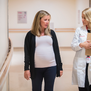
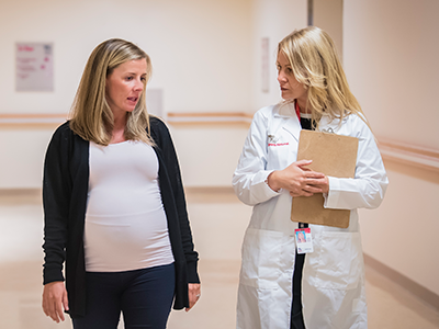
A clinical team joined forces with a research team at Children’s National Hospital to help advance treatments that can improve a child’s development caused by fetal alcohol syndrome disorder (FASDs), which is a group of conditions that can occur in a person who was exposed to alcohol before birth. This boost in collaboration between the bench and clinical hopes to optimize and develop treatments that can reverse epigenetic changes in clinical trials, paving the way to make significant progress in the field — something that is lacking to date.
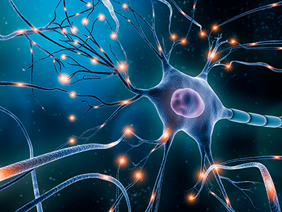
So far, Children’s National experts have published various pre-clinical studies that identified epigenetic changes caused by alcohol exposure during pregnancy. These changes observed in the pre-clinical models created neuropsychiatric problems like patients with fetal alcohol syndrome disorder. Now, they want to bring such potential treatments effective in pre-clinical models to the bedside.
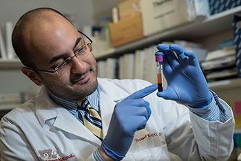
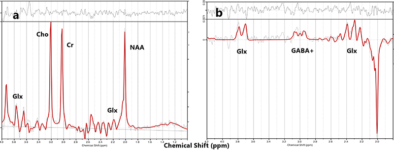
“As a first step, we are going to test whether the epigenetic changes that were observed in pre-clinical models of FASD are also true in human patients,” said Kazue Hashimoto-Torii, Ph.D., principal investigator of the Center for Neuroscience Research at Children’s National. “We hope a small amount of blood donated by patients with FASD reveal the changes. Meanwhile, my group has also been optimizing drug candidates that reverse the epigenetic changes toward clinical trials.”
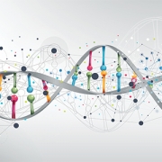
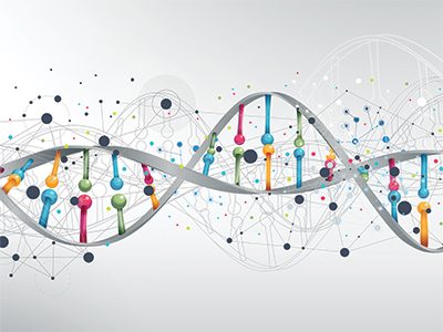
Advances in genetics and genomics have led to discoveries about the timing of exposure and developmental outcomes and genetic and epigenetic signatures that may be protective or harmful in terms of how in utero alcohol exposure affects developmental outcomes.
The hold-up in the field
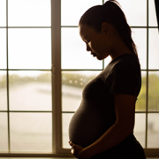
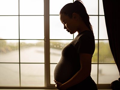
While the exact number of people with FASDs is unknown, the National Institutes of Health estimates that 1% to 5% of the population have FASDs. FASDs has a spectrum of diagnoses that represent a broad range of effects that can be manifested in an individual whose mother drank alcohol during pregnancy. These conditions can affect everyone in different ways and range from mild to severe. Individuals with mild conditions may go undiagnosed. The more affected individuals have comorbid attention-deficit/hyperactivity disorder (ADHD) and behavioral problems that become the focus of clinical encounters. The individual’s health care provider may not recognize the core features as part of FASD.
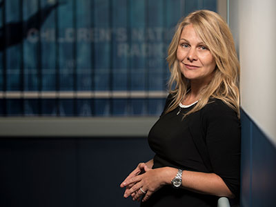
“Because there is a stigma associated with drinking while pregnant, many providers fail to get this history, and women may be reluctant to offer this information,” said Andrea Gropman, M.D., division chief of Neurodevelopmental Pediatrics and Neurogenetics at Children’s National. “There are subtle and more obvious facial dysmorphology that may help with suspicion or identification, but many individuals do not have these findings.”
The core features may be nonspecific, such as intellectual disabilities and problems with behavior and learning, difficulties with math, memory, attention, judgment and poor impulse control, which are frequent findings in ADHD, autism, learning disorders and other conditions.
“Unless history is taken and FASD is in the differential diagnosis, the diagnosis may not be made,” said Dr. Gropman. “Individuals with FASD may feel stigmatized and opt not to participate in clinical trials.”
As mentioned by Dr. Gropman, stigma can make a patient family be reluctant to seek treatment, and thus the development of treatment for FASD cannot make significant progress to date, Hashimoto-Torii added.
Children’s National Hospital leads the way in an IRB approved study
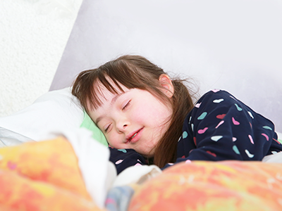
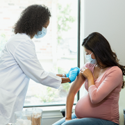
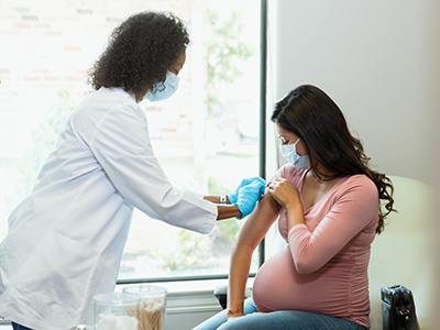
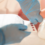
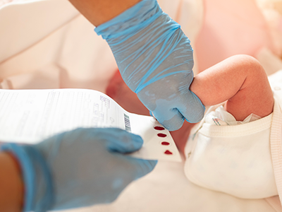
Researchers at Children’s National have identified a potential drug candidate that reverse the epigenetic changes and may lead to clinical trials. The team is seeking people to participate in an IRB approved study. The study will involve cognitive testing, filling out surveys about current functioning and cheek swab and blood sample to determine if these changes are seen in patients. To participate, subjects must be
- Children between the ages 5-12 with prenatal alcohol exposure.
- Mother of child recruited above.
For participation, please contact Grace Johnson, research coordinator at to screen for eligibility at 202-476-6034 or gjohnson3@childrensnational.org
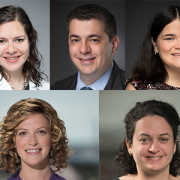
Meet the multidisciplinary team with different yet complementary skills in different fields, such as basic science, medical, social sciences, neurology and developmental disabilities, and development, who are working tirelessly to address the complex health problem.
Gropman lab:
Andrea Gropman, M.D., received her medical doctorate degree from the University of Massachusetts Medical School and specializes in neurogenetics, with a focus on mitochondrial disorders and Smith Magenis syndrome. Her latest research focuses on atypical patterns of inheritance, childhood mitochondrial disorders and other inborn errors of metabolism presenting with white matter disease.
Meira Meltzer, M.A., M.S., C.G.C., genetic counselor with a demonstrated history of working in the hospital and healthcare industry. Also skilled in molecular biology, clinical research and medical education. Strong healthcare services professional with a M.S. focused on genetic counseling from Brandeis University.
Cathy Scheiner, M.D., developmental behavioral pediatrician with a special interest in attention-deficit / hyperactivity disorder (ADHD), cerebral palsy and premature infant.
Grace Johnson, research assistant.
Hashimoto-Torii Lab:
Kazue Hashimoto-Torii, Ph.D., received her postdoctoral training in the Pasko Rakic laboratory at Yale University. Her research focuses on neurobehavior problems of children that stem from their environment during development, such as prenatal exposure to alcohol, drug and high-level glucose. A few drug candidates that her lab discovered have been patented and her lab is currently working hard to bring those medicines to bedside.
Satoshi Yamashita, M.D., Ph.D., postdoctoral research fellow skilled in developmental neurobiology. He is a pediatrician with Japanese medical license and received his Ph.D. with iPS cell research for STXBP1 encephalopathy in Japan.
Chiho Yamashita, B.N., research assistant passionate about child disease research. She is a nurse with a Japanese nursing license and worked in the pediatric department in Japan.


 Children’s National Hospital in Washington, D.C., was ranked No. 5 nationally in the U.S. News & World Report 2022-23 Best Children’s Hospitals annual rankings. This marks the sixth straight year Children’s National has made the list, which ranks the top 10 children’s hospitals nationwide. In addition, its