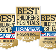
Children’s National ranked a top 10 children’s hospital and No. 1 in newborn care nationally by U.S. News
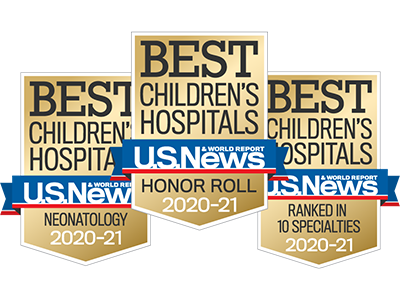
Children’s National Hospital in Washington, D.C., was ranked No. 7 nationally in the U.S. News & World Report 2020-21 Best Children’s Hospitals annual rankings. This marks the fourth straight year Children’s National has made the list, which ranks the top 10 children’s hospitals nationwide.
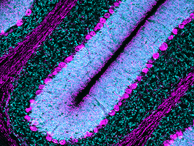
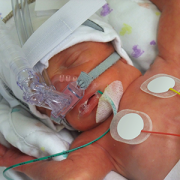
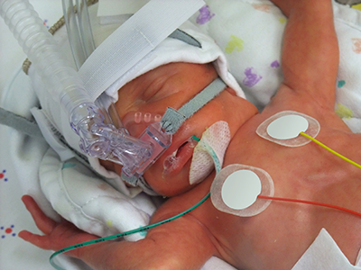
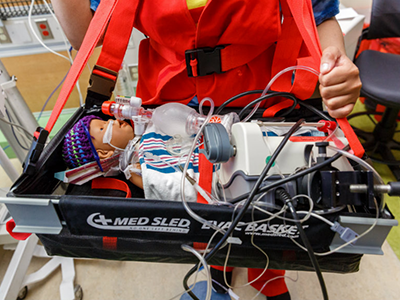
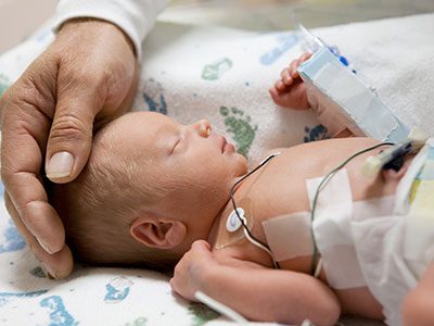
In addition, its neonatology program, which provides newborn intensive care, ranked No.1 among all children’s hospitals for the fourth year in a row.
For the tenth straight year, Children’s National also ranked in all 10 specialty services, with seven specialties ranked in the top 10.
“Our number one goal is to provide the best care possible to children. Being recognized by U.S. News as one of the best hospitals reflects the strength that comes from putting children and their families first, and we are truly honored,” says Kurt Newman, M.D., president and CEO of Children’s National Hospital.
“This year, the news is especially meaningful, because our teams — like those at hospitals across the country — faced enormous challenges and worked heroically through a global pandemic to deliver excellent care.”
“Even in the midst of a pandemic, children have healthcare needs ranging from routine vaccinations to life-saving surgery and chemotherapy,” said Ben Harder, managing editor and chief of Health Analysis at U.S. News. “The Best Children’s Hospitals rankings are designed to help parents find quality medical care for a sick child and inform families’ conversations with pediatricians.”
The annual rankings are the most comprehensive source of quality-related information on U.S. pediatric hospitals. The rankings recognize the nation’s top 50 pediatric hospitals based on a scoring system developed by U.S. News. The top 10 scorers are awarded a distinction called the Honor Roll.
The bulk of the score for each specialty service is based on quality and outcomes data. The process includes a survey of relevant specialists across the country, who are asked to list hospitals they believe provide the best care for patients with the most complex conditions.
Below are links to the seven Children’s National specialty services that U.S. News ranked in the top 10 nationally:
- Neonatology (No. 1), led by Division Chief Billie Lou Short, M.D.
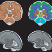
- Neurology and Neurosurgery (No. 3), led by Division Chiefs William D. Gaillard, M.D., and Robert F. Keating, M.D.
- Cancer (No. 6), led by Division Chief Jeffrey S. Dome, M.D., Ph.D.
- Nephrology (No. 7), led by Division Chief Marva Moxey-Mims, M.D., FASN
- Orthopedics (No. 9), led by Division Chief Matthew Oetgen, M.D., MBA
- Pulmonology and Lung Surgery (No.9), led by Division Chief Anastassios Koumbourlis, M.D., MPH
- Diabetes and Endocrinology (No. 10), led by Division Chief Andrew Dauber, M.D., MMSC
The other three specialties ranked among the top 50 were cardiology and heart surgery, gastroenterology and gastro-intestinal surgery, and urology.