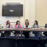
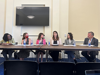
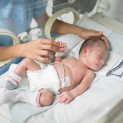
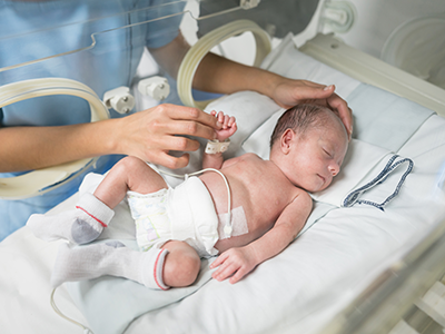
Launched 18 months ago, the city-wide DC MBW program brings together prenatal care providers, pediatricians, community-based organizations and birthing hospitals to provide essential services to mothers and babies at high risk for adverse health outcomes.
Launched 18 months ago, the city-wide DC MBW program brings together prenatal care providers, pediatricians, community-based organizations and birthing hospitals to provide essential services to mothers and babies at high risk for adverse health outcomes. Using evidence-based screening and evaluation tools, the program provides timely and targeted resources – including mental health care for mothers, developmental screening and treatment for newborns and other support – to ensure that families with the most acute social and medical needs have access to culturally relevant resources.
“Healthy moms are the foundation of healthy families and communities,” says Catherine Limperopoulos, Ph.D., the program’s director and director of the Developing Brain Institute at Children’s National. “Perinatal mood and anxiety disorders are among the most common complications of pregnancy. By offering thoughtful, individualized mental health resources – in English and Spanish – we are improving outcomes for local families facing some of the most complex and challenging health needs.”
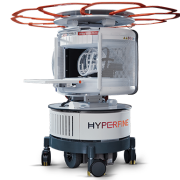
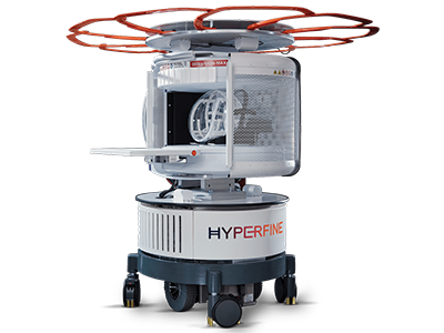
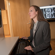
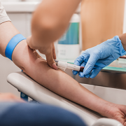
Dr. Limperopoulos has done extensive research on the profound, negative effects of expectant mothers’ stress on their unborn children. Using advanced fetal MRI technology, her published research continues to show that stress, anxiety or depression in pregnant mothers alters babies’ brain development. These mental health challenges are associated with poor obstetric outcomes and social, emotional and behavioral problems in children.
The DC MBW 1,000-referral milestone comes as Dr. Limperopoulos expands her research role at Children’s National with the creation of a new Center for Prenatal, Neonatal & Maternal Health Research that she will lead. Opened in April, the center will be a research hub for maternal health and prevalent pediatric disorders that may be present in the earliest stages of life.
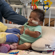
At DC MBW, Dr. Limperopoulos and her team serve mostly women of color with complex social needs that drive their anxiety, stress, depression and other mental health concerns. More than a third of the mothers referred to the wellness program are experiencing housing insecurity at intake – a number that climbs to more than 75% during their pregnancy and first year postpartum. About 30% of referred women are experiencing food insecurity. One in four are experiencing intimate partner violence, and 10% report contemplating suicide.
Dr. Limperopoulos says the team’s data indicate expectant mothers’ symptoms of depression drop dramatically within as few as six weeks of engagement with the wellness program’s services. These mothers are supported during pregnancy and beyond, as they check in with their providers during primary care visits at various Children’s National locations, participate in interactive, evidence-based play to boost child development and attend prenatal group classes that start this spring.
“It is not an overstatement to say that this program has saved lives,” says Siobhan Burke, M.D., director of Obstetrics and Gynecology at Unity Health Care. “There are so many initiatives and programs that focus on screening for depression in pregnancy, but DC MBW is actually doing the work and getting patients the treatments that they want and need. The team has designed a program that focuses on removing barriers. Whether that’s things like transportation, insurance status or language barriers, they find a way to help.”
The Centers for Disease Control and Prevention reports that the U.S maternal death rate continues to climb locally and nationally. The rates for Black mothers are significantly higher than for white and Latina mothers, making early intervention and wrap-around healthcare services even more vital to reverse this sobering trend.
The DC MBW program was underwritten by a $36 million investment from the A. James & Alice B. Clark Foundation and has hired more than a dozen full-time experts who provide care coordination, psychotherapy and patient referrals to a range of community resources. Most often, patients are referred by providers at Unity Health Care, the George Washington University Hospital, MedStar Washington Hospital Center and other leading prenatal care practices in the city.
“I credit our team’s tireless efforts for achieving this milestone so quickly. I have witnessed firsthand the dedication, time and boundless energy that they have devoted to each and every one of these people, walking side by side with them on their journey toward wellness,” Dr. Limperopoulos says. “Our research clearly supports the need for the care and resources we provide through the DC Mother-Baby Wellness initiative. Through our existing referral collaborative, we look forward to welcoming even more patients to this city-wide network as we welcome their babies into the world.”

 The hospital will advocate for the unique needs of children as part of nationwide network working to accelerate transformative health solutions.
 Children’s National Hospital was awarded nearly $7.5 million in a five-year grant to continue its leadership of an FDA-funded pediatric device consortium. Building upon a decade of previous consortium leadership, the new consortium is Alliance for Pediatric Device Innovation (APDI) and features a new and expanded roster of partners that reflects its added focus on providing pediatric innovators with expert support on evidence generation, including the use of real-world evidence (RWE), for pediatric device development.
 Children’s National Hospital in Washington, D.C., was ranked #5 in the nation on the U.S. News & World Report 2023-24 Best Children’s Hospitals annual rankings. This marks the seventh straight year Children’s National has made the Honor Roll list. The Honor Roll is a distinction awarded to only 10 children’s hospitals nationwide.


 A clinical trial testing a new drug to increase growth in children with short stature. The first ever high-intensity focused ultrasound procedure on a pediatric patient with neurofibromatosis. A low dose gene therapy vector that restores the ability of injured muscle fibers to repair. These were among the most popular articles we published on Innovation District in 2022. Read on for our full top 10 list.