Diagnostic Imaging & Radiology

Cardiology and radiology experts to participate in CMR 2018

Later this month, the international cardiovascular magnetic resonance (CMR) community will gather in Barcelona, Spain, for CMR 2018, a joint meeting organized by the European Association of Cardiovascular Imaging (EACVI) and the Society for Cardiovascular Magnetic Resonance (SCMR). Among the many attendees will be several cardiology and radiology experts from Children’s National Heart Institute:

  • Pediatric cardiology fellow Ashish Doshi, M.D., will be giving a talk titled, “Subendocardial resting perfusion defect in a case of acute fulminant myocarditis,” and will also present a poster titled, “Native T1 measurements in pediatric heart transplant patients correlate with history of prior rejection episodes.”
  • Pediatric cardiology fellow Rohan Kumthekar, M.D., will present a poster titled, “Native T1 values can identify pediatric patients with myocarditis.”
  • Cardiologist Laura Olivieri, M.D., will present two posters: “Native T1 measurements from CMR identify severity of myocardial disease over time in patients with Duchenne muscular dystrophy on therapy,” and “Feasibility of noncontrast T1 and T2 parametric mapping in assessment of acute ventricular ablation lesions in children.”
  • Pediatric cardiology fellow Neeta Sethi, M.D., will present a poster titled, “Cardiac magnetic resonance T2 mapping in the surveillance of acute allograft rejection in pediatric cardiac transplant patients.”

Additionally, Drs. Doshi and Sethi and Ileen Cronin, FNP-BC, a nurse practitioner in the Cardiac Catheterization Laboratory/Interventional Cardiac Magnetic Resonance (ICMR) Program, received travel awards to attend the conference.

CMR 2018 will be held January 31-February 3, 2018 and will focus on the theme of “Improving Clinical Value by Technical Advances.” The meeting’s emphasis will be on the common goal of improving clinical outcomes in cardiovascular disease through innovation in basic MR development and medical engineering.

Preemies’ narrowed upper airways may explain higher OSA risk

Volumetric imaging of upper airways

The airway structures of interest to the Children’s National research team included the nasopharynx (labeled red), oropharynx (labeled purple), hypopharynx (labeled green), adenoids (labeled yellow) and tonsils (labeled blue). The team displayed the volumetric imaging in three perpendicular planes and a three-dimensional model.
Credit: A. Smitthimedhin, et al, Clinical Imaging.

Infants born preterm have significantly lower nasopharyngeal and oropharyngeal volumes, compared with newborn peers carried to full term, and those lower airway volumes are independent of the infants’ gender, ethnicity or weight, according to a study published online Dec. 16, 2017 in Clinical Imaging.

According to the Centers for Disease Control and Prevention, 1 in 10 babies born in the United States is preterm, or born prior to the 37th gestational week. Premature birth leaves these children more susceptible to disordered breathing while sleeping, including obstructive sleep apnea (OSA), an ailment characterized by increased upper-airway resistance that narrows airways.

“In addition to finding some airway volumes were smaller in preterm infants, our results indicated both sets of newborns had similar hypopharyngeal volumes. This suggests that risk factors that lead to OSA are confined to the uppermost airway and do not appear to be explained by enlarged adenoids and tonsils,” says Anilawan Smitthimedhin, a Children’s National Health System radiology research fellow at the time the study was performed and lead author of the paper.

In order to diagnose OSA, clinicians now use bronchoscopy, but the method has limitations, including the need to insert a lighted instrument into the airway, which can affect pressure and resistance within the airway.

The Children’s National research team theorized that magnetic resonance imaging (MRI) could offer a non-invasive way to evaluate the upper airway, determine its anatomy and dynamic function, while shielding infants from radiation exposure that can accompany other imaging techniques.

They enrolled 96 infants who had undergone brain MRIs as part of an unrelated study about neonatal brain development. The newborns had a range of medical conditions, including suspected hypoxic ischemic encephalopathy, cardiac disease and seizures/movement disorders.

Forty-nine of the infants were born preterm; at the time of the MRI, their corrected mean gestational age was 38.4 weeks. Forty-seven of the newborns were born full term; they received MRIs at 1.7 weeks of age. The airway structures of interest included the nasopharynx (the upper part of the pharynx), oropharynx (located at the back of the mouth behind the oral cavity), hypopharynx (the entrance into the esophagus), adenoids and tonsils. The team displayed the volumetric imaging in three perpendicular planes and a three-dimensional model.

“Nasopharyngeal volume of full-term infants was 495.6 mm, compared with 221.1 mm in preterm infants. Oropharyngeal volume of full-term infants was 313.6 mm, compared with 179.3 mm in preterm infants,” Smitthimedhin says.

Aided by volumetric 3D data that more accurately measures airway and lymphoid tissue, the team proposes to study a larger group of infants to determine whether narrowing of the uppermost airways predisposes very young children to experiencing OSA later in life.

“Ultimately, our goal is to incorporate dedicated, dynamic MR imaging of the airway while children sleep, which would provide real-time, detailed information about the changes associated with sleep. This innovation holds the promise of leading to more accurate, non-invasive diagnosis of OSA in infants,” says Dorothy Bulas, M.D., chief of Diagnostic Imaging and Radiology at Children’s National.

Children’s National study co-authors include Radiologist Matthew Whitehead, M.D.; University of Maryland student Mahya Bigdeli; Pulmonologist Gustavo Nino Barrera, M.D.; Pulmonologist Geovanny Perez, M.D,; and Hansel Otero, who was at Children’s National when the research work was performed but now works at Children’s Hospital of Philadelphia.

Putting childhood epilepsy in the spotlight at American Epilepsy Society Meeting

William Gaillard

“We aim to build the evidence base for treatments that are effective specifically for children with epilepsy,” says William D. Gaillard, M.D., chief of Child Neurology, Epilepsy and Neurophysiology, and director of the Comprehensive Pediatric Epilepsy Program.

While epilepsy affects people of all ages, the unique way it manifests in infants, children and adolescents can be attributed in part to the complexities of the growing and developing brain. Researchers from the Children’s National Comprehensive Pediatric Epilepsy Program brought their expertise on the challenges of understanding and treating epilepsy in children to the recent American Epilepsy Society Annual Meeting, the largest professional gathering on epilepsy in the world.

“We aim to build the evidence base for treatments that are effective specifically for children with epilepsy,” says William D. Gaillard, M.D., chief of Child Neurology, Epilepsy and Neurophysiology, and director of the Comprehensive Pediatric Epilepsy Program. “We have learned much from studies in adult populations but technologies like functional MRI allow us to get in-depth understanding, often in non-invasive ways, of precisely how epilepsy is impacting a child.”

Dr. Gaillard was also recently elected to serve as the Second Vice President of the American Epilepsy Society. “The AES is the largest multidisciplinary professional and scientific society dedicated to the understanding, treatment and eradication of epilepsy and associated disorders, and I am honored to serve as the new Second Vice President,” he said.

The team’s presentations and poster sessions focused on several key areas in pediatric epilepsy:

Better ways to see, measure and quantify activity and changes in the brain for children with epilepsy before, during and after surgery

  • Novel applications of fMRI for children with epilepsy
    • Evaluation of an fMRI tool that tracks verbal and visual memory in children with epilepsy – one of the first to capture memory functions in this population of children using noninvasive fMRI;
    • Early study of the use of “resting-state” fMRI to map language skills before epilepsy surgery – an important first step toward noninvasively evaluating children who are too young or neurologically impaired to follow tasks in traditional MRI studies;
  • A study of whether intraoperative MRI, i.e. imaging during neurosurgery, allows for more complete removal of abnormal brain tissue associated with focal cortical dysplasia in children, which is a common cause of intractable epilepsy;
  • A preliminary case review of existing data to see if arterial spin labeling MRI, which measures blood flow to the brain, has potential to identify blood flow changes in specific locations of the brain where seizures occur;
  • An analysis of language laterality – the dominant side of the brain controlling language –  questioning the true reasons that the brains of children with epilepsy have differences in the hemisphere that predominantly controls language;
  • A review of some common assessments of language and working memory that are used pre- and post-operatively to gauge the impacts of pediatric epilepsy surgery. The study found that using multiple assessments, and studying results individually rather than as a group average, resulted in a more complete picture of the outcomes of surgery on these areas of brain function;
  • A preliminary study examining whether continuous EEG monitoring of neonates with hypoxic ischemic encephalopathy, or lack of oxygen to the brain, can be a reliable predictor of neurodevelopmental outcomes while the infant is undergoing therapeutic hypothermia.

“In order to expand our understanding of causes, impacts and outcomes, the range of research is broad given the complexity of epilepsy,” says Madison M. Berl, Ph.D. “This is the only way we can contribute to the goal of providing our colleagues and the families they serve with better resources to make informed decisions about how best to assess and treat pediatric epilepsy.”

The molecular, genetic and biological factors that contribute to onset and severity of pediatric epilepsy

  • A retrospective study of young patients with malformations in cortical development that are important causes of childhood epilepsy;
  • Investigation of a simple saliva test to effectively identify the presence of two common viral infections, human herpesvirus-6B and Epstein-Barr virus, that may be contributors to onset of epilepsy in otherwise normally functioning brains;
  • A preliminary review of the possible relationship between febrile infection-related epilepsy syndrome and the co-occurrence of another neuro-inflammatory condition – hemophagocytic lymphohistiocytosis.

Madison Berl, Ph.D., director of research in the Division of Pediatric Neuropsychology, and a pediatric neuropsychologist in the Comprehensive Pediatric Epilepsy Program, adds, “In order to expand our understanding of causes, impacts and outcomes, the range of research is broad given the complexity of epilepsy. This is the only way we can contribute to the goal of providing our colleagues and the families they serve with better resources to make informed decisions about how best to assess and treat pediatric epilepsy.”

Tracking preemies’ blood flow to monitor brain maturation

Blood is the conduit through which our cells receive much of what they need to grow and thrive. The nutrients and oxygen that cells require are transported by this liquid messenger. Getting adequate blood flow is especially important during the rapid growth of gestation and early childhood – particularly for the brain, the weight of which roughly triples during the last 13 weeks of a typical pregnancy. Any disruption to blood flow during this time could dramatically affect the development of this critical organ.

Now, a new study by Children’s National Health System researchers finds that blood flow to key regions of very premature infants’ brains is altered, providing an early warning sign of disturbed brain maturation well before such injury is visible on conventional imaging. The prospective, observational study was published online Dec. 4, 2017 by The Journal of Pediatrics.

“During the third trimester of pregnancy, the fetal brain undergoes an unprecedented growth spurt. To power that growth, cerebral blood flow increases and delivers the extra oxygen and nutrients needed to nurture normal brain development,” says Catherine Limperopoulos, Ph.D., director of the Developing Brain Research Laboratory at Children’s National and senior author of the study. “In full-term pregnancies, these critical brain structures mature inside the protective womb where the fetus can hear the mother and her heartbeat, which stimulates additional brain maturation. For infants born preterm, however, this essential maturation process happens in settings often stripped of such stimuli.”

The challenge: How to capture what goes right or wrong in the developing brains of these very fragile newborns? The researchers relied on arterial spin labeling (ASL) magnetic resonance (MR) imaging, a noninvasive technique that labels the water portion of blood to map how blood flows through infants’ brains in order to describe which regions do or do not receive adequate blood supply. The imaging work can be done without a contrast agent since water from arterial blood itself illuminates the path traveled by cerebral blood.

“In our study, very preterm infants had greater absolute cortical cerebral blood flow compared with full-term infants. Within regions, however, the insula (a region critical to experiencing emotion), anterior cingulate cortex (a region involved in cognitive processes) and auditory cortex (a region involved in processing sound) for preterm infants received a significantly decreased volume of blood, compared with full-term infants. For preterm infants, parenchymal brain injury and the need for cardiac vasopressor support both were correlated with decreased regional CBF,” Limperopoulos adds.

The team studied 98 preterm infants who were born June 2012 to December 2015, were younger than 32 gestational weeks at birth and who weighed less than 1,500 grams. They matched those preemies by gestational age with 104 infants who had been carried to term. The brain MRIs were performed as the infants slept.

Blood flows where it is needed most with areas of the brain that are used more heavily commandeering more oxygen and nutrients. Thus, during brain development, CBF is a good indicator of functional brain maturation since brain areas that are the most metabolically active need more blood.

the cerebral blood flow (CBF) maps, corresponding anatomical image aligned to the CBF map, and the regions of interest examined

This figure represents the cerebral blood flow (CBF) maps, corresponding anatomical image aligned to the CBF map, and the regions of interest examined. The scale indicates the quantitative value of the CBF map and is expressed in mL/100g/min. The data are from a preterm infant scanned at term age without evidence of brain injury. The insula (see black arrows in panel ‘D’) may be particularly vulnerable to the added stresses of the preterm infant’s life outside the womb.
Credit: M. Bouyssi-Kobar, et al., The Journal of Pediatrics.

“The ongoing maturation of the newborn’s brain can be seen in the distribution pattern of cerebral blood flow, with the greatest volume of blood traveling to the brainstem and deep grey matter,” says Marine Bouyssi-Kobar, M.S., the study’s lead author. “Because of the sharp resolution provided by ASL-MR images, our study finds that in addition to the brainstem and deep grey matter, the insula and the areas of the brain responsible for sensory and motor functions are also among the most oxygenated regions. This underscores the critical importance of these brain regions in early brain development. In preterm infants, the insula may be particularly vulnerable to the added stresses of life outside the womb.”

Of note, compromised regional brain structures in adults are implicated in multiple neurodevelopmental disorders. “Altered development of the insula and anterior cingulate cortex in newborns may represent early warning signs of preterm infants at greater risk for long-term neurodevelopmental impairments,” Limperopoulos says.

Research reported in this post was supported by the Canadian Institutes of Health Research, MOP-81116; the SickKids Foundation, XG 06-069; and the National Institutes of Health under award number R01 HL116585-01.

Congratulations to Dorothy Bulas, M.D. – 2017 RSNA Outstanding Educator recipient

Dorothy Bulas

Dorothy Bulas, M.D., section head of ultrasound and fetal imaging at Children’s National Health System, was honored with the RSNA 2017 Outstanding Educator award at the Radiological Society of North America’s (RSNA) Annual Meeting, held November 26 – December 1 in Chicago, Illinois.

The winner of the award is selected annually by the RSNA Board of Directors based on the awardee’s significant contributions and long-term commitment – 15 years or more – to radiologic education.

“In addition to being a talented clinician and an accomplished researcher, Dr. Bulas is an extraordinary teacher who has made tireless contributions to the educational programs of RSNA,” said RSNA President Richard L. Ehman, M.D. “For more than three decades, she has been a passionate and effective advocate for improving pediatric radiology worldwide – especially in poorly served countries – by participating in educational outreach.”

Liquid biopsy spots aggressive brainstem cancer earlier

Javad Nazarian

A Children’s National research team led by Javad Nazarian, Ph.D., M.S.C., tested whether circulating tumor DNA in patients’ blood and cerebrospinal fluid would provide an earlier warning that pediatric brainstem tumors were growing.

A highly aggressive pediatric brain cancer can be spotted earlier and reliably by the genetic fragments it leaves in biofluids, according to a study presented by Children’s National Health System researchers at the Society for Neuro-Oncology (SNO) 2017 Annual Meeting. The findings may open the door to non-surgical biopsies and a new way to tell if these tumors are responding to treatment.

Children diagnosed with diffuse midline histone 3 K27M mutant (H3K27M) glioma face a poor prognosis with a median survival time of only nine months after the pediatric brainstem cancer is diagnosed. Right now, clinicians rely on magnetic resonance imaging (MRI) to gauge how tumors are growing, but MRI can miss very small changes in tumor size. The Children’s research team led by Javad Nazarian, Ph.D., M.S.C., scientific director of Children’s Brain Tumor Institute, tested whether circulating tumor DNA in patients’ blood and cerebrospinal fluid would provide an earlier warning that tumors were growing. Just as a detective looks for fingerprints left at a scene, the new genetic analysis technique can detect telltale signs that tumors leave behind in body fluids.

“We continue to push the envelope to find ways to provide hope for children and families who right now face a very dismal future. By identifying these tumors when they are small and, potentially more responsive to treatment, our ultimate aim is to help children live longer,” says Eshini Panditharatna, B.A., study lead author. “In addition, we are hopeful that the comprehensive panel of tests we are constructing could identify which treatments are most effective in shrinking these deadly tumors.”

The researchers collected biofluid samples from 22 patients with diffuse intrinsic pontine glioma (DIPG) who were enrolled in a Phase I, Pacific Pediatric Neuro-Oncology Consortium clinical trial. Upfront and longitudinal plasma samples were collected with each MRI at various stages of disease progression. The team developed a liquid biopsy assay using a sensitive digital droplet polymerase chain reaction system that precisely counts individual DNA molecules.

“We detected H3K27M, a major driver mutation in DIPG, in about 80 percent of cerebrospinal fluid and plasma samples,” Panditharatna says. “Similar to adults with central nervous system (CNS) cancers, cerebrospinal fluid of children diagnosed with CNS cancers has high concentrations of circulating tumor DNA. However, after the children underwent radiotherapy, there was a dramatic decrease in circulating tumor DNA for 12 of the 15 patients (80 percent) whose temporal plasma was analyzed.”

Nazarian, the study senior author adds: “Biofluids, like plasma and cerebrospinal fluid, are suitable media to detect and measure concentrations of circulating tumor DNA for this type of pediatric glioma. Liquid biopsy has the potential to complement tissue biopsies and MRI evaluation to provide earlier clues to how tumors are responding to treatment or recurring.”

Support for this liquid biopsy study was provided by the V Foundation, Goldwin Foundation, Pediatric Brain Tumor Foundation, Smashing Walnuts Foundation, the Zickler Family Foundation, the Piedmont Community Foundation, the Musella Foundation, the Mathew Larson Foundation and Brain Tumor Foundation for Children.

Using IR imaging to improve lead apron inspection

Stanley Thomas Fricke

“When I researched how lead aprons are inspected, I learned that a combination of tactile and visual inspection is considered the gold standard. But many of the smallest holes can be missed this way,” says Stanley Thomas Fricke, Nucl. Eng., Ph.D., radiation safety officer at Children’s National Health System and study senior author.

Workers inspecting the lead aprons that shield patients from radiation during imaging tend to use tactile and visual inspections to find defects, running their fingers over the aprons since fingertips can detect even subtle changes to a surface. Yet findings from a new study could influence changes in this approach to improve inspection performance and better protect patients and inspectors.

Infrared (IR) thermal imaging is a much better detective, with 50 percent of study participants picking out all holes intentionally drilled into a test apron compared with just 6 percent of participants who detected the same defects using the tactile method, according to research published online Nov. 8, 2017 in Journal of the American College of Radiology. In addition to being a more accurate way to detect subtle defects, the IR imaging technology also reduces ionizing radiation exposure for inspectors checking the protective power of lead aprons.

“When I researched how lead aprons are inspected, I learned that a combination of tactile and visual inspection is considered the gold standard. But many of the smallest holes can be missed this way,” says Stanley Thomas Fricke, Nucl. Eng., Ph.D., radiation safety officer at Children’s National Health System and study senior author. “Unlike the fingertips, infrared light can penetrate the lead apron’s protective outer fabric and illuminate defects that are smaller than the defect size now used to reject a protective apron. This work challenges conventional wisdom and offers an inexpensive, readily available alternative.”

According to the study team, a growing number of health care settings use radiation-emitting imaging, from the operating room to the dentist’s office. Lead aprons and gonadal shields lower radiation doses experienced by health care staff and patients. In compliance with regulators, these protective devices are inspected regularly. A layer of lead inside keeps patients’ exposure to ionizing radiation at the lowest detectable level. The aprons are covered with nylon or polyester fabric for the patients’ comfort and for ease of cleaning.

“It is standard for health care institutions to use a tactile-visual approach to inspect radiation protective apparel,” Fricke says. “While increasingly common, that inspection method can allow aprons with holes and tears to slip by undetected due to the large surface area that needs to be inspected, the outer fabric that encloses the protective apron and other factors.”

Fricke recalled a news clip from years ago about an IR camera used to film swimmers at the pool that, like Superman’s powerful vision, could see through pool-goers’ clothing. The manufacturer quickly recalled the camera. But the IR technology is a perfect fit for inspectors looking for defects hidden under a lead apron’s fabric cover.

To validate this inspection alternative, the team drilled a series of nine holes ranging from 2 mm to 35 mm in diameter into a “phantom” lead apron and enclosed it within fabric that typically covers the protective shielding. The research team stapled the phantom apron to a wooden frame and placed dry wall under the frame.

Two of 31 radiation workers picked out all nine holes by touch and recorded the holes and their locations on written questionnaires.

For the IR method, the team used an infrared light to illuminate the lead apron from behind and relied on an infrared imaging camera to record 10 seconds of video from which still images were exported. Ten of 20 radiation technologists, radiology nurses and medical doctors identified all nine holes using those color photographs and recorded their entries on a questionnaire. An additional 20 percent identified eight of nine intentional defects to the phantom apron.

In both the tactile and IR groups, all participants found the largest hole and correctly recorded its location.

“Using the tactile method for inspection, most staff who work regularly with radiation-emitting devices were able to identify defects that would cause a lead apron to be rejected, which is 11 mm holes for thyroid shields and 15 mm holes for aprons,” Fricke says. “However, it is standard for these well-used aprons to develop smaller holes—which, over time, become bigger holes. Here at Children’s National, we care about every photon that touches a child.”

In the next phase of the research, the team will explore infrared flash photography, cooling the apron material and the impact of high-resolution cameras with greater depth of field.

Imaging captures obesity’s impact on the adolescent brain

Adolescent brain scan from obesity study

For the first time, a team of researchers led by Chandan Vaidya, Ph.D., chair of the Department of Psychology at Georgetown University, has used functional magnetic resonance imaging (fMRI) to capture the brain function of a small population of adolescents with obesity, both before and after bariatric surgery.

Obesity affects the whole body, from more obvious physical impacts on bones and joints to more subtle, internal impacts on organs like the brain.

For the first time, a team of researchers has used functional magnetic resonance imaging (fMRI) to capture the brain function of a small population of adolescents with obesity, both before and after bariatric surgery. The goal is to better understand the neural changes that occur when an adolescent is obese, and determine the effectiveness of interventions, such as vertical sleeve gastrectomy, at improving brain function as weight is lost.

The study, published as the November Editors’ Choice in the journal Obesity, found that executive and reward-related brain functions of study participants with obesity improved following the surgical procedure and initial weight loss.

How bariatric surgery changes the teenage brain from Research Square on Vimeo.

“We’ve known for some time that severe obesity has negative consequences on some neurocognitive function areas for adults,” says Chandan Vaidya, Ph.D., chair of the Department of Psychology at Georgetown University and a senior author of the study. “But for the first time, we’ve captured fMRI evidence in young patients, and also shown that surgical intervention and the resulting weight loss can reverse some of those deficits.”

“For me, this early evidence makes a strong case that when kids are struggling with severe obesity, we need to consider surgical intervention as an option sooner in the process,” notes Evan Nadler, M.D., director of the Bariatric Surgery Program at Children’s National Health System, who also contributed to the study. “The question that remains is whether the neurocognitive function improves more if surgery, and thus weight loss, happens earlier – and is there a time factor that should help us determine when to perform a procedure that will maximize improvements?”

The preliminary study included 36 participants and was conducted using patients recruited from the Children’s National Bariatric Surgery program, one of the first children’s hospitals to achieve national accreditation by the Metabolic and Bariatric Surgery Accreditation and Quality Improvement Program.

“We asked these questions because we know that in the kids we see, their behavioral, brain, and physical health are all very closely related to one another and have an impact on each other,” adds Eleanor Mackey, Ph.D., study senior author and co-principal investigator on the National Institute of Diabetes and Digestive and Kidney Diseases grant that funded the project. “We expected that as physical health improves, we might see corresponding improvements in brain and behavior such as cognitive and school performance.”

The study also pointed out some technical and practical challenges to studying this particular young population. Anyone with a BMI greater than 50 was not able to fit within the MR bore used in the study, preventing fMRI participation by those patients.

“In addition to future studies with a larger sample size, we’d like to see if there are neuroimaging markers of plasticity differences in a population with BMI greater than 50,” says Dr. Vaidya. “Does the severity of the obesity change how quickly the brain can adapt following surgery and weight loss?”

The abstract was selected by the journal’s editors as one that provides insights into preventing and treating obesity. It was featured at the Obesity Journal Symposium during Obesity Week 2017 in Washington, D.C., as part of the Obesity Week recognition, and a digital video abstract was also released about the findings.

Continuous EEG monitoring better predicts HIE outcomes

nurse holding newborn baby

For newborns who experience a serious complication that deprives their brain of oxygen, continuously monitoring brain activity and examining how the electrical signals evolve may be a more reliable way to identify infants most at risk for brain injury.

For newborns who experience a serious complication that deprives their brain of oxygen, continuously monitoring brain activity and examining how the electrical signals evolve may be a more reliable way to identify infants most at risk for brain injury, compared with doing evaluations at discreet intervals, according to a prospective cohort study led by Children’s National Health System research-clinicians.

Amplitude-integrated electroencephalogram (aEEG) is a bedside tool that permits clinicians to monitor the complex electrical activity of the child’s brain over time. It’s a positive sign when an aEEG shows babies beginning to sleep and wake normally by the time they are 3 days old. Conversely, severely abnormal aEEG readings in the first days of life predict poor outcomes.

The Children’s team used aEEG with infants born with hypoxic-ischemic encephalopathy (HIE), one of the most severe complications that can affect full-term infants. During pregnancy, birth or shortly after birth, a hypoxic-ischemic event can occur that impedes blood flow and oxygen delivery to the brain, resulting in destruction of brain tissue. Cooling (therapeutic hypothermia) is now standard for newborns with HIE in order to stave off life-long consequences, but deaths and neurodevelopmental disability still can occur.

“We know whole-body cooling – or lowering the body’s temperature by about 3 degrees Celsius – can help vulnerable newborns survive and can protect their brains from suffering profound injuries,” says An N. Massaro, M.D., a Children’s National neonatologist and senior author of the study published online Sept. 28, 2017 in the American Journal of Perinatology.  “What we were trying to determine with this study is whether evaluating the pattern of evolution of the aEEG as a whole provides more information compared with looking at snapshots in time.”

Eighty infants undergoing therapeutic cooling who met the inclusion criteria were enrolled in the five-year study, one of the largest such studies to date. The babies weighed more than 1,800 grams and were older than 35 weeks’ gestational age at birth, and either needed prolonged resuscitation after birth or had low APGAR scores – a measure of how well newborns fare outside the womb. Continuous recordings of EEG data occurred from the time of admission up to 12 hours after the infants’ temperatures were raised to normal and aEEG tracings were calculated.

After the therapeutic cooling blankets were removed, the infants underwent at least one magnetic resonance imaging (MRI) scan prior to discharge. During the routine follow-up check at about 18 months of age, the HIE survivors’ cognitive and motor skills were assessed using validated instruments.

Fifty-six of the infants in the study had favorable outcomes. Twenty-four infants had adverse outcomes, including 15 with severe brain injury detected by MRI and nine infants who died. These children had lower APGAR scores at five minutes, and were more likely to have severe HIE and to have experienced more frequent seizures.

“Infants whose aEEG abnormalities do not improve were at increased risk: Infants who do not reach a discontinuous background pattern by 15.5 hours of life, achieve cycling by 45.5 hours after birth and who fail to achieve continuous normal voltage by 78 hours after birth are most at risk for adverse outcomes,” Dr. Massaro says. “In addition to defining worrisome trends, we found that overall assessment of continuous aEEG readings through the course of hypothermia treatment provide the most meaningful predictive power. This means we can speak with families at the bedside with more confidence about their child’s outcomes after the infant undergoes cooling therapy.”

Related Resources

Children’s National experts contribute to new Zika guidelines

Roberta DeBiasi and Sarah Mulkey

Roberta DeBiasi, M.D., M. S., and Sarah B. Mulkey, M.D., Ph.D., members of Children’s multidisciplinary Congenital Zika Virus Program, were among the experts invited to participate in a forum held in Atlanta at CDC headquarters in late August to formulate new Zika recommendations.

The Centers for Disease Control and Prevention (CDC) on Oct. 19, 2017 updated guidelines for evaluation of women, fetuses and infants exposed to the Zika virus during pregnancy. Although only women with symptoms will now be routinely tested, asymptomatic and symptomatic infants born to these women will still be tested for the Zika virus using blood and urine tests.

Infants who appear normal, whose mothers either had negative Zika results or who had not undergone testing, will not undergo Zika testing. These infants still will undergo a standard evaluation, including a detailed physical exam, hearing screen and routine developmental assessments. The revised Zika guidance includes input from practitioners on the front lines of the Zika epidemic, including Children’s National Health System clinicians.

“These changes in the recommendations for Zika testing should not be interpreted as Zika infection risks subsiding for pregnant women and their infants in the United States. It’s simply an acknowledgement of the limitations of current testing methods – which must occur within a narrow window after Zika exposure – and the poor predictive value of Zika testing right now,” says Roberta L. DeBiasi, M.D., M.S., chief of Children’s Division of Pediatric Infectious Diseases. Dr. DeBiasi and Sarah B. Mulkey, M.D., Ph.D., members of Children’s multidisciplinary Congenital Zika Virus Program, were among the experts invited to participate in the Zika forum held in Atlanta at CDC headquarters in late August to formulate the recommendations.

While all infants will receive a standard evaluation, expanded evaluations that include an ophthalmologic assessment, more detailed hearing evaluation and ultrasound of the newborn’s head will be reserved for infants born to mothers confirmed to be Zika positive or Zika probable, or for infants born with abnormalities potentially consistent with congenital Zika syndrome, regardless of maternal status.

The majority of U.S. infants who have been exposed to Zika in the womb appeared normal at birth, according to CDC registries. Now, the next wave of these normal-appearing babies will receive standard evaluations when they are born, including a newborn hearing screening. At each well-child visit, these infants will receive:

  • A comprehensive physical examination
  • An age-appropriate vision screening
  • Developmental monitoring and screening using validated tools

“This is a natural evolution in the diagnosis and screening strategy now that the peak of the first wave of Zika transmission appears to be over,” Dr. DeBiasi says. “While we continue to evaluate new possible cases of Zika infection among pregnant women in our practice, a sizable proportion of Children’s cases are Zika-exposed infants whose physical exam and neuroimaging appeared normal at birth. Through ongoing monitoring, we hope to learn more about these children’s long-term neurodevelopment outcomes.”

Fetal MRI plus ultrasound assess Zika-related brain changes

Sarah Mulkey

Magnetic resonance imaging and ultrasound provide complementary data needed to assess ongoing changes to the brains of fetuses exposed to Zika in utero, says Sarah B. Mulkey, M.D., Ph.D.

For Zika-affected pregnancies, fetal magnetic resonance imaging (MRI) used in addition to standard ultrasound (US) imaging can better assess potential brain abnormalities in utero, according to research presented by Children’s National Health System during IDWeek 2017. In cases of abnormal brain structure, fetal MRI can reveal more extensive areas of damage to the developing brain than is seen with US.

“MRI and US provide complementary data needed to assess ongoing changes to the brains of fetuses exposed to Zika in utero,” says Sarah B. Mulkey, M.D., Ph.D., a fetal/neonatal neurologist at Children’s National Health System and lead author of the research paper. “In addition, our study found that relying on ultrasound alone would have given one mother the false assurance that her fetus’ brain was developing normally while the sharper MRI clearly pointed to brain abnormalities.”

As of Sept. 13, the Centers for Disease Control and Prevention (CDC) reported that 1,901 U.S. women were exposed to Zika at some point during their pregnancies but their infants appeared normal at birth. Another 98 U.S. women, however, gave birth to infants with Zika-related birth defects.  And eight more women had pregnancy losses with Zika-related birth defects, according to CDC registries.

The longitudinal neuroimaging study led by Children’s National enrolled 48 pregnant women exposed to the Zika virus in the first or second trimester whose infection was confirmed by reverse transcription polymerase chain reaction, which detects Zika viral fragments shortly after exposure, and/or Immunoglobulin M testing, which reveals antibodies the body produces to neutralize the virus. Forty-six of the study volunteers live in Barranquilla, Colombia, where Zika infection is endemic. Two women live in the Washington region and were exposed to Zika during travel elsewhere.

All of the women underwent at least one diagnostic imaging session while pregnant, receiving an initial MRI or US at 25.1 weeks’ gestational age. Thirty-six women underwent a second MRI/US imaging pair at roughly 31 weeks’ gestation. Children’s National radiologists read every image.

Three of 48 pregnancies, or 6 percent, were marked by abnormal fetal MRIs:

  • One fetus had heterotopias (clumps of grey matter located at the wrong place) and abnormal cortical indent (a deformation at the outer layer of the cerebrum, a brain region involved in consciousness). The US taken at the same gestational age for this fetus showed its brain was developing normally.
  • Another fetus had parietal encephalocele (an uncommon skull defect) and Chiari malformation Type II (a life-threatening structural defect at the base of the skull and the cerebellum, the part of the brain that controls balance). The US for this fetus also detected these brain abnormalities.
  • The third fetus had a thin corpus callosum (bundle of nerves that connects the brain’s left and right hemispheres), an abnormally developed brain stem, temporal cysts, subependymal heterotopias and general cerebral/cerebellar atrophy. This fetal US showed significant ventriculomegaly (fluid-filled structures in the brain that are too large) and a fetal head circumference that decreased sharply from the 32nd to 36th gestational week, a hallmark of microcephaly.

After they were born, infants underwent a follow-up MRI without sedation and US. For nine infants, these ultrasounds revealed cysts in the choroid plexus (cells that produce cerebrospinal fluid) or germinal matrix (the source for neurons and glial cells that migrate during brain development). And one infant’s US after birth showed lenticulostriate vasculopathy (brain lesions).

“Because a number of factors can trigger brain abnormalities, further studies are needed to determine whether the cystic changes to these infants’ brains are attributable to Zika exposure in the womb or whether some other insult caused these troubling results,” Dr. Mulkey says.

What Children’s has learned about congenital Zika infection

Roberta DeBiasi

Roberta DeBiasi, M.D., M.S., outlined lessons learned during a pediatric virology workshop at IDWeek2017, one of three such Zika presentations led by Children’s National research-clinicians during this year’s meeting of pediatric infectious disease specialists.

The Congenital Zika Virus Program at Children’s National Health System provides a range of advanced testing and services for exposed and infected fetuses and newborns. Data that the program has gathered in evaluating and managing Zika-affected pregnancies and births may offer instructive insights to other centers developing similar programs.

The program evaluated 36 pregnant women and their fetuses from January 2016 through May 2017. Another 14 women and their infants were referred to the Zika program for postnatal consultations during that time.

“As the days grow shorter and temperatures drop, we continue to receive referrals to our Zika program, and this is a testament to the critical need it fulfills in the greater metropolitan D.C. region,” says Roberta L. DeBiasi, M.D., M.S., chief of the Division of Pediatric Infectious Diseases and co-leader of the program. “Our multidisciplinary team now has consulted on 90 dyads (mothers and their Zika-affected fetuses/infants). The lessons we learned about when and how these women were infected and how their offspring were affected by Zika may be instructive to institutions considering launching their own programs.”

Dr. DeBiasi outlined lessons learned during a pediatric virology workshop at IDWeek2017, one of three such Zika presentations led by Children’s National research-clinicians during this year’s meeting of pediatric infectious disease specialists.

“The Zika virus continues to circulate in dozens of nations, from Angola to the U.S. Virgin Islands. Clinicians considering a strategic approach to managing pregnancies complicated by Zika may consider enlisting an array of specialists to attend to infants’ complex care needs, including experts in fetal imaging, pediatric infectious disease, physical therapists, audiologists, ophthalmologists and radiologists skilled at reading serial magnetic resonance images as well as ultrasounds,” Dr. DeBiasi says. “At Children’s we have a devoted Zika hotline to triage patient and family concerns. We provide detailed instructions for referring institutions explaining protocols before and after childbirth, and we provide continuing education for health care professionals.”

Of the 36 pregnant women possibly exposed to Zika during pregnancy seen in the program’s first year, 32 lived in the United States and traveled to countries where Zika virus was circulating. Two women had partners who traveled to Zika hot zones. And two moved to the Washington region from places where Zika is endemic. Including the postnatal cases, 89 percent of patients had been bitten by Zika-tainted mosquitoes, while 48 percent of women could have been exposed to Zika via sex with an infected partner.

Twenty percent of the women were exposed before conception; 46 percent were exposed to Zika in the first trimester of pregnancy; 26 percent were exposed in the second trimester; and 8 percent were exposed in the final trimester. In only six of 50 cases (12 percent) did the Zika-infected individual experience symptoms.

Zika infection can be confirmed by detecting viral fragments but only if the test occurs shortly after infection. Twenty-four of the 50 women (nearly 50 percent) arrived for a Zika consultation outside that 12-week testing window. Eleven women (22 percent) had confirmed Zika infection and another 28 percent tested positive for the broader family of flavivirus infections that includes Zika. Another detection method picks up antibodies that the body produces to neutralize Zika virus. For seven women (14 percent), Zika infection was ruled out by either testing method.

“Tragically, four fetuses had severe Zika-related birth defects,” Dr. DeBiasi says. “Due to the gravity of those abnormalities, two pregnancies were not carried to term. The third pregnancy was carried to term, but the infant died immediately after birth. The fourth pregnancy was carried to term, but that infant survived less than one year.”

Brain impairment in newborns with CHD prior to surgery

Catherine Limperopoulous

Children’s National researchers led by Catherine Limperopoulos, Ph.D., demonstrate for the first time that the brains of high-risk infants show signs of functional impairment before they undergo corrective cardiac surgery.

Newborns with congenital heart disease (CHD) requiring open-heart surgery face a higher risk for neurodevelopmental disabilities, yet prior studies had not examined whether functional brain connectivity is altered in these infants before surgery.

Findings from a Children’s National Health System study of this question suggest the presence of brain dysfunction early in the lives of infants with CHD that may be associated with neurodevelopmental impairments years later.

Using a novel imaging technique, Children’s National researchers demonstrated for the first time that the brains of these high-risk infants already show signs of functional impairment even before they undergo corrective open heart surgery. Looking at the newborns’ entire brain topography, the team found intact global organization – efficient and effective small world networks – yet reduced functional connectivity between key brain regions.

“A robust neural network is critical for neurons to travel to their intended destinations and for the body to carry out nerve cells’ instructions. In this study, we found the density of connections among rich club nodes was diminished, and there was reduced connectivity between critical brain hubs,” says Catherine Limperopoulos, Ph.D., director of the Developing Brain Research Laboratory at Children’s National and senior author of the study published online Sept. 28, 2017 in NeuroImage: Clinical. “CHD disrupts how oxygenated blood flows throughout the body, including to the brain. Despite disturbed hemodynamics, infants with CHD still are able to efficiently transfer neural information among neighboring areas of the brain and across distant regions.”

The research team led by Josepheen De Asis-Cruz, M.D., Ph.D., compared whole brain functional connectivity in 82 healthy, full-term newborns and 30 newborns with CHD prior to corrective heart surgery. Conventional imaging had detected no brain injuries in either group. The team used resting state functional connectivity magnetic resonance imaging (rs-fcMRI), a imaging technique that characterizes fluctuating blood oxygen level dependent signals from different regions of the brain, to map the effect of CHD on newborns’ developing brains.

The newborns with CHD had lower birth weights and lower APGAR scores (a gauge of how well brand-new babies fare outside the womb) at one and five minutes after birth. Before the scan, the infants were fed, wrapped snugly in warm blankets, securely positioned using vacuum pillows, and their ears were protected with ear plugs and ear muffs.

While the infants with CHD had intact global network topology, a close examination of specific brain regions revealed functional disturbances in a subnetwork of nodes in newborns with cardiac disease. The subcortical regions were involved in most of those affected connections. The team also found weaker functional connectivity between right and left thalamus (the region that processes and transmits sensory information) and between the right thalamus and the left supplementary motor area (the section of the cerebral cortex that helps to control movement). The regions with reduced functional connectivity depicted by rs-fcMRI match up with regional brain anomalies described in imaging studies powered by conventional MRI and diffusion tensor imaging.

“Global network organization is preserved, despite CHD, and small world brain networks in newborns show a remarkable ability to withstand brain injury early in life,” Limperopoulos adds. “These intact, efficient small world networks bode well for targeting early therapy and rehabilitative interventions to lower the newborns’ risk of developing long-term neurological deficits that can contribute to problems with executive function, motor function, learning and social behavior.”

Children’s National Fetal Medicine Institute hosts 2nd annual International Symposium on the Fetal Brain

The Children’s National Health System Fetal Medicine Institute hosted the 2nd annual International Symposium on the Fetal Brain in August 2017 in Washington, D.C.

Speakers at this year’s symposium focused on the following four areas:

  • Brain Development in an Unsupportive In Utero Environment – Diagnosis and Consequences
  • Supporting Brain Development in the Ex Utero Fetus: How Far Are We From Optimal?
  • Genomic and Epigenomic Mechanisms Underlying Differences in Brain Development
  • The Emergence of Consciousness and Pain Sensation

Adré J. du Plessis, M.B.Ch.B., M.P.H., Director of the Fetal Medicine Institute and Division Chief of Fetal and Transitional Medicine hosted the conference. In his opening remarks Dr. du Plessis noted “Our goal has been to gather together a diverse group from across the spectrum of disciplines focused on the well-being of the fetal brain and to engage all disciplines together.”

Diana-Bianchi-at-ISFB

Diana Bianchi, MD gives her keynote presentation on non-invasive fetal testing at the second annual International Symposium on the Fetal Brain.

Invited, internationally renowned speakers presented on diverse topics, including Diana Bianchi, Director of the Eunice Kennedy Shriver National Institute of Child Health and Human Development who spoke on the “Non-Invasive Fetal Testing Beyond Karyotype: What’s in it for the Fetal Brain?”

A new component to the symposium was the clinically-focused breakfast breakout sessions, created based on feedback from attendees of the 2016 Symposium. Sessions covered varied topics such as “Fetal Ultrasound: the Cornerstone of Fetal Neurodiagnosis,” “The Essentials of Neurogenetic Testing,” “Developing a Transitional Fetal-Neonatal Program” and “Using MRI to Advance Fetal Neurodiagnosis.”

The conference started with an exciting discussion by Alistair Gunn, M.B.Ch.B., Head of the Department of Physiology at the University of Auckland. His presentation “Fetal Heart Rate: What It Does and Does Not Tell Us” explored the considerable body of evidence that essentially all decelerations are mediated by chemoreflex responses to repeated hypoxia and that the parasympathetic autonomic nervous system is the critical regulator of both fetal heart rate and heart rate variation in labor.

Following a voting process from the symposium’s external speakers, the inaugural Andrea Poretti Abstract Award was presented to Katherine Ottolini for her poster titled: Breastmilk Feeds Improve Brain Microstructural Development in Very Premature Infants.

For more information about the sessions and speakers at the 2017 Symposium, please visit our website.

Reducing harm, improving quality in the NICU

Baby in the NICU

American health care is some of the most expensive in the world. To help make it more affordable, numerous efforts in all areas of medicine – from cancer care to primary care to specialized pediatrics – are focused on finding ways to improve quality and patient safety while also cutting costs.

About half a million babies born in the United States – or 10 percent to 15 percent of U.S. births – end up in the neonatal intensive care unit (NICU), most due to prematurity and very low birth weights. These vulnerable babies often need respiratory support in the form of a ventilator, which supplies oxygen to their lungs with a plastic endotracheal tube (ETT).

The typical care for these infants often involves frequent X-rays to verify the proper position of the tube. However, the American Academy of Pediatrics has counseled health care providers that ordering a daily chest X-ray simply to verify positioning of the ETT ratchets up costs without improving patient safety.

A quality-improvement initiative by Children’s National Health System’s NICU finds that these chest X-rays can be performed just twice weekly, lessening the chances of a breathing tube popping out accidentally, reducing infants’ exposure to radiation and saving an estimated $1.6 million per year.

“The new Children’s National protocol reduced the rate of chest X-rays per patient day without increasing the rate of unintended extubations,” says Michelande Ridoré, M.S., program lead in Children’s division of neonatology, who presented the research during the 2017 American Academy of Pediatrics (AAP) national conference. “That not only helps to improve patient safety – for newborns who are admitted to the NICU for longer periods, there is the additional benefit of providing significant savings to the health care system.”

Children’s NICU staff assessed how many chest X-rays were being performed per patient day before and after the protocol change, which applied to all intubated newborns in the NICU whose health condition was stable. Newborns had been undergoing a median of 0.45 chest X-rays per patient day. After the quality improvement project, that figure dropped to 0.23 chest X-rays per patient day.

When the project started in July 2015, the NICU’s monthly X-ray expenditure was $289,520. By the end of 2015, that monthly X-ray spend had fallen to $159,424 – resulting in nearly $1.6 million in annual savings.

The more restrictive strategy for ordering chest X-rays was a core component of a broader quality improvement effort aimed at lowering the number of unplanned extubations, which represent the fourth most common complication experienced by newborns in the nation’s NICUs.

“When you reduce the frequency of patients in the unit being moved, you decrease the chances of the breathing tube coming out accidentally,” Ridoré says. “By reducing unplanned extubations in the NICU, we can improve overall clinical outcomes, reduce length of stay, lower costs and improve patient satisfaction.”

When a breathing tube is accidentally dislodged, newborns can experience hypoxia (oxygen deficiency), abnormally high carbon dioxide levels in the blood, trauma to their airway, intraventricular hemorrhage (bleeding into the fluid-filled areas of the brain) and code events, among other adverse outcomes. What’s more, a patient with an unintended extubation can experience a nearly doubled hospital stay compared with the length of stay for newborns whose breathing tubes remain in their proper places. Each unplanned extubation can increase the cost of care by $36,000 per patient per admission.

To tackle this problem, Children’s National created the Stop Unintended Extubations “SUN” team. The team created a package of interventions for high-risk patients. Within one month, unintended extubations dropped from 1.18 events per 100 ventilator days to 0.59 events during the same time frame. And, within five months, that plummeted even further to 0.41 events per 100 ventilator days.

Their ultimate goal is to whittle that rate down even further to 0.3 events per 100 ventilator days, which has occurred sporadically. And the NICU notched up to 75 days between unintended extubations.

“Unintended extubation rates at Children’s National are lower than the median reported on various quality indices, but we know we can do more to enhance patient safety,” Ridoré says. ”Our SUN team will continue to address key drivers of this quality measure with the aim of consistently maintaining this rate at no more than 0.3 events per 100 ventilator days.”

Understanding the long-term consequences of prematurity

LCModel output from 32 GA baby

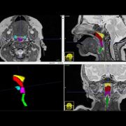
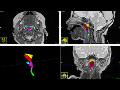
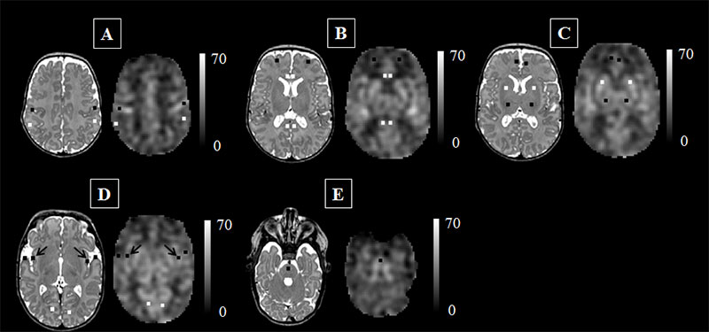
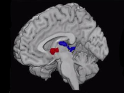
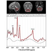
Children’s National Health System researchers processed H1-MRS data using LCModel software to calculate absolute metabolite concentrations for N-acetyl-aspartate (NAA), choline (Cho) and creatine (Cr). Preterm infants had significantly lower cerebellar NAA (p=<0.025) and higher Cho (p=<0.001) when compared with healthy term-equivalent infants. The area of the brain within the red box is the cerebellum, the region of interest for this study.

Premature birth, a condition that affects approximately 10 percent of births in the United States, often is accompanied by health problems ranging from difficulties breathing and eating to long-term neurocognitive delays and disabilities. However, the reasons for these problems have been unclear.

In a study published online Aug. 15, 2017 in Scientific Reports, a team of Children’s National Health System clinician-researchers reports that prematurity is associated with altered metabolite profiles in the infants’ cerebellum, the part of the brain that controls coordination and balance. Pre-term infants in the study had significantly lower levels of a chemical marker of nerve cell integrity and significantly higher concentrations of a chemical marker of cellular membrane turnover.

“These data suggest that interrupting the developing fetal brain’s usual growth plan during gestation – which can occur through early birth, infection or experiencing brain damage – might trigger a compensatory mechanism. The infant’s brain tries to make up for lost time or heal injured tissue by producing a certain type of cells more quickly than it normally would,” says Catherine Limperopoulos, Ph.D., director of the Developing Brain Research Laboratory at Children’s National and senior study author. “The more sensitive imaging technique that we used also revealed nerve cell damage from brain injuries extends beyond the site of injury, a finding that contrasts with what is found through conventional magnetic resonance imaging (MRI).”

It has long been clear that prematurity – birth before 37 weeks gestation – is accompanied by a number of immediate and long-term complications, from potential problems breathing and feeding at birth to impairments in hearing and sight that can last throughout an individual’s life.

Neurocognitive developmental delays often accompany pre-term birth, many of which can have long-lasting consequences. Studies have shown that children born prematurely are more likely to struggle in school, have documented learning disabilities and experience significant delays in developing gross and fine motor skills compared with children born at full-term.

Several studies have investigated the root cause of these issues in the cerebrum, the structure that takes up the majority of the brain and is responsible for functions including learning and memory, language and communication, sensory processing and movement. However, the cerebellum – a part of the brain that plays an important role in motor control – has not received as much research attention.

In the new study, Limperopoulos and colleagues used a specialized MRI technique that allowed them to parse out differences in which molecules are present in the cerebellum of full-term infants compared with premature infants. Their findings show a variety of differences that could offer clues to explain developmental differences between these two populations – and potentially identify ways to intervene to improve outcomes.

The researchers recruited 59 premature infants, born at 32 or fewer weeks’ gestation, and 61 healthy, full-term infants. Each baby received a special type of MRI known as proton magnetic resonance spectroscopy, or H1-MRS, that measures the concentrations of particular molecules in the brain. The full-term infants had these MRIs shortly after birth; the pre-term infants had them at 39 to 41 weeks gestational age, or around the time that they would have been born had the pregnancy continued to term.

Looking specifically at the cerebellum, the researchers found that the pre-term infants overall had significantly lower concentrations of N-acetyl-aspartate (NAA), a marker of the integrity of nerve cells. They also had significantly higher concentrations of choline, a marker of cell membrane integrity and membrane turnover.

Concentrations of creatine, a marker of stores of cellular energy, were about the same overall between the two groups. However, the researchers found that brain injuries, which affected 35 of the pre-term infants but none of the full-term infants, were associated with significantly lower concentrations of NAA, choline and creatine. Having a neonatal infection, which affected 21 of the pre-term infants but none of the full-term ones, was associated with lower NAA and creatine.

The findings could offer insight into exactly what’s happening in the brain when infants are born pre-term and when these vulnerable babies develop infections or their brains become injured – conditions that convey dramatically higher risks for babies born too early, Limperopoulos says. The differences between the full-term babies and the pre-term ones reflect disturbances these cells are experiencing at a biochemical level, she explains.

Limperopoulos and colleagues note that more research will be necessary to connect these findings to what is already known about developmental problems in pre-term infants. Eventually, she says, scientists might be able to use this knowledge to develop treatments that might be able to change the course of brain development in babies born too early, getting them on track with infants born at term.

“We know that the bodies of pre-term infants demonstrate a remarkable ability to catch up with peers who were born at full-term, in terms of weight and height. Our challenge is to ensure that preemies’ brains also have an opportunity to develop as normally as possible to ensure optimal long-term outcomes,” Limperopoulos says.

A closer look at the placenta to predict FGR

Catherine Limperopoulos

Using three-dimensional magnetic resonance imaging, a Children’s National research team that included Catherine Limperopoulos, Ph.D., characterized the shape, volume, morphometry and texture of placentas during pregnancy and, using a novel framework, predicted with high accuracy which pregnancies would be complicated by fetal growth restriction.

Early in development, cells from the fertilized egg form the placenta, a temporary organ that serves as an interface between the mother and her growing offspring. When things go right, as occurs in the vast majority of pregnancies, the placenta properly delivers nutrients from the mother’s diet and oxygen from the air she breathes to the developing fetus while siphoning away its waste products. This organ also plays important immune-modulating and endocrine roles.

However, in a number of pregnancies, the placenta does not do an adequate job. Unable to effectively serve the fetus, a variety of adverse conditions can develop, including preeclampsia, fetal growth restriction (FGR), preterm birth and even fetal death.

Despite the key role that the placenta plays in fetal health, researchers have few non-invasive ways to assess how well it works during pregnancy. In fact, placental disease might not be suspected until very late.

In a new study, a team of Children’s National Health System research scientists is beginning to provide insights into the poorly understood placenta.

Using three-dimensional (3D) magnetic resonance imaging (MRI), the research team characterized the shape, volume, morphometry and texture of placentas during pregnancy and, using a novel framework, predicted with high accuracy which pregnancies would be complicated by FGR.

“When the placenta fails to carry out its essential duties, both the health of the mother and fetus can suffer and, in extreme cases, the fetus can die. Because there are few non-invasive tools that reliably assess the health of the placenta during pregnancy, unfortunately, placental disease may not be discovered until too late – after impaired fetal growth already has occurred,” says Catherine Limperopoulos, Ph.D., co-director of research in the Division of Neonatology at Children’s National Health System and senior author of the study published online July 22 in Journal of Magnetic Resonance Imaging. “Identifying early biomarkers of placental disease that may impair fetal growth and well-being open up brand-new opportunities to intervene to protect vulnerable fetuses.”

The Children’s research team acquired 124 fetal scans from 80 pregnancies beginning at the 18th gestational week and continuing through the 39th gestational week. Forty-six women had normal pregnancies and healthy fetuses while 34 women’s pregnancies were complicated by FGR, defined by estimated fetal weight that fell below the 10th percentile for gestational age. The placenta was described by a combination of shape and textural features. Its shape was characterized by three distinct 3D features: Volume, thickness and elongation. Its texture was evaluated by three different sets of textural features computed on the entire placenta.

“The proposed machine learning-based framework distinguished healthy pregnancies from FGR pregnancies with 86 percent accuracy and 87 percent specificity. And it estimated the birth weight in both healthy and high-risk fetuses throughout the second half of gestation reasonably well,” says the paper’s lead author, Sonia Dahdouh, Ph.D., a research fellow in Children’s Developing Brain Research Laboratory.

“We are helping to pioneer a very new frontier in fetal medicine,” Limperopoulos adds. “Other studies have developed prediction tools based on fetal brain features in utero. To our knowledge, this would be the first proposed framework for semi-automated diagnosis of FGR and estimation of birth weight using structural MRI images of the placental architecture in vivo. This has the potential to address a sizable clinical gap since we lack methods that are both sufficiently sensitive and specific to reliably detect FGR in utero.”

The research team writes that its findings underscore the importance of future studies on a larger group of patients to expand knowledge about underlying placenta mechanisms responsible for disturbed fetal growth, as well as to more completely characterize other potential predictors of fetal/placental development in high-risk pregnancies, such as genetics, physiology and nutrition.

Spectral data shine light on placenta

preemie baby

A research project led by Subechhya Pradhan, Ph.D., aims to shed light on metabolism of the placenta, a poorly understood organ, and characterize early biomarkers of fetal congenital heart disease.

The placenta serves as an essential intermediary between a pregnant mother and her developing fetus, transporting in life-sustaining oxygen and nutrients, ferrying out waste and serving as interim lungs, kidneys and liver as those vital organs develop in utero.

While the placenta plays a vital role in supporting normal pregnancies, it remains largely a black box to science. A research project led by Subechhya Pradhan, Ph.D., and partially funded by a Clinical and Translational Science Institute Research Award aims to shed light on placenta metabolism and characterize possible early biomarkers of impaired placental function in fetal congenital heart disease (CHD), the most common type of birth defect.

“There is a huge information void,” says Pradhan, a research faculty member of the Developing Brain Research Laboratory at Children’s National Health System. “Right now, we do not have very much information about placenta metabolism in vivo. This would be one of the first steps to understand what is actually going on in the placenta at a biochemical level as pregnancies progress.”

The project Pradhan leads will look at the placentas of 30 women in the second and third trimesters of healthy, uncomplicated pregnancies and will compare them with placentas of 30 pregnant women whose fetuses have been diagnosed with CHD. As volunteers for a different study, the women are already undergoing magnetic resonance imaging, which takes detailed images of the placenta’s structure and architecture. The magnetic resonance spectroscopy scans that Pradhan will review show the unique chemical fingerprints of key metabolites: Choline, lipids and lactate.

Choline, a nutrient the body needs to preserve cellular structural integrity, is a marker of cell membrane turnover. Fetuses with CHD have higher concentrations of lactate in the brain, a telltale sign of a shortage of oxygen. Pradhan’s working hypothesis is that there may be differing lipid profiles and lactate levels in the placenta in pregnancies complicated by CHD.  The research team will extract those metabolite concentrations from the spectral scans to describe how they evolve in both groups of pregnant women.

“While babies born with CHD can undergo surgery as early as the first few days (or sometimes hours) of life to correct their hearts, unfortunately, we still see a high prevalence of neurodevelopmental impairments in infants with CHD. This suggests that neurological dysfunctional may have its origin in fetal life,” Pradhan says.

Having an earlier idea of which fetuses with CHD are most vulnerable has the potential to pinpoint which pregnancies need more oversight and earlier intervention.

Placenta spectral data traditionally have been difficult to acquire because the pregnant mother moves as does the fetus, she adds. During the three-minute scans, the research team will try to limit excess movement using a technique called respiratory gating, which tells the machine to synchronize image acquisition so it occurs in rhythm with the women’s breathing.

Panel: Significant Zika risks linger for pregnant women and developing fetuses in US

Roberta DeBiasi

The threat from Zika “is not over. It is just beginning for the families who are affected by this,” says Roberta L. DeBiasi, M.D., M.S., chief of the Division of Pediatric Infectious Diseases and co-director of the Congenital Zika Virus Program at Children’s National Health System.

The Zika virus epidemic may have fallen off the radar for many media outlets, but significant risks continue to linger for pregnant women and developing fetuses, a panel of experts told staff working for U.S. Congressional leaders.

“The threat of this virus is real, and the threat continues,” Margaret Honein, Ph.D., M.P.H., of the Centers for Disease Control and Prevention’s (CDC) pregnancy and birth defects task force, said during the July 13 briefing held in the Russell Senate Office Building.

Dr. Honein told about 100 attendees that more than 200 Zika-affected babies have been born in the United States suffering from serious birth defects, such as rigid joints, inconsolable distress that causes them to cry continuously and difficulties swallowing. Some of these infants experience seizures that cause further brain damage.

Predicting what Zika will do next in the United States is very difficult, Dr. Honein said, adding that local outbreaks could occur “at any time.” A map she displayed showed Zika’s impact in shades of blue, with Zika infections documented in nearly every state and the highest number of infections – and deepest shade of blue­ – for California, Florida and Texas.

The threat from Zika “is not over. It is just beginning for the families who are affected by this,” agreed Roberta L. DeBiasi, M.D., M.S., chief of the Division of Pediatric Infectious Diseases and co-director of the Congenital Zika Virus Program at Children’s National Health System.

Since Children’s National launched its Zika program in May 2016, the multidisciplinary team has consulted on 65 mother-fetus/infant pairs, Dr. DeBiasi said. Because in utero Zika infection can result in a wide range of side effects, the Children’s team includes pediatric infectious diseases experts, fetal/neonatal neurologists to consult on seizures, audiologists to assess hearing, physical therapists and orthopaedists to contend with limb contractures, pulmonologists to relieve breathing problems and ophthalmologists to diagnose and treat vision disorders – among other specialists.

“You really need a program that has all of these areas of expertise available for a family,” Dr. DeBiasi told attendees. “It is not possible for a family to organize 27 different appointments if you have a child with these needs.”

Children’s Zika experts also collaborate with researchers in Colombia to gauge the ability of magnetic resonance imaging to produce earlier Zika diagnoses, to assess the role of viral load as biomarkers and to document Zika’s long-term impact on children’s neurodevelopment. The Colombia study has enrolled an additional 85 women/infant pairs.

In one presentation slide, Dr. DeBiasi showed sharp magnetic resonance imaging scans from their research study of a fetal brain at 18 and 22 weeks gestation that indicated clear abnormalities, including abnormal cortical folding. Ultrasound images taken at the exact same time points did not detect these abnormalities, she said.

Asked for advice by an attendee whose clinic treats women who regularly travel between California and Mexico, Dr. DeBiasi underscored the fact that Zika infection poses a risk to developing fetuses even if the pregnant woman has no symptoms of infection. “Whether or not they’re symptomatic, the risk is the same. It’s hard for people to understand that. That is No. 1,” she said.

Another challenge is for women who scrupulously follow the CDC’s guidance on lowering their infection risk while traveling. Upon return, those women may be unaware that they could still be exposed to Zika through unprotected sex with their partner who also has travelled, for as long as six months after travel.

Children’s National earns five awards at the 2017 SPR Annual Meeting

Radiology PULSE Suite

Several technologists, fellows and faculty in the Division of Diagnostic Imaging and Radiology at Children’s National Health System were recognized at the 2017 Society of Pediatric Radiology (SPR) Annual Meeting in Vancouver, Canada, May 16-20. Each year, the international conference recognizes society members for outstanding research and education in pediatric care on the topics of imaging and image-guided care. Out of 15 major awards, Children’s National staff earned five, including two Caffey Awards – SPR’s most prestigious awards for academic excellence.

The awards received are as follows:

The Society of Pediatric Radiology Caffey Award for Best Clinical Research Paper went to attending radiologist, Dorothy Bulas, M.D., for her clinical research paper titled, “CXR Reduction Protocol in the Neonatal Intensive Care Unit (NICU) – Lessons Learned,” which highlighted collaboration with the NICU team to reduce the reliance on x-rays to monitor neonates. This method decreases the radiation dose with no risk to the patient.

The Society of Pediatric Radiology Caffey Award for Educational Exhibit was given to Benjamin Smith, M.D., a pediatric radiology fellow, for his educational poster “Sonographic Evaluation of Diaphragmatic Motion: A Practical Guide to Performance and Interpretation.” The exhibit displayed a unique technique for examining the motion of the diaphragm using ultrasound to make an accurate diagnosis of diaphragm paralysis or motion. Dr. Smith’s exhibit was also recognized by The American Academy of Pediatrics and was given the Outstanding Clinical Education Poster Award along with radiologist Hansel Otero, M.D.; sonographer Tara K. Cielma, R.D.M.S, R.D.C.S, R.V.T.; and faculty member Anjum N. Bandarkar, M.D.

The Society of Pediatric Radiology Radiographer Best Poster Award was given to Dr. Bandarkar for her poster titled, “Infantile Hypertrophic Pyloric Stenosis: Value of measurement technique to avoid equivocal exam.” The World Federation of Pediatric Imaging also awarded Dr. Bandarkar, Adebunmi O. Adeyiga, M.D. and Tara Cielma the 2017 Outstanding Radiographer Educational Poster Award for their collaborative poster on, “A Sonographic Walk‐Through: Infantile Hypertrophic Pyloric Stenosis.”

Division Chief of Diagnostic Imaging and Radiology, Raymond Sze, M.D., remarked, “This is a major win not only for the department but also for the entire hospital. The support and collaboration of our Children’s National colleagues across many departments allowed us to advance the field of pediatric imaging and earn national recognition for the high-quality and impactful research and education that’s happening at our institution.”