Tag Archive for: epilepsy

illustration of neurons with electrical impulses

Children’s National at the American Epilepsy Society Annual Meeting

illustration of neurons with electrical impulsesSeveral experts from Children’s National Hospital will be sharing their knowledge at the upcoming American Epilepsy Society Annual Meeting in Orlando, December 1-5. Here’s a sample of what you can expect.

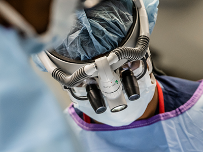
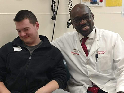
  • Chima Oluigbo, M.D., a pediatric neurosurgeon, will be on panel with other surgeons discussing different surgical techniques and approaches related to epilepsy surgery followed by hands-on practice at teaching stations. He will focus on extra-temporal epilepsy scenarios and will be presenting on Nuances of Temporal Lobe Surgery in the Pediatric Population at the Neurosurgery Symposium highlighting Surgical Controversies in Temporal Lobe Epilepsies.
  • Ersida Buraniqi, M.D., a child neurologist, will be part of a special interest group on critical care and discuss advances in electroencephalography (EEG) and multimodal neuro-monitoring for seizures in the intensive care unit (ICU). Dr. Buranigui will be doing a special presentation on EEG features to predict electrographic seizures and mortality in the pediatric intensive care unit (PICU).
  • Dana Harrar, M.D., director of Pediatric Stroke Program and co-director of Critical Care Neurology, is presenting at an invitation-only resident EEG course, providing an interactive structured curriculum on pediatric and adult EEG. Dr. Harrar will be focusing on doing an ICU-EEG nomenclature overview.
  • Madison Berl, Ph.D., director of Neuropathy Research and of the Intellectual and Developmental Disabilities Research Center Program, will be presenting during the AES Annual Course. The topic “It’s About Time” will focus on the critical importance the timing in epilepsy care plays in patient outcome. Dr. Berl will be presenting on neuropsych outcomes.
  • Leigh Sepeta, Ph.D., director of Inpatient Neuropsychology, is the vice-chair of the special interest group on neuropsychology. Additionally, Freya Prentice, M.Sc., will be doing a presentation during this session on functional mapping of the cognitive memory circuit in pediatric epilepsy.
Date Time Presenter(s) Title
12/2/23 8:00 am Chima Oluigbo, M.D., FRCSC, FAANS Skills Workshop | Epilepsy Surgery Workshop: Techniques and Clinical Scenarios
12/2/23 5:30 pm Chima Oluigbo, M.D., FRCSC, FAANS SIG | Epilepsy Surgery: Homunculus Revisited: Managing Central Lobe Epilepsies
12/2/23 5:30 pm Ersida Buraniqi, M.D. SIG | Critical Care: Advances in EEG and Multimodal Neuro-monitoring for Seizures in the ICU
12/2/23 7:00am Dana Harrar M.D. Resident EEG Course
12/3/23 9:00 am Chima Oluigbo, M.D., FRCSC, FAANS Neurosurgery Symposium | Surgical Controversies in Temporal Lobe Epilepsies
12/3/23 8:45 am Madison Berl, Ph.D. Annual Course | It’s About Time: Timing in Epilepsy Evaluation and Treatment
12/4/23 7:00 am Leigh Sepeta, M.D. SIG | Neuropsychology: Mapping Cognition in Epilepsy: From the Lab to the Clinic
12/4/23 7:00 am Freya Prentice, M.D. SIG | Neuropsychology: Mapping Cognition in Epilepsy: From the Lab to the Clinic
12/5/23 7:00 am Dana Harrar M.D. SIG | Epilepsy Education: Epilepsy Education Throughout the Training Pipeline


illustration of a brain

Inducing strokes in newborns to treat hemimegalencephaly

“The number one thing people are perplexed by is how well these babies recover and how they can only live with half a brain,” said Tayyba Anwar, M.D., neonatal neurologist and co-director of the Hemimegalencephaly Program at Children’s National Hospital. “People think if a child has half a brain that’s damaged or dysplastic, how are they functioning? But babies are so resilient. It still amazes me.”

The big picture

Children’s National experts have pioneered a novel approach of inducing strokes to stop seizures and improve neurodevelopmental outcomes in newborns under three months old with hemimegalencephaly (HME).

The procedure, called an endovascular embolic hemispherectomy, can be safely used to provide definitive treatment of HME-related epilepsy in neonates and young infants, according to a study in the Journal of NeuroInterventional Surgery.

Prior to this approach, the standard treatment was an anatomic hemispherectomy — surgical removal of the affected half of the brain. But infants had to be at least three months old to undergo such a complex surgery. Delaying surgery meant the persistent seizures compromised the development of the healthy half of the brain.

What they’re saying

In this video, Dr. Anwar and Panagiotis Kratimenos, M.D., Ph.D., neonatologist and co-director of Research in Neonatology at Children’s National, discuss the critically important neonatal care provided to babies who undergo endovascular embolic hemispherectomy and how protocols have evolved with each case to make this less invasive approach a feasible early alternative to surgical hemispherectomy.

Drs. Anwar and Kratimenos are part of the multidisciplinary team of neonatal neurologists, neurointerventional radiologistsneonatologists and neurosurgeons performing endovascular hemispherectomies.

Illustration of brain and brainwaves

Effective treatment for children with hemimegalencephaly

Illustration of brain and brainwaves

Anatomic or functional hemispherectomy are established neurosurgical treatment options and are recommended for effective seizure control and improved neurodevelopmental outcome in patients with HME.

Endovascular hemispherectomy can be safely used to provide definitive treatment of hemimegalencephaly (HME) related epilepsy in neonates and young infants when intraprocedural events are managed effectively, a new study finds.

The authors of the study, which published in the Journal of NeuroInterventional Surgery, add that this less invasive novel approach should be considered a feasible early alternative to surgical hemispherectomy.

Why it matters

Anatomic or functional hemispherectomy are established neurosurgical treatment options and are recommended for effective seizure control and improved neurodevelopmental outcome in patients with HME. Hemispherectomy in the neonate, however, is associated with high surgical risks and most neurosurgeons defer surgical hemispherectomy until the patient is at least 8 weeks old. This delay comes at a significant neurocognitive cost as the uncontrolled seizures during this time of deferred surgery have a deleterious effect on future neurocognitive outcome.

Why we’re excited

“The procedure we have developed, endovascular hemispherectomy by transarterial embolization, acutely stops seizures and this cessation of seizures has been sustained in each of the treated patients,” says Monica Pearl, M.D., director of the Neurointerventional Radiology Program at Children’s National Hospital and the study’s lead author.

This treatment option – performed early in life – provides hope and a better quality of life for these patients post procedure.

What’s been the hold-up in the field?

Currently, the only effective treatment option is hemispherectomy. With the patient population of neonates and young infants, hemispherectomy has a very high mortality and complication rate resulting in most neurosurgeons deferring treatment until at least 8 weeks. This leaves neonates and young infants without effective treatment options and on multiple antiseizure medications in an effort to control the seizures

How does this work move the field forward?

“Embolization provides a highly effective treatment option that acutely stops seizures during a time period of critical neurodevelopment and one in which traditional open neurosurgical procedures are not viable options,” Dr. Pearl says. “Specifically, we can consider and perform embolization in children as young as one or two weeks of age rather than waiting until at least 8 weeks of age. The impact of earlier intervention – acutely stopping the seizures, reducing the dose and number of antiseizure medications and avoiding more invasive surgical procedures (hemispherectomy, shunt placement) – appears to be dramatic in our recent series. We are conducting long term studies to assess this effect on neurodevelopmental outcome.”

How is Children’s National leading in this space?

Dr. Pearl and the late Taeung Chang, M.D., neurologist at Children’s National, pioneered this concept and treatment pathway. The multidisciplinary team is led by Dr. Pearl, who has performed all the embolization procedures (transarterial embolization/endovascular hemispherectomy) and Tayyba Anwar, M.D., Co-Director, Hemimegalencephaly Program at Children’s National Hospital. Our epilepsy team, neonatology team and neurosurgery team work collaboratively managing the patients before and after each procedure.

Illustration of brain and brainwaves

Risk factors for pharmacoresistant pediatric epilepsy

Illustration of brain and brainwaves

New study evaluates risk factors for the timing and development of drug-resistant pediatric epilepsy.

Focal cortical dysplasia (FCD) is the most common cause of surgically-treatable epilepsy in children. In a new study published in Neurology, researchers evaluated 143 children with confirmed FCD risk factors for the timing and development of pharmacoresistant epilepsy.

What this means

The current definition of pharmacoresistance requires failure of two appropriately-dosed and selected antiseizure medications before being able to be considered for epilepsy surgery.

“We found that the failure of just one antiseizure medication is associated with an enormous increased incidence and earlier development of pharmacoresistance,” says Nathan Cohen, M.D., neurologist at Children’s National and lead author of the study. “Our data supports the redefinition of pharmacoresistant epilepsy to the failure of just one antiseizure medication in this population, which would potentially allow these patients to benefit from earlier curative surgery.”

Why it matters

The findings showed that in children with FCD the failure of just one antiseizure medication is associated with an enormous risk and earlier incidence of pharmacoresistance. Therefore, the authors advocate for its redefinition in FCD-related epilepsy to the failure of just one antiseizure medication.

“This will allow children to be considered much earlier for potentially curative epilepsy surgery,” adds Dr. Cohen. “We find that the majority of FCD patients develop epilepsy and that the majority of those with epilepsy develop pharmacoresistance.”

In a multivariate analysis, the authors show that the FCD cortical lobar location, pathologic subtype, and age of seizure onset are not important factors in the development of pharmacoresistance.

What’s next

This data supports operational re-definition of pharmacoresistance for surgical planning in FCD-related epilepsy to the failure of one antiseizure medication, and support early, potentially curative surgery to improve outcomes in this patient population.

You can read the full study, Prevalence and Risk Factors for Pharmacoresistance in Children With Focal Cortical Dysplasia–Related Epilepsy, in Neurology.

Digital background depicting innovative technologies in (AI) artificial systems, neural interfaces and internet machine learning technologies

AI algorithm that detects brain abnormalities could help cure epilepsy

Digital background depicting innovative technologies in (AI) artificial systems, neural interfaces and internet machine learning technologies

A new AI algorithm can detect subtle brain abnormalities that cause epileptic seizures.

An artificial intelligence (AI) algorithm that can detect subtle brain abnormalities that cause epileptic seizures has been developed by a UCL-led team of international researchers, including Children’s National Hospital.

To do this, the team quantified features from MRI scans, such as how thick or folded the brain was at nearly 300,000 locations in each case.

They then trained the AI algorithm using examples labelled by expert radiologists as either a healthy brain or one with focal cortical dysplasia (FCD) based on their patterns and features.

The results, published in Brain, showed that in the main cohort of 538 patients, the algorithm was able to detect the FCD in 67% of cases.

“We put an emphasis on creating an AI algorithm that was interpretable and could help doctors make decisions. Showing doctors how the Multicentre Epilepsy Lesion Detection project (MELD) algorithm made its predictions was an essential part of that process,” said Mathilde Ripart, research assistant at UCL and the study’s co-first author.

Around 1% of the population have epilepsy and, of these, 20-30% do not respond to medications.

“We are excited to collaborate with MELD on ways to improve the treatment of pharmacoresistant epilepsy,” said Nathan Cohen, M.D., neurologist at Children’s National Hospital and co-author of the study. “This advanced imaging platform is open source and demonstrates the benefit of team science at the broadest scale.”

In children who have had surgery to control their epilepsy, FCD is the most common cause, and in adults it is the third most common cause.

Additionally, of patients who have epilepsy that have an abnormality in the brain that cannot be found on MRI scans, FCD is the most common cause.

You can read the full UCL press release here.

William Gaillard

William D. Gaillard, M.D., named as first endowed Professor of Epilepsy and Neurophysiology

William GaillardChildren’s National Hospital named William D. Gaillard, M.D., as the hospital’s first endowed Professor of Epilepsy and Neurophysiology.

Dr. Gaillard serves as Division Chief of Child Neurology, Epilepsy and Neurophysiology, Director of the Comprehensive Pediatric Epilepsy Program and Associate Director of the Center for Neuroscience Research at Children’s National Hospital. He also is Professor of Pediatrics and Neurology at the George Washington University School of Medicine, Professor of Neurology at Georgetown University and Adjunct Professor of Hearing and Speech Sciences at the University of Maryland, College Park.

About the award

Dr. Gaillard joins a distinguished group of 42 Children’s National physicians and scientists who are endowed chairs. Professorships at Children’s National advance groundbreaking work on behalf of children and their families and foster new discoveries and innovations in pediatric medicine. These prestigious appointments carry honor and reflect the recipient’s achievements and donor’s forethought to advance and sustain knowledge.

Dr. Gaillard is an internationally recognized expert in advanced structural and functional imaging, with a focus on examining the effects of epilepsy on brain structure and function. His pioneering work with functional imaging has changed clinical practice for patients with epilepsy, impacted structural and functional imaging methods and provided fundamental insights on brain plasticity. Dr. Gaillard’s research is improving patient outcomes and the lives of children with epilepsy.

Donors Betsy Williams and Tom Moore, through their vision and generosity, will ensure that Dr. Gaillard and future holders of this professorship can launch bold, new initiatives to rapidly advance the field of pediatric epilepsy and neurophysiology, elevate our leadership and improve the lifetimes of children with epilepsy and seizure disorders.

“This Professorship is a great honor and opportunity for our team and for the future of pediatric epilepsy medicine,” says Dr. Gaillard. “Thanks to Betsy, Tom and the Hess Foundation’s generosity, we can continue to invest in people and programs that show promise. We can have the flexibility to respond to the needs of our current patient families and prepare for those that will need us tomorrow. We are forever grateful.”

About the donors

Betsy and Tom are parents, members of the Epilepsy Council at Children’s National and long-time supporters. To acknowledge their gratitude and respect for Dr. Gaillard and the comprehensive team he’s built, the Professorship will be named for Dr. Gaillard in the future.

model of the brain

Treating newborns with hemimegalencephaly by inducing strokes

model of the brain

Experts at Children’s National Hospital have pioneered a novel approach using controlled strokes to stop seizures and improve neurodevelopmental outcomes in newborns under three months born with hemimegalencephaly (HME). They now consider it their new standard of care for babies in this age group with HME and refractory epilepsy.

Asking a physician to induce strokes in newborns is asking her to do something contrary to her training. But over the past eight years, experts at Children’s National Hospital have pioneered a novel approach using controlled strokes to stop seizures and improve neurodevelopmental outcomes in newborns under three months born with hemimegalencephaly (HME). They now consider it their new standard of care for babies in this age group with HME and refractory epilepsy.

“We have demonstrated the ability to intervene and stop the intractable seizures during a critical time of neurodevelopment in which no other effective medical or surgical option exists. That is extremely rewarding,” said Monica Pearl, M.D., director of the Neurointerventional Radiology Program at Children’s National. Children’s National is the only center in the world currently offering this treatment. A multi-disciplinary team led by Dr. Pearl; Taeun Chang, M.D., director of the Neonatal Neurology and Neonatal Neurocritical Care Program; neurophysiologist and neonatal neurologist Tammy Tsuchida, M.D., Ph.D.; and other experts has now successfully treated seven patients using this minimally-invasive approach.

“We want patients and providers to understand this is a better alternative to a delayed hemispherectomy, the standard of care currently offered to newborns with HME,” said Dr. Chang.

The best treatment for newborns with hemimegalencephaly

HME, a rare congenital condition occurring in a handful of newborns each year, is characterized by abnormal growth and enlargement of half of the brain which leads to intractable seizures. The seizures often result in severe cognitive delays and hemiparesis. The standard treatment is an anatomic hemispherectomy — surgical removal of the affected half of the brain, allowing the remaining half of the brain to develop and function without constant seizures.

Such a large and complex surgery poses serious risks for infants younger than three months, leaving doctors with the difficult choice to delay surgery until these newborns grow bigger and stronger, even as they are experiencing seizures. These persistent seizures compromise the development of the healthy half of the brain. One study reports as much as a drop of 10 to 20 IQ points with each month’s delay in surgical hemispherectomy.

“I was willing to consider performing these procedures because there is a clear, unmet medical need and these babies are in dire circumstances,” Dr. Pearl said. “Waiting for curative hemispherectomy means more than just lost time; uncontrolled seizures and anti-seizure medications have detrimental effects on the ‘normal,’ unaffected parts of the brain. We needed a better option for these patients.” Dr. Pearl said that complete embolization of the affected hemisphere as both primary and definitive treatment had never been described. They could only find one example in the literature – a paper from 1995 – suggesting embolization as an adjunct to surgery, and nothing suggesting it as a primary modality.

About the care received

Dr. Pearl is one of only a handful of dedicated pediatric neurointerventionalists across the country with neurovascular expertise in people of all ages, in particular neonates and young infants. For these procedures to be performed safely, the neurointerventionalist must be proficient in obtaining femoral arterial access and navigating small caliber cervicocerebral blood vessels that are less than one millimeter in diameter.

Additionally, one needs a neonatal neurocritical care service and NICU that can medically manage large strokes and their potential complications in newborns. Dr. Chang has developed a specialized protocol based on decades of managing strokes and other acute brain injuries in newborns. She created the neonatal neurocritical care service at Children’s National, the only one in the region and the largest in the world.

“Our teams are fortunate in that we each respectively have extensive prior experience in treating and managing neonates and very young infants for various cerebrovascular disorders,” Dr. Pearl said. “We relied on this collective experience to make this hemispheric embolization pathway possible.”

How it happens

To perform the embolizations, Dr. Chang and her team first optimizes control of the seizures using medications. Dr. Pearl places a sheath in the femoral artery using ultrasound guidance – a delicate task in a neonate whose femoral artery diameter is only two to three millimeters. She then navigates a catheter up the aorta and selects the targeted carotid artery using radiographic guidance. What follows is a set of intricate navigations to direct the microcatheter through small blood vessels in the brain, often less than one millimeter.

Using x-ray guidance, Dr. Pearl injects contrast through the microcatheter to visualize the arterial anatomy and advance the microcatheter into position for embolization. She uses glue that hardens when exposed to blood, blocking off the blood supply to the seizure-inducing areas. The process is repeated until the blood supply to the entire affected hemisphere is occluded. Meanwhile, Dr. Chang and her team monitor the brain’s electrical activity using an electroencephalogram (EEG) to watch how the brain responds to each stroke. The surgical epilepsy, neonatal neurocritical care and neonatology teams are all in constant communication throughout the procedure.

Together, they have to contend with the same symptoms patients have immediately following a stroke, most notably brain swelling that can cause bleeding and herniation. The resultant brain swelling is complicated further by the already enlarged hemisphere of the brain. Using neuroprotective strategies learned from treating over a thousand newborns with perinatal brain injury, Dr. Chang and her team and the NICU coordinate to minimize brain swelling and protect the healthy half of the brain by tightly controlling the brain temperature, glucose, sodium levels, and blood pressure. Over the course of a few weeks, Dr. Pearl performs three to four embolization sessions to halt blood supply to the seizing half of the brain.

“The risks of intracranial vasospasm and hemorrhage during embolization are higher in this distinct group of patients compared to other neonates requiring embolization, such as in vein of Galen malformations. These events must be controlled immediately to prevent complications and I know I only have seconds to react,” Dr. Pearl said.

“Here, we have the cultivation of brain-centric neonatal care, a large level IV tertiary NICU with expertise in keeping critically ill babies alive and rare pediatric neurologic subspecialists like Dr. Pearl and myself. All of this is what makes this level of innovation possible,” Dr. Chang said. Now, they wish this minimally invasive approach to be available to all newborns with HME and refractory epilepsy.

“This is not a fluke. This is not a one-time thing. Our team at Children’s National has been perfecting this method for close to a decade,” Dr. Chang said. As for proof, her answer is clear.

brain network illustration

Changing the surgical evaluation of epilepsy

brain network illustrationThe choice between stereoelectroencephalography (SEEG) and subdural evaluation is not mutually exclusive, according to a new opinion piece published in JAMA Neurology.

In their article, Chima Oluigbo, M.D., pediatric epilepsy neurosurgeon, William D. Gaillard, M.D., division chief of Epilepsy and Neurophysiology and Neurology, both at Children’s National Hospital, and Mohamad Z. Koubeissi, M.D., M.A., from The George Washington University Hospital, discuss how the practicing epileptologist requires a profound understanding of the roles of different technologies. It also looks at how to integrate both traditional and emerging paradigms to optimize seizure control. This issue is particularly relevant to choosing the best method of invasive intracranial electroencephalography monitoring in individual cases.

Noting that despite the dramatic increase in SEEG use in recent years, the authors talk about how many patients still benefit from invasive monitoring using subdural grids. Therefore, it is important to define the considerations that should guide decision-making on the choice of SEEG versus subdural monitoring in each patient. The authors expand on their statement explaining that it is critical to define the roles of SEEG vs subdural grid investigation in each patient as subdural grid evaluations are still indicated in specific circumstances.

Additionally combined hybrid deployment of both techniques may be indicated in specific situations. Accommodation should be made to allow customization of the technique chosen to available technical expertise and equipment as well as patient preference.

2021 neurology infographic

2021 at a glance: Neurology and Neurosurgery at Children’s National

2021 neurology infographic

illustration of Research & Innovation Campus

NIH awards $6.7M to build additional lab space at Children’s National Research & Innovation Campus

Children’s National Hospital today announced a $6.7 million award from the National Institute of Health (NIH) for the new Children’s National Research & Innovation Campus (RIC). The funds will help transform a historic building on the former site of Walter Reed Army Medical Center into new research labs. The NIH construction grant marks the first secured grant funding for Phase II of the campus project, signaling continued momentum for the first-of-its-kind pediatric research and innovation hub.

The funding was announced as D.C. Mayor Muriel Bowser, D.C. Deputy Mayor for Planning and Economic Development John Falcicchio and D.C. Council Chair Phil Mendelson took their first tour of the already-renovated Phase I of the RIC. The campus began opening in early 2021 and brings together Children’s National with top-tier research and innovation partners: Johnson & Johnson Innovation – JLABS @ Washington, DC and Virginia Tech. They come together with a focus on driving discoveries and innovation that will save and improve the lives of children.

“This NIH award is the latest confirmation that we are creating something very special at the Children’s National Research & Innovation Campus,” said Kurt Newman, M.D., president and CEO of Children’s National. “Only the D.C. region can offer this proximity to federal science agencies and policy makers. When you pair our location with these incredible campus partners, I know the RIC will be a truly transformational space where we develop new and better ways to care for kids everywhere.”

The campus is an enormous addition to the BioHealth Capital Region, the fourth largest research and biotech cluster in the U.S., with the goal of becoming a top-three hub by 2023. The RIC exemplifies the city’s commitment to building the partnerships necessary to drive discoveries, create jobs, promote economic growth, treat underserved populations, improve health outcomes, and keep D.C. at the forefront of innovation and change.

“We are proud to officially welcome the Children’s National Research & Innovation Campus to the District and to the Ward 4 community,” said Mayor Bowser, after touring the campus. “This partnership pairs a world-class hospital with a top university and a premier business incubator – right here in the capital of inclusive innovation. Not only will our community benefit from the jobs and opportunities on this campus, but the ideas and innovation that are born here will benefit children and families right here in D.C. and all around the world.”

The NIH grant funding announced today will go toward the expansion and relocation of the DC Intellectual and Developmental Disabilities Research Center (DC-IDDRC). This research center will increase the efforts to improve the understanding and treatment of children with developmental disabilities, including autism, cerebral palsy, epilepsy, inherited metabolic disorders and intellectual disability.

The space where the new lab will be built used to be the Armed Forces Institute of Pathology Building, a portion of the Walter Reed Army Medical Center. The site closed and Children’s National secured 12 acres in 2016, breaking ground on Phase I construction in 2018.

The new space will offer highly cost-effective services and unique state-of-the-art research cores that are not available at other institutions, boosting the interdisciplinary and inter-institutional collaboration between Children’s National, George Washington University, Georgetown University and Howard University. Investigators from the four institutions will access the center, which includes hoteling laboratory space for investigators whose laboratories are not on-site but are utilizing the core facilities — Cell and Tissue Microscopy, Genomics and Bioinformatics, and Inducible Pluripotent Stem Cells.

“While we have explored outsourcing some of these cores, especially genomics, we found that expertise, management, training and technical support needed for pediatric research requires on-site cores,” said Vittorio Gallo, Ph.D., interim chief academic officer, interim director of the Children’s National Research Institute, and principal investigator for the DC-IDDRC. “The facility is designed to support pediatric studies that are intimately connected with our community. We operate in a highly diverse environment, addressing issues of health equity through research.”

The RIC provides graduate students, postdocs and trainees with unique training opportunities, expanding the workforce and talent of new investigators in the D.C. area. Young investigators will have job opportunities as research assistants and facility managers as well. The new labs will support these researchers so they can tackle pressing questions in pediatric research by integrating pre-clinical and clinical models.

Phase II will place genetic and neuroscience research initiatives of the DC-IDDRC at the forefront to treat a variety of pediatric developmental disorders. Other Children’s National research centers will also benefit from this additional space. The clinical and research campuses will be physically and electronically integrated with new informatics and video-communication systems.

The total projected cost of Phase II is $180 million, with design and construction to take up to three years to complete once started.

illustration of Research & Innovation Campus

Phase II will place genetic and neuroscience research initiatives of the DC-IDDRC at the forefront to treat a variety of pediatric developmental disorders. Other Children’s National research centers will also benefit from this additional space. The clinical and research campuses will be physically and electronically integrated with new informatics and video-communication systems.

3d illustration of blood cells, plasmodium causing malaria disease

International projects spearheaded by Children’s National Neurology leaders

NIH approves grant for clinical trial on pediatric cerebral malaria in Malawi

3d illustration of blood cells, plasmodium causing malaria disease

Cerebral malaria, when patients lapse into coma after developing a malaria infection, is the most severe neurological complication of infection with Plasmodium falciparum.

The National Institutes of Health (NIH) approved a $5.8 million grant for a Phase I/IIa randomized clinical trial of 6-diazo-5-oxo-L-norleucine (DON), a new medication for pediatric cerebral malaria. Douglas Postels, M.D., neurologist at Children’s National Hospital, will serve as the trial’s principal investigator. The clinical trial will enroll participants in Blantyre, Malawi.

More than 400,000 people die each year from malaria. Cerebral malaria, when patients lapse into coma after developing a malaria infection, is the most severe neurological complication of infection with Plasmodium falciparum. Many children who survive are left suffering from neurological complications because of the disease, leaving some unable to walk, see or go to school. Dr. Postels and others are seeking to initiate this clinical trial with the primary goal to save lives and improve the quality of life for children who survive the disease.

“The purpose of this study is to see if DON is safe in the Malawian population,” Dr. Postels said, noting that adult participants will be enrolled in the first year and children subsequently. “Once the medication has proven to be safe, our intention is to expand this research elsewhere in Africa allowing us to enroll more children and evaluate whether DON decreases the likelihood of death or neurological disability in pediatric cerebral malaria.”

DON was originally tested 50 years ago as an anti-cancer agent but was recently repurposed by the National Institute of Allergy and Infectious Diseases (NIAID) for pediatric cerebral malaria. The current clinical trial is a collaborative project with the NIAID scientists who performed the pre-clinical testing with DON.

“There are currently no adjunctive treatments, used in combination with intravenous anti-malarial medications, that decrease death or disability in pediatric cerebral malaria,” Dr. Postels said. “Our hope is that DON will be the “magic bullet” that helps these critically ill children.”

Improving access to epilepsy care in Ethiopia

Over the next three years, Tesfaye Zelleke, M.D., neurophysiologist at Children’s National Hospital, the Comprehensive Pediatric Epilepsy Program team and the Children’s National Global Health Initiative will create a sustainable program to reduce the epilepsy treatment gap in Ethiopia in collaboration with the Ethiopian Ministry of Health.

In a three-tier approach, the program is looking to help children in the country benefit from the increased access to the treatment and care for epilepsy, the most common neurologic disorder affecting about 1% of the population.

Ethiopia is one of the poorest countries in Africa with very limited access to epilepsy care — there are a handful of pediatric neurologists for a population of over 120 million. Only few referral hospitals have neurology clinics and those clinics are largely concentrated in Addis Ababa, the capital city. Improving access to epilepsy care in resource poor countries like Ethiopia would require utilizing non-neurologist providers, a task-shifting model.

“In the first year, we will focus on creating an epilepsy center of excellence, training of trainers (local non-neurologist providers), create treatment guidelines for epilepsy, and produce health education material for families and public,” said Dr. Zelleke. “In the subsequent years, we plan to expand to other areas outside of Addis Ababa — the Ethiopian capital — and collaborate with epilepsy advocacy groups to continue to increase access to care.”

After the three years, Dr. Zelleke and the team have envisioned working closely with the country’s Ministry of Health to further the impact of the project at a national level.

newborn

Predicting risk for infantile spasms after acute symptomatic neonatal seizures

newborn

Infantile spasms (IS) is a severe epilepsy in early childhood. Early treatment of IS provides the best chance of seizure remission and favorable developmental outcome.

Taeun Chang, M.D., director of the Neonatal Neurology and Neurocritical Care Program at Children’s National Hospital, participated in a study with other national pediatric experts which aimed to develop a prediction rule to accurately predict which neonates with acute symptomatic seizures will develop IS.

The group of researchers found that multiple potential predictors were associated with IS, including Apgar scores, EEG features, seizure characteristics, MRI abnormalities and clinical status at hospital discharge. The final model born from this work included three risk factors: (a) severely abnormal EEG or ≥3 days with seizures recorded on EEG, (b) deep gray or brainstem injury on MRI and (c) abnormal tone on discharge exam.

The significance of these findings is that IS risk after acute symptomatic neonatal seizures can be stratified using commonly available clinical data. No child without risk factors, vs >50% of those with all three factors, developed IS. This risk prediction rule may be valuable for clinical counseling as well as for selecting participants for clinical trials to prevent post‐neonatal epilepsy. This tailored approach may lead to earlier diagnosis and treatment and improve outcomes for a devastating early life epilepsy.

Read the full study in Epilepsia.

EEG with electrical activity of abnormal brain

Speckle tracking echo reveals possible biomarker for SUDEP risk

EEG with electrical activity of abnormal brain

A study published in the journal Epilepsia used speckle tracking echocardiography to detect subtle changes in heart function found in pediatric patients with refractory epilepsy when compared to controls. Children with refractory epilepsy had impaired systolic ventricular strain compared to controls, not correlated to epilepsy history. These differences in ventricular function may be a biomarker that can indicate someone with epilepsy is at higher risk for Sudden Unexpected Death in Epilepsy (SUDEP).

Speckle tracking echocardiography is a non-invasive technique where software automatically identifies and tracks individual “speckles” of the myocardial wall on a routine echocardiogram in order to directly quantify the extent of contraction.

The study’s first authors, John Schreiber, M.D., medical director of Electroencephalography (EEG) and director of the Epilepsy Genetics program, and Lowell Frank, M.D., advanced imaging cardiologist and director of the Cardiology Fellowship Training program, both at Children’s National Hospital, answered some questions about the study findings.

Why is this important work?

Sudden unexpected death in epilepsy (SUDEP) is a rare but devastating consequence of epilepsy. Some of the proposed mechanisms of SUDEP implicate brain stem, cardiac and respiratory pathways.

This study identified alterations in ventricular function that may serve as one potential biomarker for SUDEP risk that can be evaluated non-invasively and regularly.

How will this work benefit patients?

Identification of children or adults with markedly impaired ventricular strain or diastolic function may provide the opportunity to implement a targeted treatment or monitoring strategy to prevent SUDEP.

What did you find that excites you? What are you hoping to discover?

These differences in cardiac strain were true for all patients with refractory epilepsy as a whole, not one particular group. This suggests that refractory convulsive epilepsy itself, rather than other patient-specific factors, produces these changes. Thanks in part to a grant from the Dravet Syndrome Foundation, the team is currently examining a cohort of patients with epilepsy due to pathogenic variants in sodium channel genes, SCN1A and SCN8A, to determine if these patients have greater degrees of impaired cardiac strain. SCN1A and SCN8A are also expressed in the heart, and patients have a considerably higher risk of SUDEP. It will be particularly exciting to examine for differences in specific genetic epilepsies.

How is this work unique?

Strain has been evaluated in many disease states in adult and pediatric populations and may be more sensitive to early myocardial damage than traditional measures of systolic and diastolic function. Children’s National Hospital has been an innovator in using speckle tracking echocardiography and similar techniques to evaluate subtle changes in heart function. This study is a great example of collaboration between The Comprehensive Pediatric Epilepsy Program and the Children’s National Heart Institute that is driving innovative research at Children’s National Hospital.

MRI of the patient's head close-up

Madison Berl, Ph.D., receives 2020 PERF award for Infrastructure/Registry Research

MRI of the patient's head close-up

The Pediatric Epilepsy Research Foundation Grant (PERF) has awarded Madison Berl, Ph.D., neuropsychologist at Children’s National Hospital, the 2020 PERF award for Infrastructure/Registry Research. The funds will support her work on researching neuropsychological outcomes of children being considered for pediatric epilepsy surgery.

This grant, which provides $200,000 of research funding, will allow Dr. Berl to systematically collect data outcomes and create robust prediction models that are critical to achieving precision medicine that allows for selecting the most effective surgical treatment for an individual child.

“While seizures are a critical outcome, there is increasing recognition that outcomes beyond seizure control is critical to children and their families when evaluating and treating the impact of epilepsy and its treatments,” said Dr. Berl.

Guidelines and consensus statements related to pediatric epilepsy surgery are uniformly lacking high quality published outcome data to support clinical decisions that impact likelihood of seizure freedom and optimizing outcomes beyond seizures (e.g., neuropsychological functioning, quality of life, improved sleep). Despite recognition of the need for standardized collection of data on a multi-institutional basis, the efforts that exist are limited in scope.

Moreover, as new techniques – such as laser ablation and brain stimulation – are approved for pediatric patients, there is little information available to determine which children will benefit from which intervention.

“This project fundamentally is a multi-site registry for epilepsy surgery outcomes,” Dr. Berl added.

“However, this type of infrastructure also fosters growth and active collaboration within a network of pediatric epilepsy clinicians. I am excited because if successful, this will be the start of long-term collaborative effort.”

Neurology infographic

2020 at a glance: Neurology and Neurosurgery at Children’s National


The Children’s National Division of Neurology and Neurosurgery is consistently recognized by U.S. News & World Report as one of the top neurology programs in the nation and is currently #3 in the nation.

Drs. Oluigbo and Myseros

Spotlight on Children’s National Hospital Neurosurgery

Drs. Oluigbo and Myseros

Our neurosurgery team is among the most experienced in the nation. We have performed thousands of surgeries and are dedicated to giving the best possible care. The Children’s National Hospital Division of Neurosurgery consistently ranks among the country’s top programs according to U.S. News & World Report.

Patients travel to us from all over the world because we have the resources and expertise necessary to care for their neurological conditions through multidisciplinary programs such as:

  • Spine Disorders
  • Deep Brain Stimulation Program
  • Neuro Intensive Care Unit (Neuro ICU)
  • Neuro-ophthalmology
  • Spina Bifida Program
  • Brain and Spinal Cord Tumors
  • Craniofacial Disorders
  • Chiari Malformations
  • Epilepsy
  • Brachial Plexus Injury
  • Spasticity Program
  • Neurovascular diseases such as AVM’s and Moyamoya

Minimally invasive surgery

The Children’s National Hospital Division of Neurosurgery is among the first in the country to develop new techniques and adopt the latest technologies that make minimally invasive neurosurgery possible by utilizing state of the art equipment and developing new techniques, including:

  • ROSA surgical robot / SEEG placement
  • Surgical Theater with virtual reality visualization
  • Visualase® magnetic resonance imaging (MRI)-guided laser ablation
  • 5T intra-operative MRI (iMRI)
  • Deep brain stimulation
  • Neuropace epilepsy control

Advanced treatment and cutting edge research

Children’s National is involved in cutting edge scientific research offering new hope for our patients and new methods of treatment. Our doctors have developed some of the most advanced treatments and clinics for our patients including:

  • Multidisciplinary skull base neurosurgery program
  • Participating in the 1st generation of genetic modulation trials
  • CAR T-Cell Therapy research
  • Ehlers-Danlos syndrome (EDS) /Hypermobility Program
  • Pseudotumor Cerebri Multidisciplinary panel
  • Leader in open and endoscopic craniosynostosis surgery

Ranked No. 5 in the nation

U.S. News & World Report ranks our neurosurgery program number five in the nation, reflecting our commitment to excellence in care for our patients and families.

Level 1 surgery verification

Children’s National is one of only 12 children’s hospitals in the country to attain Level 1 Surgery Verification from the American College of Surgeons.

doctor performing neurosurgery

Successful outcomes

Children with rare and medically complex conditions, such as brain tumors, craniofacial disorders, Chiari malformations, vascular disorders and brachial plexus palsy, to name a few, achieve exceptional outcomes at Children’s National. Our patients experience fewer complications, go home sooner and maintain long-term symptom relief.

Specialized expertise

Our entire team is dedicated to meeting your child’s unique needs. Our Neuro-Intensive Care Unit nurses recognize signs of pain and complications your child may not be able to explain.

Pioneering new treatments

Children’s National is at the forefront of new device-based treatments that not only fix neurologic problems, but also restore brain function. We are one of the few pediatric programs in the country offering dedicated pediatric deep brain stimulation, which uses a pacemaker-like device to significantly reduce the burden of movement disorders and difficult-to-control epilepsy, as well as Neuropace implantation to help with seizures in eloquent areas of the brain.

Training the next generation of top neurosurgeons

We are proudly training the next generation of pediatric neurosurgeons through residency programs and fellowships in conjunction with several area medical schools.

William Gaillard

William D. Gaillard, M.D., elected president of the American Epilepsy Society

William Gaillard

“The AES, is one of the oldest neurological professional organizations in the country dedicated to the scientific investigation, exchange of clinical information and eradication of epilepsy and associated disorders, and I’m honored to serve as the new president,” Dr. Gaillard said.

In early December 2019, William D. Gaillard, M.D., chief of the Divisions of Child Neurology, Epilepsy and Neurophysiology at Children’s National Hospital, began his term as president of the American Epilepsy Society (AES) at the annual meeting in Baltimore. The AES is a medical and scientific society with over 4,000 members consisting of clinicians, scientists investigating basic and clinical aspects of epilepsy, and other professionals interested in seizure disorders.

“The AES, is one of the oldest neurological professional organizations in the country dedicated to the scientific investigation, exchange of clinical information and eradication of epilepsy and associated disorders, and I’m honored to serve as the new president,” Dr. Gaillard said.

Dr. Gaillard’s clinical research focuses on the use of advanced imaging to investigate the effect of childhood epilepsy on brain structure and function with an emphasis on cognitive systems. His group also develops and implements imaging strategies to improve epilepsy outcomes.

In addition, Dr. Gaillard, an active participant in AES activities, has served as treasurer and as chair of the Clinical Investigator Workshop and Pediatric Content Committees. He also serves as an associate editor for the journal Epilepsy Research, and as a regular reviewer on AES and Epilepsy Foundation study sections.

As division chief of Child Neurology, Epilepsy and Neurophysiology, Dr. Gaillard directs a team of pediatric specialists who see thousands of patients each year. Dr. Gaillard has worked throughout his career to care for children and young adults with epilepsy from the onset of seizures through novel therapeutic interventions, medication trials and, when appropriate, surgery. Treatment options at Children’s National addresses the full range of the condition, including problems of difficult-to-control epilepsy. Additionally, treatment includes the concurrent social, education and emotional issues faced by children with the condition and their families.

His academic appointments include Professor of Pediatrics and Neurology at George Washington University, Professor of Neurology at Georgetown University, and Professor (adjunct), Hearing and Speech, University of Maryland, College Park.

Epilepsy infographic

At a glance: Comprehensive Pediatric Epilepsy Program

Epilepsy is one of the most common neurological conditions that lasts a lifetime, and, in extreme cases, can lead to death. It affects one out of every 26 people across their lifetime, and 8% of children will have a seizure before leaving childhood. One in 10 children with epilepsy is a candidate for surgery.

Children’s National has one of the largest and most experienced multidisciplinary epilepsy programs in the country with a range of programs specializing in new onset epilepsy, the ketogenic diet, intractable epilepsy, neuroinflammation, neurogenetics, epilepsy surgery, epileptic encephalopathy and more.

The Children’s National epilepsy program is continuously working to improve care for patients through clinical innovation, growing our team and expanding access in locations throughout the region.

Epilepsy infographic

To refer a patient or learn more about our program, call 202-476-3611 or visit ChildrensNational.org/Epilepsy.

brain network illustration

$2.5M to protect the brain from metabolic insult

brain network illustration

The brain comprises only 2% of the body’s volume, but it uses more than 20% of its energy, which makes this organ particularly vulnerable to changes in metabolism.

More than 30 million Americans have diabetes, with the vast majority having Type 2 disease. Characterized by insulin resistance and persistently high blood sugar levels, poorly controlled Type 2 diabetes has a host of well-recognized complications: compared with the general population, a greatly increased risk of kidney disease, vision loss, heart attacks and strokes and lower limb amputations.

But more recently, says Nathan A. Smith, MS, Ph.D., a principal investigator in Children’s National Research Institute’s Center for Neuroscience Research, another consequence has become increasingly apparent. With increasing insulin resistance comes cognitive damage, a factor that contributes significantly to dementia diagnoses as patients age.

The brain comprises only 2% of the body’s volume, but it uses more than 20% of its energy, Smith explains – which makes this organ particularly vulnerable to changes in metabolism. Type 2 diabetes and even prediabetic changes in glucose metabolism inflict damage upon this organ in mechanisms with dangerous synergy, he adds. Insulin resistance itself stresses brain cells, slowly depriving them of fuel. As blood sugar rises, it also increases inflammation and blocks nitric oxide, which together narrow the brain’s blood vessels while also increasing blood viscosity.

When the brain’s neurons slowly starve, they become increasingly inefficient at doing their job, eventually succumbing to this deprivation. These hits don’t just affect individual cells, Smith adds. They also affect connectivity that spans across the brain, neural networks that are a major focus of his research.

While it’s well established that Type 2 diabetes significantly boosts the risk of cognitive decline, Smith says, it’s been unclear whether this process might be halted or even reversed. It’s this question that forms the basis of a collaborative Frontiers grant, $2.5 million from the National Science Foundation split between his laboratory; the lead institution, Stony Brook University; and Massachusetts General Hospital/Harvard Medical School.

Smith and colleagues at the three institutions are testing whether changing the brain’s fuel source from glucose to ketones – byproducts from fat metabolism – could potentially save neurons and neural networks over time. Ketones already have shown promise for decades in treating some types of epilepsy, a disease that sometimes stems from an imbalance in neuronal excitation and inhibition. When some patients start on a ketogenic diet – an extreme version of a popular fat-based diet – many can significantly decrease or even stop their seizures, bringing their misfiring brain cells back to health.

Principal Investigator Smith and his laboratory at the Children’s National Research Institute are using experimental models to test whether ketones could protect the brain against the ravages of insulin resistance. They’re looking specifically at interneurons, the inhibitory cells of the brain and the most energy demanding. The team is using a technique known as patch clamping to determine how either insulin resistance or insulin resistance in the presence of ketones affect these cells’ ability to fire.

They’re also looking at how calcium ions migrate in and out of the cells’ membranes, a necessary prerequisite for neurons’ electrical activity. Finally, they’re evaluating whether these potential changes to the cells’ electrophysiological properties in turn change how different parts of the brain communicate with each other, potentially restructuring the networks that are vital to every action this organ performs.

Colleagues at Athinoula A. Martinos Center for Biomedical Imaging at Massachusetts General Hospital and Harvard Medical School, led by Principal Investigator Eva-Maria Ratai, Ph.D.,  will perform parallel work in human subjects. They will use imaging to determine how these two fuel types, glucose or ketones, affect how the brain uses energy and produces the communication molecules known as neurotransmitters. They’re also investigating how these factors might affect the stability of neural networks using techniques that investigate the performance of these networks both while study subjects are at rest and performing a task.

Finally, colleagues at the Laufer Center for Physical and Quantitative Biology at Stony Brook University, led by Principal Investigator Lilianne R. Mujica-Parodi, Ph.D., will use results generated at the other two institutions to construct computational models that can accurately predict how the brain will behave under metabolic stress: how it copes when deprived of fuel and whether it might be able to retain healthy function when its cells receive ketones instead of glucose.

Collectively, Smith says, these results could help retain brain function even under glucose restraints. (For this, the research team owes a special thanks to Mujica-Parodi, who assembled the group to answer this important question, thus underscoring the importance of team science, he adds.)

“By supplying an alternate fuel source, we may eventually be able to preserve the brain even in the face of insulin resistance,” Smith says.

Jake and Dr. Oluigbo

Doctors at Children’s National give Jake his life back

Jake and Dr. Oluigbo

At the age of 17, Jake underwent surgery led by neurosurgeon Chima Oluigbo, M.D., where he conducted a temporal lobe resection, also called temporal lobectomy, that works to lower the number of seizures, make them less severe or stop them completely. The surgery ended up being successful and it worked to greatly improve his overall quality of life.

Since 1969, November has been considered Epilepsy Awareness Month to highlight the importance of recognizing a seizure and promoting seizure first aid. At Children’s National Hospital, doctors in the division of neurology are committed to finding treatments for epilepsy and have done just that by helping Jacob Yates, an 18-year-old patient, get his life back.

For many families the holidays are meant for spending time with loved ones and enjoying the seasonal festivities. However, the holidays were not always a joyous occasion for Jake and his family. Since he was a baby, many of his holidays were spent in a bed due to a brain disorder that caused him to have developmental delays and, at times, up to 17 seizures a day.

“The holidays were always a tough time for the family because Jake would get so excited around Christmas that it would overwhelm his system and induce seizures that took him days to recover from,” says his mom, Jennifer.

Jake was born a preemie and hours after he was born, doctors at his local hospital had identified that he was having trouble breathing. By coincidence, the Children’s National transport team was on-site to take another patient to Children’s National, but once they looked at Jake they immediately took him instead by SkyBear Air Transport, the hospital’s rapid helicopter transport service.

During his stay at Children’s National, Jake was in the neonatal intensive care unit (NICU) for 11 days and was supported by breathing machines to help with respiratory distress and other issues stemming from him being born prematurely.

“If it wasn’t for the Children’s National transport team coincidentally being at our local hospital, Jake wouldn’t have survived staying at that location,” said Jennifer.

After he was taken care of at Children’s National, he was discharged 11 days later, but at the age of three months Jake was still experiencing respiratory issues and was taken back to his local hospital in Charles County.

“When he first arrived back at the University of Maryland Charles Regional Medical Center, the doctors thought he may have had cystic fibrosis, but it came back that perhaps he was suffering from reflux and they put him on medication,” Jennifer recalls. Unfortunately, this was not the cause and it would not be the family’s last visit to the hospital.

By the age of six months, Jake had his first seizure and he was flown back to Children’s National. Over the next year he was repeatedly admitted to the hospital as his seizures had caused him to stop breathing.

Between the ages of 4 to 6 years old, Jake became a patient of William D. Gaillard, M.D., division chief of epilepsy and neurophysiology and Roger Packer, M.D., senior vice president at the Center of Neuroscience and Behavioral Health at Children’s National. After his visit, both doctors recommended surgery, but Dr. Packer recommended that Jake receive an electroencephalogram (EEG), magnetic resonance imaging (MRI) and go through a sleep study first to identify the specific causes of his seizures.

Now on a new medication, his seizures were maintained for the most part, but doctors were still recommending that it was time for surgery. When Jake was 15, his parents re-evaluated the surgery and learned that their son had a 76% chance of being seizure and medication free.

At the age of 17, Jake underwent surgery led by Chima Oluigbo, M.D., neurosurgeon at Children’s National, where he conducted a temporal lobe resection, also called temporal lobectomy, that works to lower the number of seizures, make them less severe or stop them completely. The surgery ended up being successful and it worked to greatly improve his overall quality of life.

Before the surgery, Jake didn’t speak much, experienced anxiety and had difficulty expressing his emotions. He had never told his mother that he loved her. After the surgery, Jake looked at his mother and said, “I love you babe.”

According to Jennifer, since the surgery her son is a completely different person and states that he has been seizure free for over a year. Equally, Jake and the family can now all look forward to the holidays.

“We’re so excited to have him share the holidays,” Jennifer says. “He feels better and it shows through his attitude and the way he responds to things. Words can’t express the gratitude we have for the doctors at Children’s National Hospital. They gave my son his life back.”