Urology

Billie Lou Short and Kurt Newman at Research and Education Week

Research and Education Week honors innovative science

Billie Lou Short and Kurt Newman at Research and Education Week

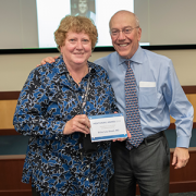
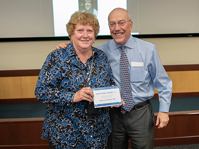
Billie Lou Short, M.D., received the Ninth Annual Mentorship Award in Clinical Science.

People joke that Billie Lou Short, M.D., chief of Children’s Division of Neonatology, invented extracorporeal membrane oxygenation, known as ECMO for short. While Dr. Short did not invent ECMO, under her leadership Children’s National was the first pediatric hospital to use it. And over decades Children’s staff have perfected its use to save the lives of tiny, vulnerable newborns by temporarily taking over for their struggling hearts and lungs. For two consecutive years, Children’s neonatal intensive care unit has been named the nation’s No. 1 for newborns by U.S. News & World Report. “Despite all of these accomplishments, Dr. Short’s best legacy is what she has done as a mentor to countless trainees, nurses and faculty she’s touched during their careers. She touches every type of clinical staff member who has come through our neonatal intensive care unit,” says An Massaro, M.D., director of residency research.

For these achievements, Dr. Short received the Ninth Annual Mentorship Award in Clinical Science.

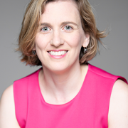
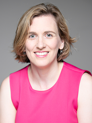
Anna Penn, M.D., Ph.D., has provided new insights into the central role that the placental hormone allopregnanolone plays in orderly fetal brain development, and her research team has created novel experimental models that mimic some of the brain injuries often seen in very preterm babies – an essential step that informs future neuroprotective strategies. Dr. Penn, a clinical neonatologist and developmental neuroscientist, “has been a primary adviser for 40 mentees throughout their careers and embodies Children’s core values of Compassion, Commitment and Connection,” says Claire-Marie Vacher, Ph.D.

For these achievements, Dr. Penn was selected to receive the Ninth Annual Mentorship Award in Basic and Translational Science.

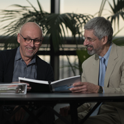
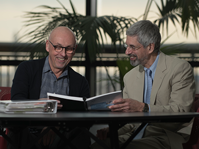
The mentorship awards for Drs. Short and Penn were among dozens of honors given in conjunction with “Frontiers in Innovation,” the Ninth Annual Research and Education Week (REW) at Children’s National. In addition to seven keynote lectures, more than 350 posters were submitted from researchers – from high-school students to full-time faculty – about basic and translational science, clinical research, community-based research, education, training and quality improvement; five poster presenters were showcased via Facebook Live events hosted by Children’s Hospital Foundation.

Two faculty members won twice: Vicki Freedenberg, Ph.D., APRN, for research about mindfulness-based stress reduction and Adeline (Wei Li) Koay, MBBS, MSc, for research related to HIV. So many women at every stage of their research careers took to the stage to accept honors that Naomi L.C. Luban, M.D., Vice Chair of Academic Affairs, quipped that “this day is power to women.”

Here are the 2019 REW award winners:

2019 Elda Y. Arce Teaching Scholars Award
Barbara Jantausch, M.D.
Lowell Frank, M.D.

Suzanne Feetham, Ph.D., FAA, Nursing Research Support Award
Vicki Freedenberg, Ph.D., APRN, for “Psychosocial and biological effects of mindfulness-based stress reduction intervention in adolescents with CHD/CIEDs: a randomized control trial”
Renee’ Roberts Turner for “Peak and nadir experiences of mid-level nurse leaders”

2019-2020 Global Health Initiative Exploration in Global Health Awards
Nathalie Quion, M.D., for “Latino youth and families need assessment,” conducted in Washington
Sonia Voleti for “Handheld ultrasound machine task shifting,” conducted in Micronesia
Tania Ahluwalia, M.D., for “Simulation curriculum for emergency medicine,” conducted in India
Yvonne Yui for “Designated resuscitation teams in NICUs,” conducted in Ghana
Xiaoyan Song, Ph.D., MBBS, MSc, “Prevention of hospital-onset infections in PICUs,” conducted in China

Ninth Annual Research and Education Week Poster Session Awards

Basic and Translational Science
Faculty:
Adeline (Wei Li) Koay, MBBS, MSc, for “Differences in the gut microbiome of HIV-infected versus HIV-exposed, uninfected infants”
Faculty: Hayk Barseghyan, Ph.D., for “Composite de novo Armenian human genome assembly and haplotyping via optical mapping and ultra-long read sequencing”
Staff: Damon K. McCullough, BS, for “Brain slicer: 3D-printed tissue processing tool for pediatric neuroscience research”
Staff: Antonio R. Porras, Ph.D., for “Integrated deep-learning method for genetic syndrome screening using facial photographs”
Post docs/fellows/residents: Lung Lau, M.D., for “A novel, sprayable and bio-absorbable sealant for wound dressings”
Post docs/fellows/residents:
Kelsey F. Sugrue, Ph.D., for “HECTD1 is required for growth of the myocardium secondary to placental insufficiency”
Graduate students:
Erin R. Bonner, BA, for “Comprehensive mutation profiling of pediatric diffuse midline gliomas using liquid biopsy”
High school/undergraduate students: Ali Sarhan for “Parental somato-gonadal mosaic genetic variants are a source of recurrent risk for de novo disorders and parental health concerns: a systematic review of the literature and meta-analysis”

Clinical Research
Faculty:
Amy Hont, M.D., for “Ex vivo expanded multi-tumor antigen specific T-cells for the treatment of solid tumors”
Faculty: Lauren McLaughlin, M.D., for “EBV/LMP-specific T-cells maintain remissions of T- and B-cell EBV lymphomas after allogeneic bone marrow transplantation”

Staff: Iman A. Abdikarim, BA, for “Timing of allergenic food introduction among African American and Caucasian children with food allergy in the FORWARD study”
Staff: Gelina M. Sani, BS, for “Quantifying hematopoietic stem cells towards in utero gene therapy for treatment of sickle cell disease in fetal cord blood”
Post docs/fellows/residents: Amy H. Jones, M.D., for “To trach or not trach: exploration of parental conflict, regret and impacts on quality of life in tracheostomy decision-making”
Graduate students: Alyssa Dewyer, BS, for “Telemedicine support of cardiac care in Northern Uganda: leveraging hand-held echocardiography and task-shifting”
Graduate students: Natalie Pudalov, BA, “Cortical thickness asymmetries in MRI-abnormal pediatric epilepsy patients: a potential metric for surgery outcome”
High school/undergraduate students:
Kia Yoshinaga for “Time to rhythm detection during pediatric cardiac arrest in a pediatric emergency department”

Community-Based Research
Faculty:
Adeline (Wei Li) Koay, MBBS, MSc, for “Recent trends in the prevention of mother-to-child transmission (PMTCT) of HIV in the Washington, D.C., metropolitan area”
Staff: Gia M. Badolato, MPH, for “STI screening in an urban ED based on chief complaint”
Post docs/fellows/residents:
Christina P. Ho, M.D., for “Pediatric urinary tract infection resistance patterns in the Washington, D.C., metropolitan area”
Graduate students:
Noushine Sadeghi, BS, “Racial/ethnic disparities in receipt of sexual health services among adolescent females”

Education, Training and Program Development
Faculty:
Cara Lichtenstein, M.D., MPH, for “Using a community bus trip to increase knowledge of health disparities”
Staff:
Iana Y. Clarence, MPH, for “TEACHing residents to address child poverty: an innovative multimodal curriculum”
Post docs/fellows/residents:
Johanna Kaufman, M.D., for “Inpatient consultation in pediatrics: a learning tool to improve communication”
High school/undergraduate students:
Brett E. Pearson for “Analysis of unanticipated problems in CNMC human subjects research studies and implications for process improvement”

Quality and Performance Improvement
Faculty:
Vicki Freedenberg, Ph.D., APRN, for “Implementing a mindfulness-based stress reduction curriculum in a congenital heart disease program”
Staff:
Caleb Griffith, MPH, for “Assessing the sustainability of point-of-care HIV screening of adolescents in pediatric emergency departments”
Post docs/fellows/residents:
Rebecca S. Zee, M.D., Ph.D., for “Implementation of the Accelerated Care of Torsion (ACT) pathway: a quality improvement initiative for testicular torsion”
Graduate students:
Alysia Wiener, BS, for “Latency period in image-guided needle bone biopsy in children: a single center experience”

View images from the REW2019 award ceremony.

Beth Tarini

Getting to know SPR’s future President, Beth Tarini, M.D., MS

Beth Tarini

Quick. Name four pillar pediatric organizations on the vanguard of advancing pediatric research.

Most researchers and clinicians can rattle off the names of the Academic Pediatric Association, the American Academy of Pediatrics and the American Pediatric Society. But that fourth one, the Society for Pediatric Research (SPR), is a little trickier. While many know SPR, a lot of research-clinicians simply do not.

Over the next few years, Beth A. Tarini, M.D., MS, will make it her personal mission to ensure that more pediatric researchers get to know SPR and are so excited about the organization that they become active members. In May 2019 Dr. Tarini becomes Vice President of the society that aims to stitch together an international network of interdisciplinary researchers to improve kids’ health. Four-year SPR leadership terms begin with Vice President before transitioning to President-Elect, President and Past-President, each for one year.

Dr. Tarini says she looks forward to working with other SPR leaders to find ways to build more productive, collaborative professional networks among faculty, especially emerging junior faculty. “Facilitating ways to network for research and professional reasons across pediatric research is vital – albeit easier said than done. I have been told I’m a connector, so I hope to leverage that skill in this new role,” says Dr. Tarini, associate director for Children’s Center for Translational Research.

“I’m delighted that Dr. Tarini was elected to this leadership position, and I am impressed by her vision of improving SPR’s outreach efforts,” says Mark Batshaw, M.D., Executive Vice President, Chief Academic Officer and Physician-in-Chief at Children’s National. “Her goal of engaging potential members in networking through a variety of ways – face-to-face as well as leveraging digital platforms like Twitter, Facebook and LinkedIn – and her focus on engaging junior faculty will help strengthen SPR membership in the near term and long term.”

Dr. Tarini adds: “Success to me would be leaving after four years with more faculty – especially junior faculty – approaching membership in SPR with the knowledge and enthusiasm that they bring to membership in other pediatric societies.”

SPR requires that its members not simply conduct research, but move the needle in their chosen discipline. In her research, Dr. Tarini has focused on ensuring that population-based newborn screening programs function efficiently and effectively with fewer hiccups at any place along the process.

Thanks to a heel stick to draw blood, an oxygen measurement, and a hearing test, U.S. babies are screened for select inherited health conditions, expediting treatment for infants and reducing the chances they’ll experience long-term health consequences.

“The complexity of this program that is able to test nearly all 4 million babies in the U.S. each year is nothing short of astounding. You have to know the child is born – anywhere in the state – and then between 24 and 48 hours of birth you have to do testing onsite, obtain a specific type of blood sample, send the blood sample to an off-site lab quickly, test the sample, find the child if the test is out of range, get the child evaluated and tested for the condition, then send them for treatment. Given the time pressures as well as the coordination of numerous people and organizations, the fact that this happens routinely is amazing. And like any complex process, there is always room for improvement,” she says.

Dr. Tarini’s research efforts have focused on those process improvements.

As just one example, the Advisory Committee on Heritable Disorders in Newborns and Children, a federal advisory committee on which she serves, was discussing how to eliminate delays in specimen processing to provide speedier results to families. One possible solution floated was to open labs all seven days, rather than just five days a week. Dr. Tarini advocated for partnering with health care engineers who could help model ways to make the specimen transport process more efficient, just like airlines and mail delivery services. A more efficient and effective solution was to match the specimen pick-up and delivery times more closely with the lab’s operational times – which maximizes lab resources and shortens wait times for parents.

Conceptual modeling comes so easily for her that she often leaps out of her seat mid-sentence, underscoring a point by jotting thoughts on a white board, doing it so often that her pens have run dry.

“It’s like a bus schedule: You want to find a bus that not only takes you to your destination but gets you there on time,” she says.

Dr. Tarini’s current observational study looks for opportunities to improve how parents in Minnesota and Iowa are given out-of-range newborn screening test results – especially false positives – and how that experience might shake their confidence in their child’s health as well as heighten their own stress level.

“After a false positive test result, are there parents who walk away from newborn screening with lingering stress about their child’s health? Can we predict who those parents might be and help them?” she asks.

Among the challenges is the newborn screening occurs so quickly after delivery that some emotionally and physically exhausted parents may not remember it was done. Then they get a call from the state with ominous results. Another challenge is standardizing communication approaches across dozens of birthing centers and hospitals.

“We know parents are concerned after receiving a false positive result, and some worry their infant remains vulnerable,” she says. “Can we change how we communicate – not just what we say, but how we say it – to alleviate those concerns?”

Dr. Kurt Newman in front of the capitol building

Kurt Newman, M.D., shares journey as a pediatric surgeon in TEDx Talk

Kurt Newman, M.D., president and chief executive officer of Children’s National, shares his poignant journey as a pediatric surgeon, offering a new perspective for approaching the most chronic and debilitating health conditions. In this independently-organized TEDx event, Dr. Newman also shares his passion for Children’s National and the need to increase pediatric innovations in medicine.

Parasite collage

Which micro-organisms lurk within urine?


schistosome blood fluke

Therapy derived from parasitic worms downregulates proinflammatory pathways

schistosome blood fluke

A therapy derived from the eggs of the parasitic Schistosoma helps to protect against one of chemotherapy’s debilitating side effects by significantly downregulating major proinflammatory pathways, reducing inflammation.

A therapy derived from the eggs of parasitic worms helps to protect against one of chemotherapy’s debilitating side effects by significantly downregulating major proinflammatory pathways and reducing inflammation, indicates the first transcriptome-wide profiling of the bladder during ifosfamide-induced hemorrhagic cystitis.

The experimental model study findings were published online Feb. 7, 2019, in Scientific Reports.

With hemorrhagic cystitis, a condition that can be triggered by anti-cancer therapies like the chemotherapy drug ifosfamide and other oxazaphosphorines, the lining of the bladder becomes inflamed and begins to bleed. Existing treatments on the market carry their own side effects, and the leading therapy does not treat established hemorrhagic cystitis.

Around the world, people can become exposed to parasitic Schistosoma eggs through contaminated freshwater. Once inside the body, the parasitic worms mate and produce eggs; these eggs are the trigger for symptoms like inflammation. To keep their human hosts alive, the parasitic worms tamp down excess inflammation by secreting a binding protein with anti-inflammatory properties.

With that biological knowledge in mind, a research team led by Michael H. Hsieh, M.D., Ph.D., tested a single dose of IPSE, an Interleukin-4 inducing, Schistosoma parasite-derived anti-inflammatory molecule and found that it reduced inflammation, bleeding and urothelial sloughing that occurs with ifosfamide-related hemorrhagic cystitis.

In this follow-up project, experimental models were treated with ifosfamide to learn more about IPSE’s protective powers.

The preclinical models were given either saline or IPSE before the ifosfamide challenge. The bladders of the experimental models treated with ifosfamide had classic symptoms, including marked swelling (edema), dysregulated contraction, bleeding and urothelial sloughing. In contrast, experimental models “pre-treated” with IPSE were shielded from urothelial sloughing and inflammation, the study team found.

Transcriptional profiling of the experimental models’ bladders found the IL-1-B TNFa-IL-6 proinflammatory cascade via NFkB and STAT3 pathways serving as the key driver of inflammation. Pretreatment with IPSE slashed the overexpression of Il-1b, Tnfa and Il6 by 50 percent. IPSE drove significant downregulation of major proinflammatory pathways, including the IL-1-B TNFa-IL-6 pathways, interferon signaling and reduced (but did not eliminate) oxidative stress.

“Taken together, we have identified signatures of acute-phase inflammation and oxidative stress in ifosfamide-injured bladder, which are reversed by pretreatment with IPSE,” says Dr. Hsieh, a urologist at Children’s National Health System and the study’s senior author. “These preliminary findings reveal several pathways that could be therapeutically targeted to prevent ifosfamide-induced hemorrhagic cystitis in humans.”

When certain chemotherapy drugs are metabolized by the body, the toxin acrolein is produced and builds up in urine. 2-mercaptoethane sulfonate Na (MESNA) binds to acrolein to prevent urotoxicity. By contrast, IPSE targets inflammation at the source, reversing inflammatory changes that damage the bladder.

“Our work demonstrates that there may be therapeutic potential for naturally occurring anti-inflammatory molecules, including pathogen-derived factors, as alternative or complementary therapies for ifosfamide-induced hemorrhagic cystitis,” Dr. Hsieh adds.

In addition to Dr. Hsieh, study co-authors include Lead Author Evaristus C. Mbanefo and Rebecca Zee, Children’s National; Loc Le, Nirad Banskota and Kenji Ishida, Biomedical Research Institute; Luke F. Pennington and Theodore S. Jardetzky, Stanford University; Justin I. Odegaard, Guardant Health; Abdulaziz Alouffi, King Abdulaziz City for Science & Technology; and Franco H. Falcone, University of Nottingham.

Financial support for the research described in this report was provided by the Margaret A. Stirewalt Endowment, the National Institute of Diabetes and Digestive and Kidney Diseases under award R01DK113504, the National Institute of Allergy and Infectious Diseases under award R56AI119168 and a Urology Care Foundation Research Scholar Award.

mitochondria

Treating nephrotic-range proteinuria with tacrolimus in MTP

mitochondria

Mitochondria are the cell’s powerplants and inside them the MTP enzymatic complex catalyzes three steps in beta-oxidation of long-chain fatty acids.

In one family, genetic lightning struck twice. Two sisters were diagnosed with mitochondrial trifunctional protein (MTP) deficiency. This is a rare condition that stops the body from converting fats to energy, which can lead to lactic acidosis, recurrent breakdown of muscle tissue and release into the bloodstream (rhabdomyolysis), enlarged heart (cardiomyopathy) and liver failure.

Mitochondria are the cell’s powerplants and inside them the MTP enzymatic complex catalyzes three steps in beta-oxidation of long-chain fatty acids. MTP deficiency is so rare that fewer than 100 cases have been reported in the literature says Hostensia Beng, M.D., who presented an MTP case study during the American Society of Nephrology’s Kidney Week.

The 7-month-old girl with known MTP deficiency arrived at Children’s National lethargic with poor appetite. Her laboratory results showed a low corrected serum calcium level, elevated CK level and protein in the urine (proteinuria) at a nephrotic range. The infant was treated for primary hypoparathyroidism and rhabdomyolysis.

Even though the rhabdomyolysis got better, the excess protein in the girl’s urine remained at worrisome levels. A renal biopsy showed minimal change disease and foot process fusion. And electron microscopy revealed shrunken, dense mitochondria in visceral epithelial cells and endothelium.

“We gave her tacrolimus, a calcineurin inhibitor that we are well familiar with because we use it after transplants to ensure patient’s bodies don’t reject the donated organ. By eight months after treatment, the girl’s urine protein-to-creatinine (uPCR) ratio was back to normal. At 35 months, that key uPCR measure rose again when tacrolimus was discontinued. When treatment began again, uPCR was restored to normal levels one month later,” Dr. Beng says.

The girl’s older sister also shares the heterozygous deletion in the HADHB gene, which provides instructions for making MTP. That missing section of the genetic how-to guide was predicted to cause truncation and loss of long-chain-3-hydroxyacl CoA dehydrogenase function leading to MTP deficiency.

The older sister was diagnosed with nephrotic syndrome and having scar tissue in the kidney’s filtering unit (focal segmental glomerulosclerosis) when she was 18 months old. By contrast, she developed renal failure and progressed to end stage renal disease at 20 months of age.

“Renal involvement has been reported in only one patient with MTP deficiency to date, the older sister of our patient,” Dr. Beng adds.

Podocytes are specialized cells in the kidneys that provide a barrier, preventing plasma proteins from leaking into the urine. Podocytes, however, need energy to function and are rich in mitochondria.

“The proteinuria in these two sisters may be related to their mitochondrial dysfunction. Calcineurin inhibitors like tacrolimus have been reported to reduce proteinuria by stabilizing the podocyte actin cytoskeleton. Tacrolimus was an effective treatment for our patient, who has maintained normal renal function, unlike her sister,” Dr. Beng says.

American Society of Nephrology’s Kidney Week presentation

  • “Treatment of nephrotic-range proteinuria with tacrolimus in mitochondrial trifunctional protein deficiency

Hostensia Beng, M.D., lead author; Asha Moudgil, M.D., medical director, transplant, and co-author; Sun-Young Ahn, M.D., MS, medical director, nephrology inpatient services, and senior author, all of Children’s National Health System.

Dr. Michael Hsieh's clay shield

Innovative urologist Michael Hsieh takes unbeaten path

Dr. Michael Hsieh's clay shield

For an elementary school art project, Michael H. Hsieh, M.D., Ph.D., was instructed to fashion a coat of arms out of clay. In addition to panels for truth, justice and Taiwan, in the shield’s M.D. panel, a snake twists around a rod, like the staff for Asclepius, a Greek god associated with healing.

Children’s urologist Michael H. Hsieh, M.D., Ph.D., knew from age 10 that he would become a doctor. Proof is at his parents’ home. For an elementary school art project, students were instructed to fashion a coat of arms out of clay. In addition to panels for truth, justice and Taiwan, in the shield’s M.D. panel, a snake twists around a rod, like the staff for Asclepius, a Greek god associated with healing.

“I liked science. When I can use it to help patients, that is very rewarding,” says Dr. Hsieh, the first doctor in his family.

These days, Dr. Hsieh’s Twitter profile serves as a digital coat of arms, describing him as “tinker, tailor,” #UTI #biologist, epithelial #immunologist, helminthologist and #urologist.

Tinker/tailor is shorthand for the mystery drama, “Tinker Tailor Solider Spy,” he explains, adding that the “tinker” part also refers “to the fact that I am always questioning things, and science is about experimentation, trying to seek answers to questions.”

While still in medical school during a rotation Dr. Hsieh saw a bladder operation on a young child and thought it was “amazing.” That experience in part inspired Dr. Hsieh to become a urologist and bladder scientist. His training in immunology and study of the bladder naturally led him to study urinary tract infections and parasitic worms that affect the urinary tract. In addition, thanks to R01 funding from the National Institutes of Health (NIH), Dr. Hsieh is co-principal investigator with Axel Krieger, University of Maryland, and Jin U. Kang, Johns Hopkins, on a project to develop imaging robots for supervised autonomous surgery on soft tissue.

The $1 million in NIH funding pushes the boundaries on amazing by using multi-spectral imaging technology and improved techniques to reduce surgical complications.

Anastomosis is a technique used by surgeons to join one thing to another, whether it’s a vascular surgeon suturing blood vessels, an orthopedic surgeon joining muscles or a urologist stitching healthy parts of the urinary tract back together. Complications can set in if their stitching is too tight, prompting scar tissue to form, or too loose, letting fluid seep out.

“The human eye can see a narrow spectrum of electromagnetic radiation. These multi-spectral imaging cameras would see across greater set of wavelengths,” he says.

The project has three aims: figuring out the best way to place sutures using multi-spectral imaging, accurately tracking soft tissue as they model suturing and comparing the handicraft of a robot against anastomosis hand-sewn by surgeons.

“I like challenges, and I like new things. I am definitely not interested in doing permutations of other people’s work,” Dr. Hsieh explains. “I would much rather go on a path that hasn’t been tread. It is more difficult in some ways, but on a day-to-day basis, I know I am making a contribution.”

In another innovative research project, Dr. Hsieh leveraged a protein secreted by a parasitic worm, Schistosoma haematobium, that suppresses inflammation in hosts as a new therapeutic approach for chemotherapy-induced hemorrhagic cystitis, a form of inflammation of the bladder.

Watching his first surgery nearly 30 years ago, he had no idea robots might one day vie to take over some part of that complicated procedure, or that parasite proteins could be harnessed as drugs. However, he has a clear idea which innovations could be on the horizon for urology in the next three decades.

“My hope is 30 years from now, we will have a solid UTI vaccine and more non-antibiotic therapies. UTIs are the second-most common bacterial infection in childhood and, in severe cases, can contribute to kidney failure,” he says.

Globally, parasitic worms pose an ongoing challenge, affecting more than 1 billion worldwide – second only to malaria. People persistently infected by schistosome worms fail to reach their growth potential, struggle academically and lack sufficient energy for exercise or work.


“There is a feeling that the infection prevalence might be decreasing globally, but not as quickly as everyone hopes. In 30 years perhaps with more mass drug administration and additional drugs – including a vaccine – we’ll have it close to eliminated globally. It would become more like polio, casting a slim shadow with small pockets of infection here or there, rather than consigning millions to perpetual poverty.”

Lactobacillis-Bacteria

Does ZIP code factor into genitourinary system health?

Lactobacillis-Bacteria

Clinicians suspect that taking probiotics, such as lactobacillus supplements, and making changes to diet may prevent urinary diseases that occur commonly among pediatric patients. A research team led by Children’s faculty is exploring whether changes in the built environment also affect the urinary microbiome.

Emerging evidence suggests that the variety and volume of bacteria that reside in the bladder – the urinary microbiome – significantly impact whether people’s genitourinary systems remain healthy or become susceptible to disease.

Already, clinicians suspect that taking probiotics and making changes to diet may prevent urinary diseases that occur commonly among pediatric patients. A research team led by Children’s faculty is exploring whether changes in the built environment also affect the urinary microbiome.

Using experimental models, they looked at how stable the urinary microbiome was over time. Then, they measured the potential effect of changing the built environment on the urinary microbiome of preclinical models.

They did this by following six C57BL/6 experimental models for five months, starting from when they were nine weeks old. They collected urine specimens when the study began and repeated sample collections each month. The multidisciplinary team isolated microbial DNA from these specimens to determine the makeup of the bacterial community present in their urinary tracts.

All of the experimental models shared a single cage, drank the same water and ate the exact same chow. At four months, however, they moved the preclinical models to a different facility within the same county. Their chow and bedding remained unchanged, but the water source changed since they received tap water at both locations.

“There were no changes in the proportion of specific bacteria in the urinary microbiomes from month zero through month five, which means the urinary microbiomes of healthy experimental models remain stable over time,” says Michael Hsieh, M.D., Ph.D., a urologist at Children’s National Health System and senior author of the work presented during the Pediatric Urology Fall Conference. “However, the convergence of the Shannon Diversity Index, the clustering seen on Principal coordinate analyses and changes in functional analyses taken as a whole suggest that an overall shift of the urinary microbiome occurred due to a change in the physical environment.”

This work suggests that where patients live could influence which bacteria grow in the urinary tracts, including during urinary tract infections.

The Societies for Pediatric Urology’s Pediatric Urology Fall Conference

  • “Effects of time and the built environment on the stability of the mouse urinary microbiome: implications for clinical utility.”

Catherine S. Forster, M.D., MS, pediatric hospitalist, Children’s National; James Cody, Ph.D., Biomedical Research Institute; Nirad Banskota, MS, Biomedical Research Institute; Crystal Stroud, MS, Children’s National; Ljubica Caldovic, Ph.D., principal investigator, Children’s National; and Michael Hsieh, M.D., Ph.D., urologist, Children’s National.

E coli bacteria

Urinary bacteria in spinal cord injury cases may tip balance toward UTIs

E coli bacteria

Patients with spinal cord injuries nearly universally have bacteria present in their urine regardless of whether they have a urinary tract infection.

The fallout from spinal cord injury doesn’t end with loss of mobility: Patients can have a range of other issues resulting from this complex problem, including loss of bladder control that can lead to urine retention. One of the most serious implications is urinary tract infections (UTIs), the most common cause of repeat hospitalization in people with spinal cord injuries, explains Hans G. Pohl, M.D., associate chief in the division of Urology at Children’s National Health System.

Diagnosing UTIs in people with spinal cord injuries is trickier than in people who are otherwise healthy, Dr. Pohl explains. Patients with spinal cord injuries nearly universally have bacteria present in their urine regardless of whether they have a UTI. It’s unclear whether these bacteria are innocent bystanders or precursors to UTIs in patients who don’t yet show symptoms. And although antibiotics can wipe out this bacterial population, these drugs can have undesirable side effects and frequent use can promote development of antibiotic-resistant bacteria.

Although clinical dogma has long promoted the idea that “healthy” urine is sterile, Dr. Pohl and colleagues have shown that a variety of bacteria live in urine, even in people without symptoms. These microorganisms, like the intestinal microbiome, live in harmony with their hosts and may even help promote health. However, it’s unclear what this urinary microbiome might look like for patients with spinal cord injury before, during and after UTIs.

To start investigating this question, Dr. Pohl and co-authors recently reported a case study they published online Sept. 21, 2018, in Spinal Cord Series and Cases. The case report about a 55-year-old man who had injured the thoracic segment of his spinal cord—about the level of the bottom of his shoulder blades—in a skiing accident when he was 19 was selected as “Editor’s Choice” for the journal’s October 2018 issue.  The patient had a neurogenic bladder, which doesn’t function normally due to impaired communication with the spinal cord. To compensate for this loss of function, this patient needed to have urine removed every four to six hours by catheterization.

Over eight months Dr. Pohl, the study’s senior author, and colleagues collected 12 urine samples from this patient:

  • One was collected at a time the patient didn’t show any symptoms of a UTI
  • Nine were collected when the patient had UTI symptoms, such as bladder spasticity
  • Two samples were collected when the patient had finished antibiotic treatment for the UTI.

The researchers split each sample in half. One part was put through a standard urinalysis and culture, much like what patients with a suspected UTI would receive at the doctor’s office. The other part was analyzed using a technique that searched for genetic material to identify bacteria that might be present and to estimate their abundance.

The researchers found a variety of different bacteria present in these urine samples. Regardless of the patient’s health status and symptoms, the majority of these bacterial species are known to be pathogenic or potentially pathogenic. By contrast, this patient’s urine microbiome appeared to largely lack bacterial species known to be either neutral or with potentially probiotic properties, such as Lactobacillus.

All of the bacteria that grew in culture also were identified by their genetic material in the samples. However, genetic sequencing also identified a possible novel uropathogenic species called Burkholderia fungorum that didn’t grow in the lab in five of the samples. This bacterium is ubiquitous in the environment and has been identified in soil- and plant-based samples. It also has been discovered in the respiratory secretions of patients with cystic fibrosis, in patients with a heart condition called infectious endocarditis, in the vaginal microbiota of patients with bacterial vaginosis, and in the gut of patients with HIV who have low T-cell counts. Dr. Pohl says it’s unclear whether this species played an infectious role in this patient’s UTI or whether it’s just part of his normal urine flora.

“Consistent with our previous work, this case report demonstrates that rather than healthy urine being sterile, there is a diverse urine bacterial ecosystem during various states of health and disease,” Dr. Pohl says. “Rather than UTIs resulting from the growth or overgrowth of a single organism, it’s more likely that a change in the healthy balance of the urine ecosystem might cause these infections.”

By monitoring the relative abundance of different bacteria types present in the urine of patients with spinal cord injury and combining this information with a patient’s symptoms, Dr. Pohl says doctors may be able to make more accurate UTI diagnoses in this unique population.

In addition to Dr. Pohl, study co-authors include Marcos Pérez-Losada, Ljubica Caldovic, Ph.D., Bruce Sprague and Michael H. Hsieh, M.D., Children’s National; Emma Nally, Suzanne L. Groah and Inger Ljungberg, MedStar National Rehabilitation Hospital; and Neel J. Chandel, Montefiore Medical Center.

AlgometRX

Breakthrough device objectively measures pain type, intensity and drug effects

AlgometRX

Clinical Research Assistant Kevin Jackson uses AlgometRx Platform Technology on Sarah Taylor’s eyes to measure her degree of pain. Children’s National is testing an experimental device that aims to measure pain according to how pupils react to certain stimuli. (AP Photo/Manuel Balce Ceneta)

Pediatric anesthesiologist Julia C. Finkel, M.D., of Children’s National Health System, gazed into the eyes of a newborn patient determined to find a better way to measure the effectiveness of pain treatment on one so tiny and unable to verbalize. Then she realized the answer was staring back at her.

Armed with the knowledge that pain and analgesic drugs produce an involuntary response from the pupil, Dr. Finkel developed AlgometRx, a first-of-its-kind handheld device that measures a patient’s pupillary response and, using proprietary algorithms, provides a diagnostic measurement of pain intensity, pain type and, after treatment is administered, monitors efficacy. Her initial goal was to improve the care of premature infants. She now has a device that can be used with children of any age and adults.

“Pain is very complex and it is currently the only vital sign that is not objectively measured,” says Dr. Finkel, who has more than 25 years of experience as a pain specialist. “The systematic problem we are facing today is that healthcare providers prescribe pain medicine based on subjective self-reporting, which can often be inaccurate, rather than based on an objective measure of pain type and intensity.” To illustrate her point, Dr. Finkel continues, “A clinician would never prescribe blood pressure medicine without first taking a patient’s blood pressure.”

The current standard of care for measuring pain is the 0-to-10 pain scale, which is based on subjective, observational and self-reporting techniques. Patients indicate their level of pain, with zero being no pain and ten being highest or most severe pain. This subjective system increases the likelihood of inaccuracy, with the problem being most acute with pediatric and non-verbal patients. Moreover, Dr. Finkel points out that subjective pain scores cannot be standardized, heightening the potential for misdiagnosis, over-treatment or under-treatment.

Dr. Finkel, who serves as director of Research and Development for Pain Medicine at the Sheikh Zayed Institute for Pediatric Surgical Innovation at Children’s National, says that a key step in addressing the opioid crisis is providing physicians with objective, real-time data on a patient’s pain level and type, to safely prescribe the right drug and dosage or an alternate treatment.,

She notes that opioids are prescribed for patients who report high pain scores and are sometimes prescribed in cases where they are not appropriate. Dr. Finkel points to the example of sciatica, a neuropathic pain sensation felt in the lower back, legs and buttocks. Sciatica pain is carried by touch fibers that do not have opioid receptors, which makes opioids an inappropriate choice for treating that type of pain.

A pain biomarker could rapidly advance both clinical practice and pain research, Dr. Finkel adds. For clinicians, the power to identify the type and magnitude of a patient’s nociception (detection of pain stimuli) would provide a much-needed scientific foundation for approaching pain treatment. Nociception could be monitored through the course of treatment so that dosing is targeted and personalized to ensure patients receive adequate pain relief while reducing side effects.

“A validated measure to show whether or not an opioid is indicated for a given patient could ease the health care system’s transition from overreliance on opioids to a more comprehensive and less harmful approach to pain management,” says Dr. Finkel.

She also notes that objective pain measurement can provide much needed help in validating complementary approaches to pain management, such as acupuncture, physical therapy, virtual reality and other non-pharmacological interventions.

Dr. Finkel’s technology, called AlgometRx, has been selected by the U.S. Food and Drug Administration (FDA) to participate in its “Innovation Challenge: Devices to Prevent and Treat Opioid Use Disorder.” She is also the recipient of Small Business Innovation Research (SBIR) grant from the National Institute on Drug Abuse.

Bladder cancer’s unique bacterial “fingerprint”

Michael H. Hsieh, M.D., Ph.D.

Michael H. Hsieh, M.D., Ph.D.

Decades ago, researchers thought that the native bacteria scattered throughout the human body—such as in the gut, the oral cavity and the skin—served little useful purpose. This microbiota, whose numbers at least match those of the cells in the body they live on and in, were considered mostly harmless hitchhikers.

More recently, research has revealed that these natural flora play key roles in maintaining and promoting health. In addition, studies have shown that understanding what a “typical” microbiome looks like and how it might change over time can provide an early warning system for some health conditions, including cancer.

Now, a small, multi-institutional study conducted in experimental models suggests that as bladder cancer progresses, it appears to be associated with a unique bacterial fingerprint within the bladder—a place thought to be bacteria-free except in the case of infection until just a few years ago. The finding opens the possibility of a new way to spot the disease earlier.

Bladder cancer is the fourth-most common malignancy among U.S. men but, despite its prevalence, mortality rates have remained stubbornly high. Patients often are diagnosed late, after bladder cancer has advanced. And, it remains difficult to discern which patients with non-invasive bladder cancer will go on to develop muscle-invasive disease.

Already, researchers know that patients with grade 4 oral squamous cell carcinoma, women with increasingly severe grades of cervical cancer and patients with cirrhosis who develop liver cancer have altered oral, vaginal and gut microbiomes, respectively.

New technological advances have led to identification of a diverse community of bacteria within the bladder, the urinary microbiome. Leveraging these tools, a research team that includes Children’s National Health System investigators studied whether an experimental model’s urinary bacterial community changed as bladder cancer progressed, evolving from a microbiome into a urinary “oncobiome.”

To test the hypothesis, the research team led by Michael H. Hsieh, M.D., Ph.D., a Children’s urologist, exposed an experimental model of bladder cancer to a bladder-specific cancer-causing agent, n-butyl-n-(4-hydroxybutyl) nitrosamine (BBN). Bladder cancers induced by BBN closely resemble human cancers in tissue structure at the microscopic level and by gene expression analyses. Ten of the preclinical models received a .05 percent concentration of BBN in their drinking water over five months and were housed together. Ten other experimental models received regular tap water and shared a separate, adjacent cage.

Researchers collected urine samples ranging from 10 to 100 microliters at the beginning of the longitudinal study, one week after it began, then once monthly. They isolated microbial DNA from the urine and quantified it to determine how much DNA was microbial. All of the bladders from experimental models exposed to BBN and two bladders from the control group were analyzed by a pathologist trained in bladder biology.

According to the study published online July 5, 2018, by the biology preprint server Biorxiv, they found a range of pathologies:

  • Five of the experimental models that received BBN did not develop cancer but had histology consistent with inflammation. Three had precancer on histology: urothelial dysplasia, hyperplasia or carcinoma in situ. Two developed cancer: invasive urothelial carcinomas, one of which had features of a squamous cell carcinoma.
  • The experimental model that developed invasive carcinoma had markedly different urinary bacteria at baseline, with Rubellimicrobium, a gram negative organism found in soil that has not been associated with disease previously, Escherichia and Kaistobacter, also found in soil, as the most prominent bacteria. By contrast, in the other experimental models the most common urinary bacteria were Escherichia, Prevotella, Veillonella, Streptococcus, Staphylococcus and Neisseria.
  • By month four, the majority of experimental models exposed to BBN had significantly higher proportion of Gardnerella and Bifidobacterium compared with their control group counterparts.

“Closely analyzing the urinary bacterial community among experimental models exposed to BBN, we saw distinct differences in microbial profiles by month four that were not present in earlier months,” Dr. Hsieh says. “While Gardnerella is associated with the development of cancer, Bifidobacterium has been shown to exert antitumor immunity, and its increasing abundance points to the need for additional research to understand its precise role in oncogenesis.”

Dr. Hsieh adds that although the study is small, its findings are of significance to children who are prone to developing urinary tract infections (UTIs), including children with spina bifida, due to the association between UTIs and bladder cancer. “This work is important because it not only suggests that the urinary microbiome could be used to diagnose bladder cancer, but that it could also perhaps predict cancer outcomes. If the urinary microbiome contributes to bladder carcinogenesis, it may be possible to favorably change the microbiome through antibiotics and/or probiotics in order to treat bladder cancer.”

In addition to Dr. Hsieh, co-authors include Catherine S. Forster, M.D., M.S., and Crystal Stroud, of Children’s National; James J. Cody, Nirad Banskota, Yi-Ju Hsieh and Olivia Lamanna, of the Biomedical Research Institute; Dannah Farah and Ljubica Caldovic, of The George Washington University; and Olfat Hammam, of Theodor Bilharz Research Institute.

Research reported in this news release was supported by the National Institutes of Health under award number R01 DK113504 and the Margaret A. Stirewalt Endowment.

Making the grade: Children’s National is nation’s Top 5 children’s hospital

Children’s National rose in rankings to become the nation’s Top 5 children’s hospital according to the 2018-19 Best Children’s Hospitals Honor Roll released June 26, 2018, by U.S. News & World Report. Additionally, for the second straight year, Children’s Neonatology division led by Billie Lou Short, M.D., ranked No. 1 among 50 neonatal intensive care units ranked across the nation.

Children’s National also ranked in the Top 10 in six additional services:

For the eighth year running, Children’s National ranked in all 10 specialty services, which underscores its unwavering commitment to excellence, continuous quality improvement and unmatched pediatric expertise throughout the organization.

“It’s a distinct honor for Children’s physicians, nurses and employees to be recognized as the nation’s Top 5 pediatric hospital. Children’s National provides the nation’s best care for kids and our dedicated physicians, neonatologists, surgeons, neuroscientists and other specialists, nurses and other clinical support teams are the reason why,” says Kurt Newman, M.D., Children’s President and CEO. “All of the Children’s staff is committed to ensuring that our kids and families enjoy the very best health outcomes today and for the rest of their lives.”

The excellence of Children’s care is made possible by our research insights and clinical innovations. In addition to being named to the U.S. News Honor Roll, a distinction awarded to just 10 children’s centers around the nation, Children’s National is a two-time Magnet® designated hospital for excellence in nursing and is a Leapfrog Group Top Hospital. Children’s ranks seventh among pediatric hospitals in funding from the National Institutes of Health, with a combined $40 million in direct and indirect funding, and transfers the latest research insights from the bench to patients’ bedsides.

“The 10 pediatric centers on this year’s Best Children’s Hospitals Honor Roll deliver exceptional care across a range of specialties and deserve to be highlighted,” says Ben Harder, chief of health analysis at U.S. News. “Day after day, these hospitals provide state-of-the-art medical expertise to children with complex conditions. Their U.S. News’ rankings reflect their commitment to providing high-quality care.”

The 12th annual rankings recognize the top 50 pediatric facilities across the U.S. in 10 pediatric specialties: cancer, cardiology and heart surgery, diabetes and endocrinology, gastroenterology and gastrointestinal surgery, neonatology, nephrology, neurology and neurosurgery, orthopedics, pulmonology and urology. Hospitals received points for being ranked in a specialty, and higher-ranking hospitals receive more points. The Best Children’s Hospitals Honor Roll recognizes the 10 hospitals that received the most points overall.

This year’s rankings will be published in the U.S. News & World Report’s “Best Hospitals 2019” guidebook, available for purchase in late September.

Schistosoma haematobium egg

For hemorrhagic cystitis, harnessing the power of a parasite

Schistosoma haematobium egg

“Urogenital Schistosoma infestation, which is caused by S. haematobium, also causes hemorrhagic cystitis, likely by triggering inflammation when the parasite’s eggs are deposited in the bladder wall or as eggs pass from the bladder into the urinary stream. S. haematobium eggs secrete proteins, including IPSE, that ensure human hosts are not so sickened that they succumb to hemorrhagic cystitis,” says Michael H. Hsieh, M.D., Ph.D.

Every year, hundreds of thousands of U.S. patients – and even more throughout the world – are prescribed cyclophosphamide or ifosfamide. These two chemotherapy drugs can be life-saving for a wide range of pediatric cancers, including leukemias and cancers of the eyes and nerves. However, these therapies come with a serious side effect: Both cause hemorrhagic cystitis in up to 40 percent of patients. This debilitating condition is characterized by severe inflammation in the bladder that can cause tremendous pain, life-threatening bleeding, and frequent and urgent urination.

Infection with a parasitic worm called Schistosoma haematobium also causes hemorrhagic cystitis, but this organism has a fail-safe: To keep its host alive, the parasite secretes a protein that suppresses inflammation and the associated pain and bleeding.

In a new study, a Children’s-led research team harnessed this protein to serve as a new therapy for chemotherapy-induced hemorrhagic cystitis.

“Urogenital Schistosoma infestation, which is caused by S. haematobium, also causes hemorrhagic cystitis, likely by triggering inflammation when the parasite’s eggs are deposited in the bladder wall or as eggs pass from the bladder into the urinary stream. S. haematobium eggs secrete proteins, including IPSE, that ensure human hosts are not so sickened that they succumb to hemorrhagic cystitis,” says Michael H. Hsieh, M.D., Ph.D., senior author of the study published April 3, 2018, by The FASEB Journal. “This work in an experimental model is the first published report of exploiting an uropathogen-derived host modulatory molecule in a clinically relevant model of bladder disease, and it points to the potential utility of this as an alternate treatment approach.”

S. mansoni IPSE binds to Immunoglobulin E (IgE), an antibody produced by the immune system that is expressed on the surface of basophils, a type of immune cell; and mast cells, another immune cell that mediates inflammation; and sequesters chemokines, signaling proteins that alert white cells to infection sites. The team produced an ortholog of the uropathogen-derived protein. A single IV dose proved superior to multiple doses of 2-Mercaptoethane sulfonate sodium (MESNA), the current standard of care, in suppressing chemotherapy-induced bladder hemorrhaging in an experimental model. It was equally potent as MESNA in dampening chemotherapy-induced pain, the research team finds.

“The current array of medicines we use to treat hemorrhagic cystitis all have shortcomings, so there is a definite need for novel therapeutic options,” says Dr. Hsieh, a Children’s National Health System urologist. “And other ongoing research projects have the potential to further expand patients’ treatment options by leveraging other urogenital parasite-derived, immune-modulating molecules to treat inflammatory bowel diseases and autoimmune disorders.”

Future research will aim to describe the precise molecular mechanisms of action, as well as to generate other orthologs that boost efficacy while reducing side effects.

In addition to Dr. Hsieh, Children’s study co-authors include Lead Author, Evaristus C. Mbanefo; Loc Le and Luke F. Pennington; Justin I. Odegaard and Theodore S. Jardetzky, Stanford University; Abdulaziz Alouffi, King Abdulaziz City for Science and Technology; and Franco H. Falcone, University of Nottingham.

Financial support for this research was provided by National Institutes of Health under award number RO1-DK113504.

Staphylococcus

How our bladder’s microbiota affect health

Staphylococcus

The presence of bacteria such as Staphylococcus in the urine is linked to the incidence and severity of urge urinary incontinence as well as treatment success.

About half of the cells in our bodies aren’t really “ours” at all. They’re the microbiota: The vast array of microorganisms that live in our gut, skin, oral cavity and other places. Decades ago, researchers thought that these organisms simply happened to colonize these areas, playing only a tangential role in health, for example, helping to break down food in the intestines or causing cavities. More recent work has revealed the incredibly complex role they play in diseases ranging from diabetes and schizophrenia.

The bladder is no exception. Just a single decade ago, the bladder was thought to be a sterile environment. But that view has shifted radically, with more sensitive cultivation methods and precise 16S rRNA gene-sequencing techniques revealing a significant bladder microbiome that could have an enormous impact on pediatric urologic diseases. These findings have opened brand new fields of research aimed at clarifying the role that the bladder’s microbiome plays in common urological diseases that affect children, according to a review article published online Feb. 22, 2018, by Current Urology Reports.

“There is a growing appreciation for the role of diverse bacteria in contributing to improved health as well as triggering disease processes or exacerbating illness,” says Michael H. Hsieh, M.D., Ph.D., director of the Clinic for Adolescent and Adult Pediatric Onset Urology (CAPITUL) at Children’s National Health System and study senior author. “Already, we know that probiotics and dietary modifications have the potential to play powerful roles in preventing urinary diseases that commonly occur among pediatric patients,” Dr. Hsieh says. This underscores the importance of conducting even more studies to improve our understanding and to identify new therapies for health conditions that resist current treatment options.”

The review conducted by Dr. Hsieh and co-authors highlights the effects of the microbiome on a number of urologic diseases that affect children, including:

  • Urinary tract infection A number of studies point to the association between decreased microbial diversity and the incidence of what is commonly called urinary tract infection (UTI) or “dysbiosis.” This relationship suggests that using probiotics to replace or supplement antibiotics could favorably alter the urinary microbiome. Future research will focus on the pathophysiological role of the microbiome to determine whether it can be manipulated to prevent or treat UTIs.
  • Urge urinary incontinence While data vary by study, the presence of bacteria in the urine, especially certain bacterial species – such as Gardnerella, Staphylococcus, Streptococcus, Actinomyces, Aerococcus, Corynebacterium and Oligella – are linked to the incidence and severity of urge urinary incontinence (UUI) as well as treatment success. Most studies find an association between greater genitourinary biodiversity and reduced incidence and lessened severity of UUI as well as improved treatment response. Future research will focus on further clarifying this relationship.
  • Urolithiasis Calcium oxalate stones, the most common type of kidney stone, have a microbiome that differs from the urinary microbiome leading researchers to question whether the stone’s own bacterial makeup could help to predict recurrence of future kidney stones. What’s more, Oxalobacter formigenes, a gram-negative bacterium, lowers oxalate levels in the blood and are associated with a 70 percent reduction in the risk of kidney stones forming. In an experimental model, fecal transplants with the full microbiome represented had a pronounced and persistent effect on oxalate production. Patients who receive some antibiotics often have reduced rates of formigenes colonization. However, the bacteria are resistant to amoxicillin, augmentin, ceftriaxone and vancomycin, which could point to preferential use of these antibiotics to stave off disease and ward off kidney stone formation.

Additional authors include Daniel Gerber, study lead author, The Georgetown University School of Medicine and Health Sciences; and Catherine Forster, M.D., study co-author, Children’s National.

Rebecca Zee

Children’s urology fellow wins best basic science award

Rebecca Zee

Rebecca Zee, a Children’s urology fellow, was awarded the best basic science prize at the Societies for Pediatric Urology annual meeting for her abstract describing a novel treatment to prevent ischemia reperfusion injury following testicular torsion.

Occurring in 1 in 4,000 males, testicular torsion occurs when the testis twists along the spermatic cord, limiting blood supply to the testicle. Despite prompt surgical intervention and restoration of blood flow, up to 40 percent of patients experience testicular atrophy due to a secondary inflammatory response, or ischemia reperfusion injury. Cytisine, a nicotine analog that the Food and Drug Administration approved for smoking cessation, recently has been found to activate a novel anti-inflammatory cascade, limiting the post-reperfusion inflammatory response.

“Administration of cytisine was recently found to limit inflammation and preserve renal function following warm renal ischemia,” Zee says. “We hypothesized that cytisine would similarly prevent ischemia reperfusion injury and limit testicular atrophy following testicular torsion.”

Using an established experimental model, Zee and colleagues induced unilateral testicular torsion by anesthetizing the adult male experimental models and rotating their right testicles by 720 degrees for two hours. In the treatment arm, the preclinical models were given cytisine as a 1.5 mg/kg injection one hour before or one hour after creating the testicular torsion. Eighteen hours after blood flow was restored to the right testis, total leukocyte infiltration and inflammatory gene expression were evaluated. Thirty days later, the researchers measured testicular weight and evaluated pro-fibrotic genes.

“We found that the administration of cytisine significantly decreases long-term testicular atrophy and fibrosis following testicular torsion,” says Daniel Casella, M.D., a urologist at Children’s National Health System and the study’s senior author. “What is particularly exciting is that we found similar long-term outcomes in the group that was given cytisine one hour after the creation of testicular torsion. This scenario is much more clinically applicable, given that we would not be able to treat patients until they present with testicular pain,” Dr. Casella adds.

Additional research is needed to determine the optimal cytisine dosing and administration regimen, however the researchers are hopeful that they can transition their findings to a pilot clinical trial in the near future.

In addition to Zee and Dr. Casella, the multi-institutional team included Children’s co-authors Nazanin Omidi, Christopher Bayne, Michael Hsieh, M.D., and Evaristus Mbanefo, in addition to Elina Mukherjee and Sunder Sims-Lucas, Ph.D., from the University of Pittsburgh.

Financial support for this work was provided by the Joseph E. Robert Jr. Center for Surgical Care.

Presidnet's Award for Innovation in Research

President’s Award highlights innovative work by early-career researchers

Presidnet's Award for Innovation in Research

As part of Research and Education Week 2018, two Presidential awardees were recognized for their research contributions, Catherine “Katie” Forster, M.D., M.S., and Nathan Anthony Smith, Ph.D.

Catherine “Katie” Forster, M.D., M.S., and Nathan Anthony Smith, Ph.D., received the President’s Award for Innovation in Research honoring their respective research efforts to explore an understudied part of the microbiome and to shed light on an underappreciated player in nerve cell communication.

Drs. Forster and Smith received their awards April 19, 2018, the penultimate day of Research and Education Week 2018, an annual celebration of the excellence in research, education, innovation and scholarship that takes place at Children’s National Health System. This year marks the fifth time the President’s Award honor has been bestowed to Children’s faculty.

Dr. Forster’s work focuses on preventing pediatric urinary tract infections (UTIs). Frequently, children diagnosed with illnesses like spina bifida have difficulty urinating on their own, and they often develop UTIs. These repeated infections are frequently treated with antibiotics which, in turn, can lead to the child developing antibiotic-resistant organisms.

“The majority of the time if you culture these children, you’ll grow something. In a healthy child, that culture would indicate a UTI,” Dr. Forster says. “Children with neurogenic bladder, however, may test positive for bacteria that simply look suspect but are not causing infection. Ultimately, we’re looking for better ways to diagnose UTI at the point of care to better personalize antibiotic treatment and limit prescriptions for children who do not truly need them.”

Powered by new sequencing techniques, a research group that includes Dr. Forster discovered that the human bladder hosts a significant microbiome, a diverse bacterial community unique to the bladder. Dr. Forster’s research will continue to characterize that microbiome to determine how that bacterial community evolves over time and whether those changes are predictable enough to intervene and prevent UTIs.

“Which genes are upregulated in Escherichia coli and the epithelium, and which genes are upregulated by both in response to each other? That can help us understand whether genes being upregulated are pathogenic,” she adds. “It’s a novel and exciting research area with significant public health implications.”

Smith’s work focuses on the role of astrocytes, specialized star-shaped glial cells, in modulating synaptic plasticity via norepinephrine. Conventional thinking describes astrocytes as support cells but, according to Smith, astrocytes are turning out to be more instrumental.

Norepinephrine, a neurotransmitter that plays an essential role in attention and focus, is released by a process known as volume transmission, which is a widespread release of a neurotransmitter at once, says Smith, a principal investigator in Children’s Center for Neuroscience Research. Astrocytes, which outnumber neurons in the brain, are strategically and anatomically located to receive this diffuse input and translate it into action to modulate neural networks.

“We hypothesize that astrocytes are integral, functional partners with norepinephrine in modulating cortical networks,” Smith adds. “Since astrocytes and norepinephrine have been implicated in many central nervous system functions, including learning and attention, it is critical to define mechanistically how astrocytes and norepinephrine work together to influence neural networks. This knowledge also will be important for the development of novel therapeutics to treat diseases such as attention deficit hyperactivity disorder and epilepsy.”

Eric Vilain

Exploring differences of sex development

Eric Vilain, M.D., Ph.D.

Eric Vilain, M.D., Ph.D., analyzes the genetic mechanisms of sex development to give families more answers that will help them make better treatment (or non treatment) decisions for a child diagnosed with DSD.

Eric Vilain, M.D., Ph.D., is well versed in the “world of uncertainty” that surrounds differences of sex development. Since joining Children’s National as the director of the Center for Genetic Medicine Research in 2017, he’s shared with our research and clinical faculty and staff his expertise about the ways that genetic analysis might help address some of the complex social, cultural and medical implications of these differences.

Over the summer, he gave a keynote address entitled “Disorders/Differences of Sex Development: A World of Uncertainty” during Children’s National’s Research and Education Week, an annual celebration of research, education, innovation and scholarship at Children’s National and around the world. In January 2018, he shared a more clinically oriented version of the talk at a special Children’s National Grand Rounds session.

The educational objective of these talks is to inform researchers and providers about the mechanisms of differences of sex development (DSD), which are defined as congenital conditions in which the development of chromosomal, gonadal or anatomical sex is atypical.

The primary goal, though, is to really shine light on the complexity of this hot topic, and share how powerful genetic tools can be used to provide vital, concrete information for care providers, patients and families to assist with difficult treatment (and non-treatment) decisions.

“A minority of DSD cases are able to receive a genetic diagnosis today,” he points out. “But geneticists know how important it is to come to a diagnosis and so we seek to increase the number of patients who receive a concrete genetic diagnosis. It impacts genetic counseling and reproductive options, and provides a better ability to predict long term outcomes.”

“These differences impact physiology and medicine. We want to better understand the biology of reproduction, with an emphasis on finding ways to preserve fertility at all costs, and how these variations may lead to additional complications, including cancer risk.”

At conception, he explains, both XX and XY embryos have bipotential gonads capable of differentiating into a testis or an ovary, though embryos are virtually indistinguishable from a gender perspective up until six weeks in utero.

Whether or not a bipotential gonad forms is largely left up to the genetic makeup of the individual. For example, a gene in the Y chromosome (SRY) triggers a cascade of genes that lead to testis development. If there is no Y chromosome, it triggers a series of pro-female genes that lead to ovarian development.

Dr. Vilain notes that a variation of enzymes or transcription factors can occur at any single step of sex development and alter all the subsequent steps. Depending on the genotype, an individual may experience normal gonadal development, but abnormal development of the genitalia, for example.

He also noted that these genes are critical to determining the differences between men and women in non-gonadal tissues, including differences in gene expression within the brain. One study in the lab of investigator Matt Bramble, Ph.D., investigates if gonadal hormones impact sex differences in the brain by modifying the genome.

This work is a prime example of research informing the care provided to patients and families. Dr. Vilain is also a member of the multidisciplinary clinical team of the PROUD Clinic at Children’s National, a program completely devoted to caring for patients with a wide array of genetic and endocrine issues, including urogenital disorders and variations of sex development.

Love is in the air and, for parasites, inside our bodies

Michael H. Hsieh

As featured in a PBS video, schistosome worms form lifelong bonds and females produce thousands of eggs daily only when they live inside human hosts, says Michael H. Hsieh, M.D., Ph.D.

“Love is in the air, the sea, the earth and all over and inside our bodies,” the PBS Valentine’s Day-themed video begins. As the public television station notes, what humans consider romance can look vastly different for creatures big and small, including serenading mice, spiders who wrap their gifts in silk and necking giraffes.

The “spooning” parasites segment of the video is where viewers see research conducted by Michael H. Hsieh, M.D., Ph.D., director of the Clinic for Adolescent and Adult PedIatric OnseT UroLogy at Children’s National Health System, and video filmed in his lab.

Schistosomiasis, a chronic infection with schistosome worms, is a distinctly one-sided love affair. As shown in Dr. Hsieh’s video clips, the male worm is shorter and fatter and equipped with a groove, a love canal where the longer, thinner female lodges, enabling the pair to mate for decades. This lifelong bond and the thousands of eggs it produces daily can only occur when the worms are inside the human host, Dr. Hsieh says.

While the video stresses Valentine’s Day romance, there are few rosy outcomes for humans who are the subject of the schistosome worms’ attention.

“Heavily and chronically infected individuals can have lots of problems,” Dr. Hsieh says. “This is a stunting and wasting health condition that prevents people from reaching their growth potential, impairs their academic performance and leaves them sapped of the energy needed to exercise or work. It truly perpetuates a cycle of poverty, particularly for affected children.”

Even the potential bright spot in this sobering story, the ability of the body’s immune system to fend off the parasitic worms, is only partly good news.

Schistosome worms have co-evolved with their human hosts, learning to take advantage of human vulnerabilities. Take the immune system. If it kicks too far into overdrive in trying to wall off the eggs from the rest of the body, it can interfere with organ function and trigger liver failure, kidney failure and early onset of bladder cancer, he says.

However, Dr. Hsieh and other schistosomiasis researchers are working on ways to positively harness the human immune response to schistosome worms, including developing diagnostics, drugs and vaccines. He says he and his colleagues would “love” to eliminate schistosomiasis as a global scourge.

Twitter Pediatric Urology Journal Club @pedurojc

Journal club, with a 140-character limit

Twitter Pediatric Urology Journal Club @pedurojc

@perforin & @chrbayne have launched a new journal club focused on pediatric urology via Twitter, a platform that democratizes and distills the academic discussion.

Journal club is a rite of passage for nearly everyone who works in an academic laboratory. What might sound like an exclusive group of readers and authors united by a secret handshake is actually a regular meeting of scientists – faculty members and young trainees alike – who gather to discuss a highlighted paper in their field of expertise.

Some of these gatherings might involve a handful of people from the same lab; others might include a larger group from the same institutional department or division. Typically, one person presents a paper, sharing all the relevant details about a study’s methodology and conclusions. Afterward, everyone has the chance to pose questions, make comments and thoroughly discuss conclusions.

“It’s an excellent academic opportunity in terms of teaching and training of early career scientists and clinicians, and it remains useful no matter what stage you are in your career,” says Michael Hsieh, M.D., Ph.D., a urologist who directs the Clinic for Adolescent and Adult PedIatric OnseT UroLogy (CAPITUL) at Children’s National Health System who has participated in a heavy share of journal club meetings over the years.

But, what if journal club didn’t have to adhere to this traditional format? What if this academic discussion could move to a venue more fitting for the 21st century, more inclusive of scientists in different geographic locations, with varying viewpoints and expertise?

That’s what Dr. Hsieh and others are trying to accomplish with a new pediatric urology-focused journal club on Twitter. When Christopher Bayne, a second-year fellow training in pediatric urology at Children’s National under Dr. Hsieh’s mentorship, approached him with the idea, Dr. Hsieh said that he jumped at the chance.

Traditional journal clubs, the two explain, can be hindered by several factors. One is a tendency toward “group think,” Dr. Hsieh says – members of the same lab, or even the same institution, tend to have the same training and practices, so they’re less likely to feel comfortable introducing new ideas about these areas into the discussion. Journal club discussions also are limited by uncertainties about what a study author might have had in mind with their methodology and conclusions. Study authors are rarely included in the discussion, Dr. Hsieh adds.

Michael Hsieh

“It’s an excellent academic opportunity in terms of teaching and training of early career scientists and clinicians, and it remains useful no matter what stage you are in your career,” says Michael Hsieh, M.D., Ph.D., a urologist who directs the Clinic for Adolescent and Adult PedIatric OnseT UroLogy (CAPITUL) at Children’s National Health System.

Twitter, Bayne says, offers an easy way around these barriers. Rather than including just members of the same lab, their Pediatric Urology Journal Club (PUJC) can accommodate any registered Twitter user in their discussions. That means that any interested person around the world – researchers, clinician-scientists, other health care providers, as well as patients and their families, for example – can participate in the monthly discussions.

Participation also isn’t dictated by geography. During recent PUJC meetings, individuals joined the thread from Brazil, Ireland and Turkey. The meetings, sponsored by the Journal of Pediatric Urology, take place in the first days to weeks after the selected paper has been available under “open access,” giving anyone a chance to read it – even if they lack a journal subscription. This format enables all participants to join threads, erasing the restrictions of geography or busy clinical and research schedules.

Thus far, the meetings have included papers on:

  • A comparison of the cost and complications of performing a surgery either robotically or through an open procedure to fix the tubes that connect the kidneys to the bladder in patients with a condition known as vesicouretal reflux, in which urine flows in the wrong direction.
  • The pros and cons of treating varicoceles, enlarged veins inside the scrotum that potentially cause fertility problems. The condition is asymptomatic in adolescents.
  • The importance of the diameter of the ureter, the part of the tube closest to the outside of the body that carries urine to be expelled, for resolving vesicouretal reflux, an abnormal flow of urine.

This new platform has attracted a core group of relatively young and young-at-heart devotees, Bayne says. He and other organizers have included study authors in every meeting thus far, often guiding older and Twitter-naive scientists through the process of creating an account.

And the typical 140-character limit Twitter imposes on comments known as tweets? “It might be counterintuitive,” Bayne says, “but I see the character limit as one of this journal club’s biggest strengths.” This cutoff encourages discussion members to distill their thoughts, often including two or three distinct points, into concise and deeply meaningful statements. “Participants have really latched on to the efficiency of this approach to learning about a topic and having a lively discussion.”

Thus far, their approach has been increasing in popularity. Their very first PUJC meeting in February 2017 attracted a modest number of just 24 active participants who sent 310 tweets, but generated nearly 136,000 impressions, or views.

The researchers plan to continue the monthly PUJC meetings through the Twitter handle @pedurojc. You can follow updates from Dr. Hsieh on his handle: @perforin and updates from Bayne’s on his: @chrbayne.

Boy and Mom with Doctor

Straightening out testicular torsion care

Boy and Mom with Doctor

A new collaborative accelerated care pathway for testicular torsion assessment and treatment may save critical time between diagnosis and intervention.

The clock starts ticking for a child with testicular torsion as soon as the pain starts. To increase the likelihood of successfully salvaging the twisted testicle and spermatic cord, surgical intervention – which involves restoring blood flow to the testis – should ideally occur within six hours from the onset of pain.

That’s six hours for a parent to identify that there is a problem, bring a child to the emergency department (ED) and go through all the steps required to get the child to the operating room. This process starts with an emergency physician, who probably doesn’t see many cases of this relatively rare condition, being able to identify the potential issue and contact the pediatric urologist on call. Next, diagnostic imaging orders need to be placed and actual imaging needs to occur for the diagnosis to be made. Finally, the patient needs to be moved to the pre-operative area, assessed by the anesthesia team and then taken to surgery.

In April 2016, the Division of Urology at Children’s National launched a new, accelerated care pathway for testicular torsion assessment and treatment that was developed collaboratively with the Emergency Department, Diagnostic Imaging and Radiology, the Department of Anesthesiology, and the peri-operative and operating room team.

“What stood out to us when we looked at the total time from identifying the problem to getting to surgery, was the length of time from when the diagnosis was made in the emergency department to the operating room,” says Tanya Davis, M.D., a pediatric urologist who led this new initiative along with Harry Rushton, Jr., M.D., chief of the Division of Urology. “It was an area where we could easily identify and streamline the process to accelerate the time for a patient to get from arrival in the ED to the surgical suite.”

Now, when a patient presents in the emergency department with the symptoms of testicular torsion, there is a straightforward path mapped out for the physician. “Who you need to talk to, how to reach them, relevant phone numbers, details on when to communicate to the attending physician, the ideal order of activities, the ability for residents to quickly transport the patient rather than waiting for hospital transport to surgery, and, most important, making it clear to everyone involved that this condition is a true emergency when every second matters,” Dr. Davis adds.

Torsion ED to OR Graph

Analysis of the streamlined care pathway, which emphasizes communication that the condition is a true emergency, has improved time from ED to OR within target ranges.

Since the initiative’s launch, 21 cases, from referrals and direct diagnosis, have come into the ED. The new protocol is working efficiently, reducing the mean time from the ED to the OR by more than an hour, now averaging below the team’s target goal of less than 2.5 hours from ED arrival to the OR.

Though salvage rates have not improved yet, the team will continue to collect data and monitor the impact of the accelerated pathway. Additionally, Dr. Davis says that a significant need remains for referring emergency and primary care physicians, as well as parents, to understand the condition and its need for urgent treatment. Children’s National urologists are developing handouts for both physicians and families to help raise awareness.

The hope is that more general knowledge of testicular torsion will allow parents, primary care doctors and emergency department staff to expedite diagnosis when a child complains of scrotal pain or has visible discoloration, further reducing the time from onset of pain to successful intervention. With such a short window of time for treatment, the accelerated care pathway is showing promising results.