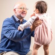
Marc Levitt, M.D., reflects on the Colorectal & Pelvic Reconstruction Program

Dr. Levitt shares insights into the program’s journey, key successes and future goals.
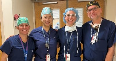
This September, the Colorectal and Pelvic Reconstruction Program at Children’s National proudly marks five years of commitment to patient-centered care for children and their families. Under the guidance of Division Chief Marc Levitt, M.D., the program has achieved remarkable advancements in clinical care, improving patient outcomes and enhancing family experiences.
Dr. Levitt shares insights into the program’s journey, key successes and future goals, highlighting the importance of collaboration among specialists and the continuous pursuit of innovative treatments.
Q: What was your vision for the program when it first launched? Has the vision changed?
A: When the Colorectal Program first launched, we aimed to establish a comprehensive program for colorectal patient care. This included integrating specialists across multiple disciplines, such as pediatric general surgery, urology, gynecology, gastroenterology, nursing, neurosurgery, orthopedics, pathology, radiology, anesthesia, psychology and pelvic floor therapy. Our efforts have exceeded expectations; the team has developed a cohesive and collaborative dynamic where each specialist contributes effectively to patient care. This integrated approach enhances outcomes, as patients and their families are at the center of our model, surrounded by a network of dedicated caregivers and coordinators, all focused on improving the patients’ quality of life.
Q: Can you highlight some key successes or achievements of the program?
A: The creation of our integrated colorectal care program at Children’s National has been a significant success, bolstered by strong institutional support. While this model requires considerable effort, it ultimately attracts patients and leads to outstanding outcomes. We’ve received patient inquiries from 48 states (AK,AL, AR, AZ, CA, CO, CT, D.C., DE, FL, GA, HI, IA, ID, IL, IN, KS, KY, LA, MA, MD, MI, MN, MO, MS, MT, NC, ND, NE, NH, NJ, NM, NV, NY, OH, OK, OR, PA, RI, SC, TN, TX, UT, VA, VT, WA, WI, WV) and 68 countries (Australia, Bahamas, Bangladesh, Bulgaria, Canada, Chile, Croatia, Cyprus, Dominican Republic, Ecuador, Egypt, England, Ethiopia, France, Germany, Greece, Guatemala, Haiti, Hong Kong, India, Ireland, Israel, Jamaica, Jordan, Kazakhstan, Kenya, Kuwait, Lithuania, Maldives, Mauritania, Mexico, Monaco, Morocco, Myanmar, The Netherlands, New Zealand, Nepal, Nigeria, Northern Ireland, Norway, Oman, Palestine, Pakistan, Paraguay, Papua New Guinea, Peru, Philippines, Portugal, Romania, Russia, Saint Lucia, Saudi Arabia, Scotland, Serbia, Singapore, Slovakia, Slovenia, Spain, Sri Lanka, South Korea, St. Kitts & Nevis, Sudan, Sweden, Ukraine, Uzbekistan, United Arab Emirates, Venezuela, Wales).
Some key successes include the development of innovative surgical procedures for conditions such as imperforate anus and anorectal malformations, advanced repair techniques for cloacal malformations, integrated care strategies for colonic dysmotility, and novel approaches for the newborn care of patients with cloacal exstrophy. All these new approaches were developed at Children’s National over the past five years.
In addition to clinical innovations, we have significantly enhanced our academic environment. Over the last five years, our team has authored more than one hundred articles and three well-regarded books, helping health care professionals around the world improve the care they provide to their patients. We have also trained surgical fellows and nurses in colorectal care and welcomed clinicians from over 30 countries for specialized training. This outreach is especially fulfilling, as it enables us to influence the care of children around the globe whom we may never meet, by sharing valuable skills and knowledge with these practitioners.
Q: Can you share examples of particularly challenging cases or high-profile cases that have influenced the program’s clinical approach?
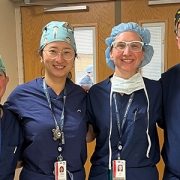
A: The way we care for a patient with a cloaca is unique – the entire team joins together in the operating room to assess the anatomy, and every member then gives their thoughts on how to approach the reconstruction. The very next day we perform that surgery, with the understanding of the anatomy in mind, and what plays out in that operating room is quite magical. This collaborative approach frequently leads to exceptional outcomes, as our diverse ideas come together to form a cohesive plan. Through this teamwork, we have developed creative ways to solve complex anatomical problems that no one individual surgeon would have thought of on their own.
Q: How have patient outcomes improved over time?
A: We have optimized the collaborative experience so that patients only need a single visit to see all the specialists they require. Our outcomes for complex colorectal surgeries have significantly improved, particularly for conditions like cloaca, Hirschsprung disease and anorectal malformations. Advances in surgical techniques and enhancements in nursing care have led to dramatically better results. We have reduced, and in some cases eliminated, complications from these surgeries, while also seeing improvements in bowel continence and kidney health. Our nursing teams — outpatient, operating room and inpatient — play a crucial role, offering unique insights into pre- and post-operative care that are vital for achieving successful outcomes.
Q: What have been the biggest challenges or barriers faced by the program and how have you addressed these challenges?
A: One of the biggest challenges we face is ensuring that patients can get to us. Many patients encounter obstacles with insurance companies that restrict travel outside their network, or they deal with the difficulties of traveling from far away for their surgery. We have worked diligently to address these issues by improving our insurance approval process and making families comfortable during their stay in Washington, D.C., including assistance with accommodations, necessary medical supplies, etc. If we can remove these barriers, we can care for more patients and make the experience easier for them and their families.
Q: How does the colorectal program collaborate with other departments or services? How has the program integrated from various specialties (e.g., urology, gynecology, GI) to enhance patient care?
A: We have specialists who are fully integrated in their roles on the colorectal and pelvic reconstruction team. General pediatric surgeons, urologists, gynecologists and gastroenterologists work both in the colorectal program and in their home program in a uniquely integrated fashion. For example, our urologists each spend half of their time in general urology and the other half in colorectal, handling the urologic aspects of colorectal patients’ care. In this way collaboration is enhanced and encouraged.
Q: What are the future goals or plans for the colorectal program?
A: Our future goals for the colorectal program are focused on expanding our impact and enhancing patient care. First and foremost, we aim to help as many patients as possible. We plan to collaborate with other colorectal centers worldwide to share knowledge and best practices. Training surgeons and nurses remains a priority, as we want to equip as many healthcare professionals as possible with the skills needed in this field. Additionally, we are committed to traveling to the developing world to provide care for children who cannot access services at Children’s National. Finally, we intend to enhance our research efforts, leveraging basic science to investigate and address conditions related to the colon, ultimately seeking to reduce or eliminate associated illnesses.
 For the last several years, the physicians, advance practice providers, and nurses within the
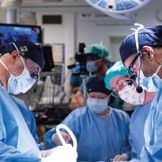

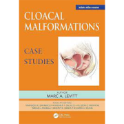
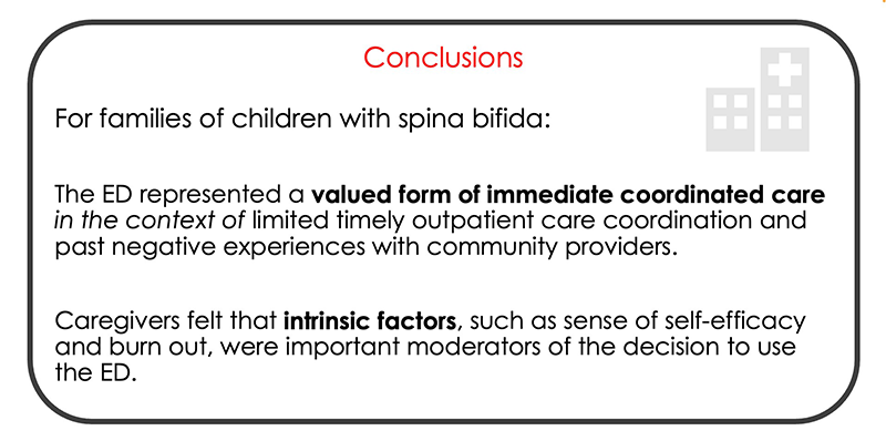
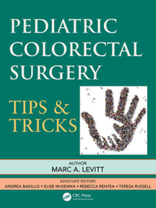
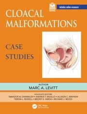 “Within the field of pediatric colorectal and pelvic reconstruction, the most complex anatomic problem a pediatric surgeon can face is that of a cloacal malformation,” writes
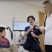
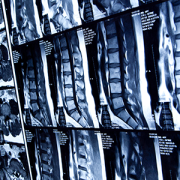
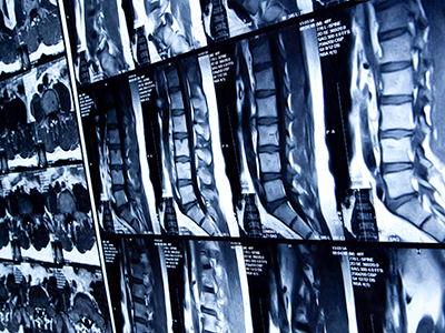

 Advanced MRI visualization techniques to follow blood flow in the hearts of cardiac patients. Gene therapy for pediatric patients with Duchenne muscular dystrophy. 3D-printed casts for treating clubfoot. These were among the most popular articles we published on Innovation District in 2023. Read on for our full list.
 Children’s National Hospital in Washington, D.C., was ranked #5 in the nation on the U.S. News & World Report 2023-24 Best Children’s Hospitals annual rankings. This marks the seventh straight year Children’s National has made the Honor Roll list. The Honor Roll is a distinction awarded to only 10 children’s hospitals nationwide.

 A clinical trial testing a new drug to increase growth in children with short stature. The first ever high-intensity focused ultrasound procedure on a pediatric patient with neurofibromatosis. A low dose gene therapy vector that restores the ability of injured muscle fibers to repair. These were among the most popular articles we published on Innovation District in 2022. Read on for our full top 10 list.