lung ct scan

With COVID-19, artificial intelligence performs well to study diseased lungs

lung ct scan

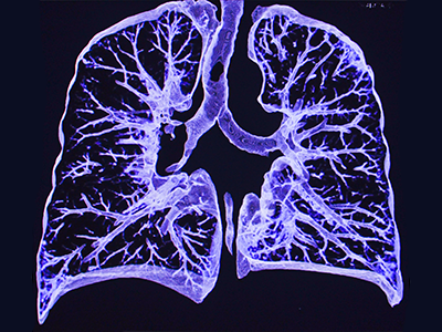
New research shows that artificial intelligence can be rapidly designed to study the lung images of COVID-19 patients.

Artificial intelligence can be rapidly designed to study the lung images of COVID-19 patients, opening the door to the development of platforms that can provide more timely and patient-specific medical interventions during outbreaks, according to research published this month in Medical Image Analysis.

The findings come as part of a global test of AI’s power, called the COVID-19 Lung CT Lesion Segmentation Challenge 2020. More than 2,000 international teams came together to train the power of machine learning and imaging on COVID-19, led by researchers at Children’s National Hospital, AI tech giant NVIDIA and the National Institutes of Health (NIH).

The bottom line

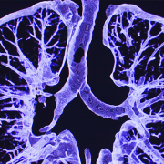
Many of the competing AI platforms were successfully trained to analyze lung lesions in COVID-19 patients and measure acute issues including lung thickening, effusions and other clinical findings. Ten leaders were named in the competition, which ran between November and December 2020. The datasets included patients with a range of ages and disease severity.

Yet work remains before AI could be implemented in a clinical setting. The AI models performed comparably to radiologists when analyzing data similar to what the algorithms had already encountered. However, the AI was less valuable when trained on fresh data from other sources during the testing phase, indicating that systems may need to study larger and more diverse data sets to meet their full potential. This is a challenge with AI that has been noted by others too.

What they’re saying

“These are the first steps in learning how we can quickly and accurately train AI for clinical use,” said Marius George Linguraru, D.Phil., M.A., M.Sc., principal investigator at the Sheikh Zayed Institute for Pediatric Surgical Innovation at Children’s National, who led the Grand Challenge Initiative. “The global interest in COVID-19 gave us a groundbreaking opportunity to address a health crisis, and multidisciplinary teams can now focus that interest and energy on developing better tools and methods.”

Holger Roth, senior applied research scientist at NVIDIA, said the challenge gave researchers around the world a shared platform for developing and evaluating AI algorithms to quickly detect and quantify COVID lesions from lung CT images. “These models help researchers visualize and measure COVID-specific lesions of infected patients and can facilitate timelier and patient-specific medical interventions to better treat COVID,” he said.

Moving the field forward

The organizers see great potential for clinical use. In areas with limited resources, AI could help triage patients, guide the use of therapeutics or provide diagnoses when expensive testing is unavailable. AI-defined standardization in clinical trials could also uniformly measure the effects of the countermeasures used against the disease.

Linguraru and his colleagues recommend more challenges, like the lung segmentation challenge, to develop AI applications in biomedical spaces that can test the functionality of these platforms and harness their potential. Open-source AI algorithms and public curated data, such as those offered through the COVID-19 Lung CT Lesion Segmentation Challenge 2020, are valuable resources for the scientific and clinical communities to work together on advancing healthcare.

“The optimal treatment of COVID-19 and other diseases hinges on the ability of clinicians to understand disease throughout populations – in both adults and children,” Linguraru said. “We are making significant progress with AI, but we must walk before we can run.”

Innovations for health equity: Food pharmacy app wins Hackathon

When families come into the endocrinology clinic, 66% of prediabetes and Type 2 diabetes patients screen positive for food insecurity. One remedy: a smartphone app envisioned by Children’s National Hospital researchers to communicate with families between visits and provide resources to help stock pantries with nutritious foods.

The Children’s National Food Pharmacy app is on its way from idea to reality, thanks to the inaugural Health Equity in Research Hackathon event at the Children’s National Research & Innovation Campus. This team-based, “Shark Tank”-like competition involved roughly 50 experts designing creative healthcare solutions that could be delivered through ubiquitous smartphones.

“It takes a village to raise a child, and we want to show that at Children’s National we are part of that community,” said food pharmacy founder and diabetes educator Alexis Richardson, M.S., R.D., L.D.N., C.D.C.E.S.

Why it matters

The rate of new-onset Type 2 diabetes increased by a staggering 182% during the first nine months of the COVID-19 pandemic. Now, the Children’s National food pharmacy provides families that screen positive for food insecurity during quarterly clinic appointments with a 50-pound bag of medically-tailored groceries.

The new app, as envisioned, would follow them home to connect them with food bank information and other nutritional resources, eliminating paper forms and other hurdles that get in the way of care.

What’s ahead

Children’s National leaders are committed to making the proposal a reality. “We are going to support today’s winner through the next steps to prepare them to enter the app development pipeline at the Sheikh Zayed Institute,” said Lisa Guay-Woodford, M.D., director for the Clinical and Translational Science Institute at Children’s National (CTSI-CN) and one of the main judges of the competition.

The app development will happen in the months ahead. Kevin Cleary, Ph.D., technical director of the Sheikh Zayed Institute of Pediatric Surgical Innovation, said the Hackathon planted the seeds. “It really depends on the drive of the individual to see the idea to fruition,” Cleary told competitors.

Other app entries were encouraged to continue their work:

  • The Surgical Checklist, led by Brian K. Reilly, M.D., co-director of the Cochlear Implant Program: this app would help patients and providers successfully navigate the often-confusing pre-operative checklist, including required physical exams, lab work, imaging and pre-procedure fasting. Reilly said the hospital handles about 15,000 cases a year, and about 10% are rescheduled, often for reasons that could be avoided with digital organization and reminders for families.
  • More than Determined, led by Pediatrician Jessica Lazerov, M.D., M.B.A.: this app aims to give time-strapped providers a platform to better understand and address social determinants of health – such as access to safe housing, education and jobs – that can promote better preventative care outcomes.

The Health Equity in Research Hackathon was created by the new Health Equity in Research Unit, a joint initiative between the CTSI-CN and the Center for Translational Research within the Children’s National Research Institute.

Dr. Lisa Guay-Woodford and the winners of the Health Equity in Research Hackathon

Dr. Lisa Guay-Woodford, director for the Clinical and Translational Science Institute, joins the winners of the inaugural Health Equity in Research Hackathon: the Children’s National Food Pharmacy. The team’s proposed app will connect families facing food insecurity with resources and guidance for nutritious eating.

echocardiogram

AI may revolutionize rheumatic heart disease early diagnosis

echocardiogram

Researchers at Children’s National Hospital have created a new artificial intelligence (AI) algorithm that promises to be as successful at detecting early signs of rheumatic heart disease (RHD) in color Doppler echocardiography clips as expert clinicians.

Researchers at Children’s National Hospital have created a new artificial intelligence (AI) algorithm that promises to be as successful at detecting early signs of rheumatic heart disease (RHD) in color Doppler echocardiography clips as expert clinicians. Even better, this novel model diagnoses this deadly heart condition from echocardiography images of varying quality — including from low-resource settings — a huge challenge that has delayed efforts to automate RHD diagnosis for children in these areas.

Why it matters

Current estimates are that 40.5 million people worldwide live with rheumatic heart disease, and that it kills 306,000 people every year. Most of those affected are children, adolescents and young adults under age 25.

Though widely eradicated in nations such as the United States, rheumatic fever remains prevalent in developing countries, including those in sub-Saharan Africa. Recent studies have shown that, if detected soon enough, a regular dose of penicillin may slow the development and damage caused by RHD. But it has to be detected.

The hold-up in the field

Diagnosing RHD requires an ultrasound image of the heart, known as an echocardiogram. However, ultrasound in general is very variable as an imaging modality. It is full of texture and noise, making it one of the most challenging to interpret visually. Specialists undergo significant training to read them correctly. However, in areas where RHD is rampant, people who can successfully read these images are few and far between. Making matters worse, the devices used in these low resource settings have their own levels of varying quality, especially when compared to what is available in a well-resourced hospital elsewhere.

The research team hypothesized that a novel, automated deep learning-based method might detect successfully diagnose RHD, which would allow for more diagnoses in areas where specialists are limited. However, to date, machine learning has struggled the same way the human eye does with noisy ultrasound images.

Children’s National leads the way

Using approaches that led to successful objective digital biometric analysis software for non-invasive screening of genetic disease, researchers at the Sheikh Zayed Institute for Pediatric Surgical Innovation, including medical imaging scientist Pooneh Roshanitabrizi, Ph.D., and Marius Linguraru, D.Phil., M.A., M.Sc., principal investigator, partnered with clinicians from Children’s National Hospital, including Craig Sable, M.D., associate chief of Cardiology and director of Echocardiography, and cardiology fellow Kelsey Brown, M.D., who are heavily involved in efforts to research, improve treatments and ultimately eliminate the deadly impacts of RHD in children. The collaborators also included cardiac surgeons from the Uganda Heart Institute and cardiologists from Cincinnati Children’s Hospital Medical Center.

Dr. Linguraru’s team of AI and imaging scientists spent hours working with cardiologists, including Dr. Sable, to truly understand how they approach and assess RHD from echocardiograms. Building the tool based on that knowledge is why this tool stands apart from other efforts to use machine-learning for this purpose. Orienting the approach to the clinical steps of diagnosis is what led to the very first deep learning algorithm that diagnoses mild RHD with similar success to the specialists themselves. After the platform was built, 2,136 echocardiograms from 591 children treated at the Uganda Heart Institute fed the learning algorithm.

What’s next

The team will continue to collect data points based on clinical imaging data to refine and validate the tool. Ultimately, researchers will look for a way that the algorithm can work directly with ultrasound/echocardiogram machines. For example, the program might be run through an app that sits on top of an ultrasound device and works on the same platform to communicate directly with it, right in the clinic. By putting the two technologies together, care providers on the ground will be able to diagnose mild cases and prescribe prophylactic treatments like penicillin in one visit.

The first outcomes from the program were showcased in a presentation by Dr. Roshanitabrizi at one of the biggest and most prestigious medical imaging and AI computing meetings — the 25th International Conference on Medical Image Computing and Computer Assisted Intervention (MICCAI).

Healthcare icons

Children’s National Innovation Day aims to advance pioneering pediatric life science projects

Healthcare icons

Children’s National Research & Innovation Campus’ Innovation Day will feature life science projects focused on improving pediatric care.

Pioneering life science projects focused on improving pediatric care will be on display at the Children’s National Research & Innovation Campus when the hospital hosts its 2022 Innovation Day on Friday, August 26. Hosted by Children’s National Innovation Ventures, the program’s goal is to showcase life sciences and healthcare projects that are mature enough to look for a co-developer, strategic partner, investor or licensing vehicle.

“For us, a successful Innovation Day means we are able to match these entrepreneurs with the strategic partner they need at this stage of their device development journey,” says Kolaleh Eskandanian, Ph.D., M.B.A., P.M.P., vice president and chief innovation officer at Children’s National Hospital and executive director of the Sheikh Zayed Institute for Pediatric Surgical Innovation.

“Data continues to show that the national Capital Region (NCR) remains one of the most robust life sciences and technology hubs in the country with no shortage of visionary leaders. There is no better place to conduct this important work as we seek more ways to advance pediatric medical technologies to improve health outcomes.”

A total of 17 projects will be showcased at the event, with each having up to 10-minute window to make their presentation. All attendees will be provided a survey, enabling valuable feedback for presenters as they look to take their next strategic step on the pathway to further development and commercialization.

In addition to presenting projects, the 2022 Innovation Day will also feature startup companies whose mission is aligned with Children’s National’s quest to bring novel pediatric medical products to patients and families. Throughout the day, attendees will have the opportunity to meet with the hospital’s researchers, academic entrepreneurs and prominent stakeholders in the life science and healthcare innovation ecosystem in the region. Interested investors and strategics can also request one-on-one meetings with startups and research teams.

Eskandanian, who also serves as the executive director of the FDA-funded National Capital Consortium for Pediatric Device Innovation (NCC-PDI), notes that accelerating this pathway to commercialization for pediatric products will bring more viable technologies to market, an important step in addressing the ongoing development disparity in innovations developed for children versus adults.

“For too long, children have been left behind in the development and commercialization of medical products and we remain committed to altering the trend,” Eskandanian says. “Children’s National’s Innovation Ventures team is working closely with the presenting innovators to provide any and all support that we can to get these products to the next stage.”

Registration for Children’s National’s 2022 Innovation Day is currently open. Those interested in attending the day-long showcase can register at https://conta.cc/3CrAfTl.

Supporting the progress of pediatric innovators is a key focus of the new Children’s National Research & Innovation Campus, a one-of-its-kind ecosystem that drives discoveries that save and improve the lives of children. On a nearly 12-acre portion of the former, historic Walter Reed Army Medical Center in Northwest Washington, D.C., Children’s National has combined its strengths with those of public and private partners, including industry, universities, federal agencies, start-up companies and academic medical centers. The campus provides a rich environment of public and private partners which, like the NCC-PDI network, helps to bolster pediatric innovation and commercialization.

NCC-PDI Finalists

Pediatric medical device competition names finalists

Five finalists have been named in the prestigious annual “Make Your Medical Device Pitch for Kids!” competition presented by the National Capital Consortium for Pediatric Device Innovation (NCC-PDI). Representing innovations in pediatric technologies that aim to address unmet medical needs for children, these five finalists now have access to a pediatric accelerator program led by MedTech Innovator and will compete for a share of $150,000 in grant funding from the U.S. Food and Drug Administration (FDA) in the final virtual pitch event in October 2022. The pediatric pitch event is part of the 10th Annual Symposium on Pediatric Device Innovation, co-located with the MedTech Conference, powered by AdvaMed.

“Addressing unmet needs across pediatric populations is critical to advancing children’s health and we are delighted to once again work with pioneering companies that seek to bridge this care gap,” says Kolaleh Eskandanian, Ph.D., M.B.A, P.M.P, vice president and chief innovation officer at Children’s National Hospital and principal investigator of NCC-PDI. “As an FDA-funded consortium, NCC-PDI serves as a critical device development resource, bringing together individuals and institutions that support viable pediatric innovations and create faster pathways to commercialization. We congratulate this year’s finalists and look forward to seeing the progress made in the coming months as they navigate the accelerator program.”

The following are the five pediatric device innovations that judges selected for the final competition:

  • CorInnova – Houston, TX – Minimally invasive biventricular non-blood contacting cardiac assist device to treat heart failure.
  • Innovation Lab – La Palma, CA – Mechanical elbow brace stabilizes tremors for pediatric ataxic cerebral palsy to improve the performance of Activities of Daily Living (ADLs).
  • Prapela – Biddeford, ME – Prapela’s incubator pad is the first innovation to improve the treatment of apnea of prematurity in over twenty years.
  • Tympanogen – Richmond, VA – Perf-Fix replaces surgical eardrum repair with a nonsurgical clinic procedure
  • Xpan – Concord, Ont. – Xpan’s universal trocar enables safest and most dynamic access and effortless upsizing in conventional/mini/robotic procedures.

Beginning in June 2022, the five finalists will participate in a pediatric-focused track of the MedTech Innovator accelerator, the world’s largest accelerator of medical devices.

NCC-PDI is one of five consortia in the FDA’s Pediatric Device Consortia Grant Program created to support the development and commercialization of medical devices for children, which lags significantly behind the progress of adult medical devices. NCC-PDI is led by the Sheikh Zayed Institute for Pediatric Surgical Innovation at Children’s National and the A. James Clark School of Engineering at the University of Maryland, with support from partners MedTech Innovator, BioHealth Innovation and design firm Archimedic.

To date, NCC-PDI has mentored nearly 200 medical device sponsors to help advance their pediatric innovations, with 16 devices having received either their FDA market clearance or CE marking.

The accelerator program is the consortium’s latest addition to a network of resources and experts that it provides in support of pediatric innovators.

Eskandanian adds that supporting the progress of pediatric innovators is a key focus of the new Children’s National Research & Innovation Campus, a one-of-its-kind ecosystem that drives discoveries that save and improve the lives of children. On a nearly 12-acre portion of the former, historic Walter Reed Army Medical Center in Northwest Washington, D.C., Children’s National has combined its strengths with those of public and private partners, including industry, universities, federal agencies, start-up companies and academic medical centers. The campus provides a rich environment of public and private partners which, like the NCC-PDI network, will help bolster pediatric innovation and commercialization.

NCC-PDI Finalists social card

NCC-PDI announcement

Medical device pitch competition returns with $150K in FDA awards

Kolaleh-Eskandanian

“This pitch competition helps to recognize and support the advancement of innovations that can specifically address the needs of pediatric patients,” says Kolaleh Eskandanian, Ph.D., M.B.A., P.M.P., vice president and chief innovation officer at Children’s National Hospital and principal investigator of NCC-PDI.

Recognizing the continued gap in the development and commercialization of medical devices for children versus adults, the National Capital Consortium for Pediatric Device Innovation (NCC-PDI), in collaboration with MedTech Innovator, is accepting applications through April 22, 2022, for its annual “Make Your Medical Device Pitch for Kids!” competition. Recognizing the wide range of unmet needs for diagnostic and therapeutic devices designed especially for children, this year’s competition is open to any innovation in medical technology that addresses a significant unmet need in pediatric medical care.

“As one of the five FDA Pediatric Device Consortia, NCC-PDI is focused on seeking out and addressing significant unmet needs in pediatric medical technology,” says Kolaleh Eskandanian, Ph.D., M.B.A., P.M.P., vice president and chief innovation officer at Children’s National Hospital and principal investigator of NCC-PDI. “While great advances are made in adult medical devices, children are often left behind because the pediatric market is small and there are not incentives to develop for pediatrics. This pitch competition helps to recognize and support the advancement of innovations that can specifically address the needs of pediatric patients.”

Using a virtual format, semi-finalists chosen from all submissions will make their first pitch on May 20, 2022. Up to six finalists selected from this first round will earn participation in a special pediatric-focused track of the MedTech Innovator accelerator program, the largest medical device accelerator in the world, beginning in June 2022. These innovators will then participate in the competition finals in the fall 2022 where judges will award up to $150,000 in FDA-sponsored grants to the devices selected as most impactful and commercially viable.

Unlike devices for adults, the development and commercialization of pediatric medical devices lags behind by approximately five to 10 years. Programs like the NCC-PDI pitch competition and MedTech Innovator accelerator program offer innovators access to expert insight and consultation to help overcome regulatory hurdles and advance the product’s development path.

NCC-PDI is one of five members in the FDA’s Pediatric Device Consortia Grant Program created to support the development and commercialization of medical devices for children. NCC-PDI is led by the Sheikh Zayed Institute for Pediatric Surgical Innovation at Children’s National and the A. James Clark School of Engineering at the University of Maryland with support from partners MedTech Innovator, BioHealth Innovation and design firm Archimedic.

Eskandanian adds that supporting the progress of pediatric innovators is a key focus of the new Children’s National Research & Innovation Campus, a one-of-its-kind ecosystem that drives discoveries that save and improve the lives of children. On a nearly 12-acre portion of the former, historic Walter Reed Army Medical Center in Northwest Washington, D.C., Children’s National has combined its strengths with those of public and private partners, including industry, universities, federal agencies, start-up companies and academic medical centers. The campus provides a rich environment of public and private partners which, like the NCC-PDI network, will help bolster pediatric innovation and commercialization.

NCC-PDI announcement

Applications for the “Make Your Medical Device Pitch for Kids!” competition are open now through April 22 for innovations that address unmet pediatric needs.


parathyroid close-up

A new imaging device with AI may reduce complications during thyroid surgery

parathyroid close-up
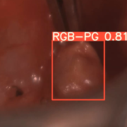
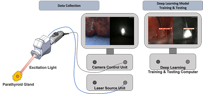
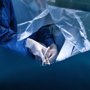
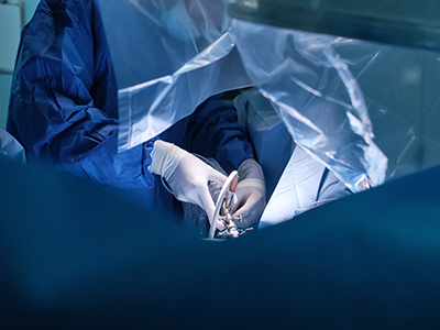
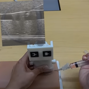
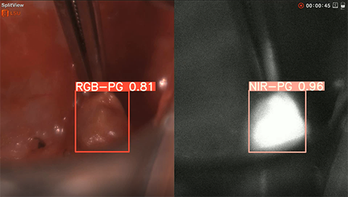
Surgeons perform approximately 150,000 thyroidectomies in the United States. Post-surgical complications from this procedure frequently occur due to the misidentification or accidental removal of healthy parathyroid glands. On average, 27% of these patients suffer from transient or permanent hypocalcemia, a condition in which the blood has too little calcium, leading to lifelong complications and socioeconomic burden.

To improve parathyroid detection during surgery, Children’s National Hospital experts developed a prototype equipped with a dual-sensor imaging device and a deep learning algorithm that accurately detects parathyroids, according to a new study published in the Journal of Biophotonics.

“What excited us in this study was that even deep-seated tissues were able to be imaged without light loss, and high resolution imaging was possible due to the unique optical design,” said Richard Jaepyeong Cha, Ph.D., council member of the International Society of Innovative Technologies for Endocrine Surgery and principal investigator for the Sheikh Zayed Institute for Pediatric Surgical Innovation at Children’s National Hospital. “Moreover, in several cases, parathyroid autofluorescence was detected even before the surgeon dissected the parathyroid gland, and while it was covered by fat and/or fascia.”

What’s unique

This is the first study that uses color RGB/NIR paired imaging-based parathyroid detection by incorporating multi-modal (both RGB light and near-infrared autofluorescence, or NIRAF, ground truth imaging) data into parathyroid identification using a deep learning algorithm.

The patient benefit

“We envision that our technology will open a new door for the digital imaging paradigm of dye-free, temporally unlimited, and precise parathyroid detection and preservation,” said Richard. “Successful translation of this technology will potentially reduce the risk of hypoparathyroidism after common thyroid surgery and improve the clinical outcomes.”

The results support the effectiveness of their novel approach despite the small sample size, which can potentially improve specificity in the identification of parathyroid glands during parathyroid and thyroid surgeries.

The hold-up in the field

It is often difficult for surgeons with naked eyes to identify parathyroid glands from thyroid tissue because of the small size, the variable position, and similar appearance to the surrounding tissues.

Since 2011, surgeons have benefited from using NIRAF, a non-invasive optical method for intraoperative real-time localization of parathyroids.

While the NIRAF technology has gained traction among endocrine surgery community, false negatives can occur with current devices that use the NIRAF technology in secondary hyperparathyroidism cases. According to Kim et al., the technology is still suboptimal, and a significant percentage of parathyroid is being missed.

Children’s National Hospital leads the way

Engineers in Children’s National are leading this field through several innovations:

  • Non-dye injected, label-free use in real-time in comparison to temporally limited ICG angiography. This technology was featured as the cover article in the journal Lasers in Surgery and Medicine 54(3), 2022.).
  • Simultaneous perfusion assessment from four glands at any time during operation.
  • Arterial flow detection from pulsatile information in well-perfused PG vasculature.
  • Quantified parathyroid detection and classification with prediction values using deep learning technique.

You can read the full study “A co-axial excitation, dual-RGB/NIR paired imaging system toward computer-aided detection (CAD) of parathyroid glands in situ and ex vivo” in the Journal of Biophotonics.

overview of parathyroid surgery procedure

doctor listening to patients heart

Children’s National leads patient-centered study of adult congenital heart disease

doctor listening to patients heart

The team will work with the 14 research institutions across the United States to collect data and then examine rates of complications or associated illnesses, as well as how adult patients have accessed health care throughout their lives.

Investigators at Children’s National Hospital and the Louisiana Public Health Institute are leading a $4.9 million research effort to study how gaps in health care affect the health and well-being of adults with congenital heart disease (CHD), supported by the Patient-Centered Outcomes Research Institute (PCORI).

The research is led by Anitha John, M.D., Ph.D., medical director of the Washington Adult Congenital Heart Program at Children’s National Hospital and Thomas Carton, Ph.D., chief data officer at the Louisiana Public Health Institute, as well as two patient co-investigators, Ruth Phillippi and Scott Leezer. The study leads also include representatives from the Adult Congenital Heart Association and Anu Agarwal, M.D., who represents the University of California – San Francisco (UCSF).

“With the increasing number of adult patients with CHD, it is important for us to understand how current recommended practices influence patient outcomes,” says Dr. John. “This project will guide us on how to best care for our patients, not just through childhood, but across their entire lifespans. Most importantly, this project will involve direct outreach to patients, incorporating patient reported outcomes as a measure of long-term outcomes.”

Together, the team will work with the 14 research institutions across the United States to collect data and then examine rates of complications or associated illnesses, as well as how adult patients have accessed health care throughout their lives. The findings may help predict which patients are at greater risk of falling out of routine health care, and when these gaps in care are likely to occur across a patient’s lifespan. The study will also correlate findings with how patients are actually feeling in their everyday lives.

The 14 participating institutions are:

  • Ochsner Health
  • Children’s Hospital of Philadelphia
  • Icahn School of Medicine at Mount Sinai
  • University of California – San Francisco
  • Nationwide Children’s Hospital
  • Duke University Health System
  • NYU Grossman School of Medicine
  • Nicklaus Children’s Hospital
  • Children’s Hospital Colorado
  • University of Miami
  • Columbia University Irving Medical Center
  • Cincinnati Children’s Hospital Medical Center
  • Weill Cornell Medical College
  • University of Florida

“This unprecedented look at the health of adults living with congenital heart disease allows us to get a full spectrum view by combining clinical data with patient-reported health data,” says Dr. Carton.

The first patient-powered registry for adults with CHD — the Congenital Heart Initiative (CHI) is a key component of this research. Launched with seed funding from Children’s National and the Heart Research Alliance at UCSF, the CHI is led by Dr. John and her team at Children’s National in addition to a broad multi-stakeholder advisory board, including patients. The CHI was co-developed with input from patients, clinicians and researchers and continues to involve these voices in the advancement of the registry. Patients who are recruited for this research will participate via enrollment in the registry, which will allow researchers to ask patients directly about health, wellness and any specific barriers to care. Learn more about CHI’s progress in their first annual report.

“Patients, like myself, are charting a new course and we desire answers to significant questions, as do our providers, about impact of lifelong specialized care, along with improved understanding of the quality-of-life patients experience,” says Leezer. “This project represents a huge step forward towards obtaining answers for the adult CHD community.”

The study also draws on the vast health data resources of PCORnet®, the National Patient-Centered Clinical Research Institute, to conduct this study more efficiently. With health records for 66 million patients available for observational studies, the PCORI-funded PCORnet provides vast scale to power research on conditions affecting even small numbers of people.

“We are confident that this research will yield critical learnings that will empower the community, align resources and spur future innovation to better meet the specialized care needs of this emerging population,” says Mark Roeder, president and chief executive officer for the Adult Congenital Heart Association.

PCORI is an independent, nonprofit organization authorized by Congress in 2010 to fund research that will provide patients, their caregivers and clinicians and other health care decision makers with the evidence-based information needed to make better-informed health care choices.

Jacob Smith

Q&A with Pediatric Surgical Innovation Fellow Jacob Smith, M.D.

Jacob Smith

Jacob Smith, M.D.

Jacob Smith, M.D. is currently a fellow with The Joseph E. Robert, Jr. Fellowship in Pediatric Surgical Innovation at Children’s National Hospital. The fellowship provides is an exciting and dynamic post-graduate research experience focused on biomedical innovation. Participants can focus their work on specific areas of interest. Here, Dr. Smith shares some information on his work with the Urology Department.

Q: How has the Joseph E. Robert, Jr., Fellows in Pediatric Surgical Innovation program allowed you to expand your experience in pediatric urology?

A: The Roberts Fellow program has provided me the ability to work with Michael Hsieh, M.D., who has years of experience in UTI and schistosomiasis research. The UTI research has been a focus of mine and we are working on ways to diagnosis and treat UTI faster in patients. One population that this can benefit are those that deal with recurrent UTIs, such as patients in our spina bifida population.

Q: Talk a little about your work on spina bifida and areas of opportunity for research.

A: Briony Varda, M.D. is heavily involved in our spina bifida program here at Children’s National. I am working with Dr. Varda to develop a database that looks at the use of the emergency department (ED) by children with spina bifida in the Washington, D.C. area. We are also conducting interviews with families and other stakeholders in this population to better understand the factors that drive this population to utilize the ED. We hope that this research can give us a better understanding of the spina bifida care in our area and inform us to provide improvement in care.

Q: How will your work to develop methods to diagnose urinary tract infections faster and how will this benefit our patient population?

A: As mentioned before, a common patient population that deals with recurrent UTIs is the spina bifida population. The protocol that we are attempting to develop would provide us quicker results than the standard urine culture and sensitivity testing that can take anywhere between 48-72 hours to result on average. We hope by providing a quicker result, we can better identify the necessary treatment these patients need to treat their infections. Another scenario that we hope this can be utilized in is for those patients that have recurrent UTI symptoms but negative urine cultures. We hope we can determine if there are viable cells that may be too few to culture but still could cause these symptoms.

Q: What are you currently working on that you are most excited about?

A: As you can tell, a lot of my research is conducted around the spina bifida population. I am excited to dive into the data we have collected on this population and determine if there are ways we could improve the care of these patients.

Q: What made you interested in specializing in pediatric urology?

A: Pediatric urology is a great field. It has a very diverse patient population. There are a multitude of problems that could arise from multiple congenital malformations to stones and voiding dysfunctions. There is also a range of cases from minor and major reconstruction, robotics, endourology and laparoscopy. One thing that does differentiate pediatric urology is that many of the problems that we repair are congenital and it is a privilege to be able to help these children and families. In fact, I was a patient myself as a child with a ureteral malformation which required surgery. The concept of helping other children with urology concerns brings an obvious “pay-it-forward” mentality that also attracted me to the field. I am excited to obtain the knowledge and skills necessary to treat my future patients.

MRI Room

Children’s National uses HIFU to perform first ever non-invasive brain tumor procedure

MRI Room

Children’s National Hospital successfully performed the first-ever high-intensity focused ultrasound (HIFU) procedure on a pediatric patient with neurofibromatosis (NF). This is the youngest patient to undergo HIFU treatment in the world. Image provided by Insightec.

Children’s National Hospital successfully performed the first-ever high-intensity focused ultrasound (HIFU) procedure on a pediatric patient with neurofibromatosis (NF). This is the youngest patient to undergo HIFU treatment in the world. The advancement of children’s medical devices in the U.S. continues to significantly lag behind adult devices. This is why this milestone marks a significant advance in making pediatric surgery more precise and less invasive.

The hospital is offering this treatment to patients under an ongoing research clinical trial. Children’s National is one of the first pediatric hospitals in the nation to use HIFU for neuro-oncology patients. It’s also the first hospital in the world to use it to treat a pediatric patient with NF. NF is a condition that occurs in approximately 1 in 3,500 births and causes tumors to form in the brain, spinal cord and nerves.

“Using HIFU to treat our pediatric patients is a quantum leap towards non-invasive surgery for kids,” said Robert Keating, M.D., division chief of Neurosurgery and co-director of the HIFU program at Children’s National. “It’s exciting because the future is now here and it’s significantly better for our kids, in terms of non-invasive surgery with lower risk of complications and no exposure to radiation.”

Focused ultrasound (FUS) is a non-invasive therapeutic technology with the potential to transform the treatment of many medical disorders by using ultrasonic thermal energy to specifically target tissue deep in the body. The technology can treat without incisions or the need of radiation.

FUS, which has been used for adult clinical trials for many decades, can be delivered through high- or low-intensity focused ultrasound (LIFU). HIFU uses non-invasive therapy that uses focused ultrasound waves to thermally ablate a focal area of tissue. Children’s National will now use HIFU to treat low-grade type tumors located in difficult locations of the brain, such as hypothalamic hamartomas and pilocytic astrocytoma, as well as for movement disorders and epilepsy.

An alternative approach, LIFU uses lower levels of energy to disrupt the blood-brain barrier. Unlike medications, which often have difficulty crossing the blood-brain barrier, LIFU can transiently open the blood-brain barrier to chemotherapy. This may allow more effective treatment of tumors and offer opportunities to treat, for the first time, the entire extent of a malignant brain tumor.

“Having focused ultrasound technology as a tool and conducting clinical trials will allow our neurologists and oncologists to offer a non-invasive treatment option to many patients who suffer from neurological conditions,” said Hasan Syed, M.D., co-director of the HIFU program at Children’s National. “The milestone of performing this first HIFU procedure will lead the way to better understanding of the effect of this technology and provide patients with more options.”

At Children’s National, the HIFU program is being led by Dr. Keating and a multidisciplinary team, including clinicians and investigators from the Sheik Zayed Institute for Pediatric Innovationradiologyoncologysurgery and orthopedics. In an effort to collaborate with the region’s adult hospitals, Children’s National will also treat adult patients on a selective basis who have movement disorders such as essential tremor and Parkinson’s. There is a scarcity of similar resources in the metro region. Many adult patients face one-year wait periods for treatment of their movement disorders, requiring many to travel out of state for treatment.

The LIFU program is scheduled to be operational in 2022. It will likely be the first in the U.S. to treat high-grade pediatric brain tumors with disruption of the blood-brain barrier and provide more effective routes for chemotherapy as well as potential immunotherapy and molecular approaches.

“The use of LIFU with microbubbles to open up the blood-brain barrier is an exciting, potentially game-changing approach for children with these tumors,” said Roger Packer, M.D., senior vice president of the Center for Neurosciences and Behavioral Medicine at Children’s National. “It should safely allow the blood-brain barrier to open and allow delivery of potentially life-saving personalized therapy to the tumor and spare the rest of the brain. It is the most exciting, new development in brain tumor therapy for these malignant midline tumors in the past 50 years.”

Children’s National continues to be a leader in pediatric HIFU use. In 2015, Children’s National doctors became the first in the U.S. to use MR-HIFU to treat pediatric osteoid osteoma – a benign, but painful bone tumor. Successful clinical trial results led to FDA approval in early 2021 for the use of the technology for this treatment. In 2020, the Focused Ultrasound Foundation also designated Children’s National as the first global pediatric Center of Excellence for using this technology to help patients with specific types of childhood tumors.

kidneys with science images

PMA-based PCR amplifies DNA from only live bacteria in urine

kidneys with science images

The question of why urinary tract infection (UTI) symptoms can persist in some patients who have been seemingly appropriately treated with antibiotics and have negative post-antibiotic urine cultures is one that urologists have long sought to answer.

Experts at Children’s National Hospital have successfully developed propidium monoazide (PMA)-based polymerase chain reaction (PCR) methods that amplify DNA from only live bacteria in urine. The study’s results, published in Frontiers, suggest that non-PMA bound DNA from live bacteria can be present in urine, even after antibiotic treatment.

PMA has been shown to differentiate between non-viable and viable bacteria in various settings. However, its effectiveness in urine has not been previously studied.

The question of why urinary tract infection (UTI) symptoms can persist in some patients who have been seemingly appropriately treated with antibiotics and have negative post-antibiotic urine cultures is one that urologists have long sought to answer.

“One theory is that very low levels of bacteria that don’t show up on cultures may be the cause,” says Michael Hsieh, M.D., director of Transitional Urology at Children’s National and senior author of the study.

Although PCR has previously been used to try and confirm this theory, the use of this method has been criticized because PCR can amplify DNA from dead bacteria (which obviously don’t cause UTI).

The authors developed a PCR test that selectively detects DNA from live bacteria. In a preclinical setting, results show that with the use of antibiotics, cultures collected can be negative but urine can contain DNA from live bacteria, as detected using the PCR test.

“We think something similar can occur in patients and we show some data in the paper confirming the PCR test can work with patient samples,” Dr. Hsieh adds. “I’m excited that we might finally have an explanation as to why some patients have persistent UTI symptoms after antibiotic treatment.”

Namely, he adds, that these patients still have a UTI. This may lead to better follow-up management of patients with UTI. The next step is to confirm the theory in patients.

surgeon doing laparoscopic surgery

Autonomous robotic laparoscopic surgery for intestinal anastomosis

surgeon doing laparoscopic surgery

Children’s National Hospital in collaboration with the University of North Carolina Wilmington and Johns Hopkins University developed an enhanced autonomous strategy for laparoscopic soft tissue surgery.

A new approach to soft tissue surgery could simplify autonomous surgical planning and enable collaborative surgery between an autonomous robot and human, a new study published in Science Robotics finds. This is the first time a robot can complete an autonomous soft tissue surgical task under laparoscopic conditions, forming the foundation for future soft tissue surgeries.

Children’s National Hospital in collaboration with the University of North Carolina Wilmington and Johns Hopkins University developed an enhanced autonomous strategy for laparoscopic soft tissue surgery. The multi-institutional effort made it possible to perform a robotic laparoscopic small bowel anastomosis in phantom and in vivo intestinal tissues. The findings further suggest that autonomous robot-assisted surgery has the potential to provide more efficacy, safety and consistency independent of an individual surgeon’s skill and experience.

The hold-up in the field

Autonomous anastomosis is known to be a challenging soft tissue surgery task. And in the laparoscopic setting, surgeries like these prove to be more challenging because of the need for high maneuverability and repeatability under motion and vision constraints – especially in pediatric patients.

“This work represents the first time autonomous soft tissue surgery has been performed using a laparoscopic technique and is the first step in bridging the gap between human and machine towards completing autonomous surgical tasks in soft tissue surgeries,” says Hamed Saeidi, Ph.D., assistant professor at University of North Carolina Wilmington and lead author of the study.

To overcome the unpredictable motions of the tissue, the experts used machine learning based techniques to track the dynamic motions of the soft tissue during the surgery. These methods also pave the way for markerless methods for tracking the tissue motion in future surgeries.

“Until now, laparoscopic autonomous surgeries were not possible in soft tissue due to the unpredictable motions of the tissue and limitations on the size and capabilities of surgical tools,” says Justin Opfermann, M.S., Ph.D., student and Johns Hopkins University and co-author.

What’s unique

Performing autonomous surgery would require the development of novel suturing tools, imaging systems and robotic controls to visualize a surgical scene, generate an optimized surgical plan and then execute that surgical plan with the highest precision.

The autonomous robot takes its skill one step further when performing surgical tasks on soft tissues by enabling a robot-human collaboration to complete more complicated surgical tasks where preoperative planning is not possible.

Additionally, the robot used in this work uses a novel shared control scheme called “conditional autonomy,” whereby the robot performs the majority of the surgical task, which the surgeon oversees.

Bottom line

“Combining all of these features into a single system is non-trivial,” Opfermann adds. “In 2016, we were the first group to demonstrate feasibility of semi-autonomous small bowel anastomosis with a robot in soft tissue, and now we can perform autonomous laparoscopic anastomosis.”

The resulting anastomosis had more consistency and achieved higher burst strength than surgeons suturing with manual technique, resulting in less anastomotic leak.

In laparoscopic surgeries – and pediatric patients especially – these challenges are even more difficult due to the small size of the patient. Robotic anastomosis is one way to ensure that surgical tasks that require high precision and repeatability can be performed with more accuracy and precision in every patient independent of surgeon skill.

“As a surgeon, I can attest to the potential benefits of improving how we perform surgery on our patients,” says Michael Hsieh, M.D., Ph.D., director of Transitional Urology at Children’s National Hospital. “Working with my engineering colleagues at Johns Hopkins, we’ve been able to develop prototypes of supervised, autonomous suturing robots that may be a step towards such improvements.”

Diego Preciado

Diego Preciado, M.D., Ph.D., named division chief of Otolaryngology

Diego Preciado

“It is the privilege of a lifetime for me to be able to continue my service to Children’s National in this leadership role,” said Dr. Preciado.

Children’s National Hospital has named Diego Preciado, M.D., Ph.D., as its new chief of the Division of Pediatric Otolaryngology. Dr. Preciado joined Children’s National in 2006 where he has practiced for the past 16 years.

“It is the privilege of a lifetime for me to be able to continue my service to Children’s National in this leadership role,” said Dr. Preciado. “Every otolaryngologist in the world knows of the extraordinary work and tradition of excellence established by my predecessor Dr. George Zalzal. I will work with the surgeon-in-chief to accelerate our mission by engaging our diverse and loyal faculty to become the global leader for complex pediatric otolaryngology care, research, education and advocacy.”

Dr. Preciado is a professor with tenure of Surgery and Pediatrics, Genomics and Precision Medicine at Children’s National and George Washington University. He holds the Joseph E. Robert Endowed Chair in Pediatric Otolaryngology, has served as the vice chief of the Division of Pediatric Otolaryngology for the past seven years, and the program director of the ACGME accredited Pediatric Otolaryngology fellowship since 2009. His clinical practice interests are primarily focused on pediatric airway reconstruction, childhood hearing loss/cochlear implants and velopharyngeal insufficiency.

Dr. Preciado’s translational research lab has been funded over the years through various awards including R01, U01 and R21 National Institutes of Health grants. He has authored over 130 peer-reviewed manuscripts, 20 book chapters, and has been an invited speaker regionally, nationally and internationally. In 2020, he received the Robert Ruben Scientific Achievement Award from the Society for Ear Nose and Throat Advances in Children.

He served as past president of the American Society of Pediatric Otolaryngology (2020-2021) and currently serves as president of the International Society for Otitis Media (2019-2023). He is a senior examiner for the American Board of Otolaryngology – Head and Neck Surgery where he also serves on the Complex Pediatric Otolaryngology Steering Committee.

“Given Dr. Preciado’s superb clinical, research and leadership background, we know the division is well-positioned for continued growth under his guidance,” said Dr. Anthony Sandler, M.D., senior vice president and surgeon-in-chief of the Joseph E. Robert Jr., Center for Surgical Care at Children’s National.

computer circuit board

Integrating clinical parameters with lung imaging to predict respiratory outcomes in premature babies

computer circuit board

The team will develop an objective framework to predict the risk and assess the severity of respiratory disease in premature babies using non-invasive low-radiation X-ray imaging biomarkers and clinical parameters from the patient bedside.

Children’s National Hospital received a $1.7M award from the National Institutes of Health (NIH) National Heart, Lung, and Blood Institute (NHLBI) to develop computational tools that integrate continuous clinical parameters with lung imaging to predict respiratory outcomes for babies born severely premature in newborn intensive care unit (NICU) settings.

The multi-disciplinary team of internationally recognized experts in quantitative imaging, machine learning and neonatal respiratory research believes they can improve clinical practice. To get there, they will develop an objective framework to predict the risk and assess the severity of respiratory disease in premature babies using non-invasive low-radiation X-ray imaging biomarkers and clinical parameters from the patient bedside.

“This computational tool will assist clinicians in making critical decisions about the course of therapy and other necessary follow-ups,” said Gustavo Nino, M.D., M.S.H.S., D’A.B.S.M., principal investigator in the Center for Genetic Medicine at Children’s National. “An objective informed decision about the severity of lung disease in prematurity will result in fewer rehospitalizations, better long-term outcomes and life-saving benefits.”

Prematurity is the largest single cause of death in children under five in the world. Lower respiratory tract infections (LRTI) are the top cause of hospitalization and mortality in premature infants. Clinical tools to predict the risk and assess the severity of LRTI in premature babies are needed to allow early interventions that can decrease the high morbidity and mortality in this patient group.

“Our new technology will provide clinicians an accurate, fast and comprehensive summary of the respiratory status of premature babies,” said Dr. Nino. “The data analysis along with the software technology will help determine if a premature baby seen in the NICU can be safely discharged or will require further monitoring and treatment.”

Predictive analytics could help in many ways. For example, there are instances where newborns in the NICU are on the right path with no risks in the future, but there are babies who will come back with severe infections.

“In the first scenario, if we can predict earlier that they’re fine, this could reduce the number of chest X-rays and extra tests, so we assess that this child can be safely sent home,” said Marius George Linguraru, D.Phil., M.A., M.Sc., principal investigator in the Sheikh Zayed Institute for Pediatric Surgical Innovation at Children’s National. “On the other hand, for kids that may come back to the hospital in the near future, we could predict earlier that they are not that well by looking at images and other continuous measurements such as supplemental oxygen.”

This approach, in essence, is a collection of continuous data from the NICU, which is very complex itself because it needs to be collected every day and fed into a machine learning model that digests the data to identify risk patterns for the health of the lung.

“If we find that there is still a risk, it does not necessarily mean that the child has to stay in the NICU any longer, but they might continue treatment, and we will have to define how this integrates into the clinical management of these patients,” said Linguraru. “If there is something in the data that we can put our finger on, we will know which kids require timely attention, hopefully reducing future adverse situations with potential comorbidities and financial burdens.”

Hans Pohl

Q&A with Hans Pohl, M.D., on the future of pediatric urology

Hans Pohl

Hans Pohl, Division Chief, Urology

The Urology team at Children’s National Hospital is led by Hans Pohl, M.D., and includes seven other fellowship-trained pediatric urologists and three nurse practitioners. Dr. Pohl has experience in treating patients with spina bifida and bladder exstrophy, in addition to the other more common diagnoses and in using laparoscopy to perform surgery through minimally invasive techniques.

Here, Dr. Pohl tells us more about the department he is leading and what it means for the future of pediatric urology patients at Children’s National.

What excites you most about current research in the field of urology?

The most cutting-edge research these days pertains to understanding the complex and diverse role played by bacteria in the urinary tract. We previously thought the urinary tract is sterile but that turns out to not be the case. Just like the friendly bacteria living in our bodies, there are bacteria that live within our urinary tracts. It is thought that when the normal function of the urinary tract is disrupted, the balance of healthy to unhealthy bacteria is disrupted. Our faculty at the Children’s National Urology Division are looking at urinary tract infection (UTI) from various aspects. Michael Hsieh, M.D., is investigating the role of bacteria in causing inflammation; Daniel Casella, M.D., has investigated how a drug called varenicline might reduce inflammation in infected kidneys; and Briony Varda, M.D., will be investigating the socioeconomic factors affecting how people living with spina bifida manage their urinary tracts and UTIs. By looking at UTIs at all levels from bacteria factors to host factors to treatment we will understand better how to reduce the impact of a very common problem on human beings.

What are some of the most valuable changes or advancements for the program you hope to see in the next couple of years?

I believe that our next step in program evolution will focus on improving the quality of life of children with urination abnormalities and UTIs. Everyone has experienced that their child has urinary symptoms at some point during childhood, some also have UTIs. Pediatric urologists have learned that normal lower urinary tract function is critically important in reducing a child’s risk for a UTI. Even if a child does not have UTIs, there can be significant social stigma amongst a child’s peers when loss of urine control happens.

What makes the Urology Division at Children’s National unique from other programs in the country?

We have grown considerably over the past several years, adding to our faculty surgeons with complimentary skills outside of the operating room. For instance, we have pediatric urologists who are also basic scientists, translational scientists, systems scientists, a clinical informaticist and minimally invasive surgeons. These varied qualities create a dynamic group of people who bring diverse perspectives to treating patients’ problems and generating creative solutions. We believe that our democratic process of complex care management where all surgeons can openly think about how to optimize patient management is unique. Patients don’t get one opinion from their surgeon, they get ten.

NCC-PDI logo

Pediatric medical device competition takes aim at congenital heart disease

NCC-PDI logo

Consistent with its mission of addressing the most pressing pediatric device needs, this year’s competition focused on innovations in electrophysiology devices that monitor and treat congenital heart disease (CHD) and arrhythmias in pediatric patients.

The National Capital Consortium for Pediatric Device Innovation (NCC-PDI) announces five awardees chosen in its prestigious annual “Make Your Medical Device Pitch for Kids!” competition to share $150,000 in grant funding from the U.S. Food and Drug Administration (FDA) to support the advancement of pediatric medical devices. In an unprecedented decision, the competition judges determined that all five finalists were deserving of a grant award and recognition for the potential patient benefit and commercial viability of their innovations.

Consistent with its mission of addressing the most pressing pediatric device needs, this year’s competition, conducted by NCC-PDI partner MedTech Innovator, focused on innovations in electrophysiology devices that monitor and treat congenital heart disease (CHD) and arrhythmias in pediatric patients. The virtual pediatric pitch event was part of the 9th Annual Symposium on Pediatric Device Innovation.

This year’s pediatric device innovation awardees are:

  • PeriCor – The Children’s Hospital at Montefiore – New York, NY, and Children’s National Hospital – PeriTorq, a catheter grip tool for use during pediatric cardiac interventional procedures;
  • Inkspace Imaging – Pleasanton, CA – a pediatric cardiac and vascular MRI coil;
  • Karios Technologies – Charlottesville, VA – Tissue Shield, a technology to prevent scar tissue formation (adhesions) on the heart after surgery;
  • Sibel – Niles, IL – ANNE One, ICU-grade wireless sensors for cardiopulmonary monitoring in neonates with congenital heart defects;
  • Starlight Cardiovascular – San Diego, CA – Project Lifeline, a less-invasive way to maintain sufficient circulation in newborns with ductal-dependent circulation that increases safety, procedural success and ease of use.

Congenital heart disease (CHD) affects six out of 1,000 babies born in the U.S. each year and is often complicated by arrhythmias, a condition where the heart beats too rapidly, too slowly or irregularly due to a misfiring of the body’s electrical impulses. While the last decade brought great advances in technologies that improve the care of adult arrhythmias, pediatric patients have been left behind, with only five devices approved for use in children in the same period. As a result, pediatric specialists are often using off-label or improvised devices to treat pediatric arrhythmias, including in the smallest newborns.

“Recognizing this unmet need, NCC-PDI opened the challenge earlier this year to select companies to enter MedTech Innovator’s pediatric accelerator program, made possible by NCC-PDI. The five companies have immensely benefited from the accelerator program and are well-positioned to compete for funding. They have the potential to advance pediatric health and provide a greater standard of care for children living with CHD,” says Kolaleh Eskandanian, Ph.D., M.B.A, P.M.P, vice president and chief innovation officer at Children’s National Hospital and principal investigator of NCC-PDI. “For too long, the unique needs of children have been overlooked in pediatric device development. Thanks to the support of the FDA, we are able to build our challenge competitions around the direst unmet needs, which are determined through a thorough needs assessment and market analysis conducted to inform each request for proposal. The funding incentivizes pediatric innovation and helps more companies navigate the path to commercialization.”

NCC-PDI is one of five consortia in the FDA’s Pediatric Device Consortia Grant Program created to support the development and commercialization of medical devices for children, which lags significantly behind the progress of adult medical devices. NCC-PDI is led by the Sheikh Zayed Institute for Pediatric Surgical Innovation at Children’s National Hospital and the A. James Clark School of Engineering at the University of Maryland, with support from partners MedTech Innovator,  BioHealth Innovation and design firm Archimedic.

A pediatric accelerator program, powered by MedTech Innovator, is the consortium’s latest addition to a network of resources and experts that NCC-PDI provides in support of pediatric innovators. All five of this year’s competition finalists had an opportunity to participate in the year-long accelerator program.

Eskandanian adds that supporting the progress of pediatric innovators is a key focus of the new Children’s National Research & Innovation Campus, a one-of-its-kind ecosystem that drives discoveries that save and improve the lives of children. On a nearly 12-acre portion of the former, historic Walter Reed Army Medical Center in Northwest Washington, D.C., Children’s National has combined its strengths with those of public and private partners, including industry, universities, federal agencies, start-up companies and academic medical centers, the campus provides a rich environment of public and private partners which, like the NCC-PDI network, will help bolster pediatric innovation and commercialization.

scientist using the HoloUS platform

New platform uses augmented reality glasses for ultrasound-guided procedures

Children’s National experts developed a novel visualization platform, called “HoloUS,” for ultrasound-guided procedures using commercially available see-through augmented reality (AR) glasses, according to a pre-clinical study published in the International Journal of Computer Assisted Radiology and Surgery. The finding suggests that the HoloUS application enhanced the user experience and simplified hand-eye coordination.

“By eliminating head-turning and back-and-forth viewing of the patient and the ultrasound screen, our solution could improve the safety, accuracy, intuitiveness, and length of ultrasound-guided procedures, while making them easier to learn and perform,” said Raj Shekhar, M.S., Ph.D., principal investigator for Sheikh Zayed Institute for Pediatric Surgical Innovation at Children’s National and senior author in the study.

Ultrasound-guided procedures demand a high degree of hand-eye coordination to advance a needle with one hand while holding the ultrasound probe with another as they visualize the live ultrasound image. With this application, the practitioner will wear the AR glasses that will allow them to visualize the ultrasound image rendered live in their field of view.

“We are excited about the possibility of improving the accuracy and efficiency from augmented reality visualization because these benefits may translate clinically in the upcoming years after conducting carefully designed clinical studies,” said Trong Nguyen, staff scientist for Sheikh Zayed Institute for Pediatric Surgical Innovation at Children’s National and lead author.

Microsoft’s HoloLense headset, often used for hands-free work and video gaming, set the stage to develop the custom application that would display information from a portable ultrasound machine connected through a wireless network in the form of a hologram. The voice controls on the HoloLense allowed scaling and movement of the ultrasound imaging while displaying in real-time.

Largely driven by video gaming and consumer applications, AR and virtual reality technologies have been growing and are expected to grow at an even faster clip in the coming decade.

“We are a handful of researchers exploring the innovative application of the technology for ultrasound visualization,” said Shekhar. “We are trying to establish ourselves as the leader in the use of AR for pediatric ultrasound procedures.”

To further advance this technology, Shekhar’s team has an IRB protocol pending that will continue to shed light on the benefits through more bench testing and an NIH grant proposal that is also pending. In the meantime, they will adopt second-generation glasses to continue to improve the technology.

AI chip illustration

How radiologists and data scientists can collaborate to advance AI in clinical practice

AI chip illustration

The scientific community continues to debate AI’s possibility of outperforming humans in specific tasks. In the context of the machine’s performance versus the clinician, Linguraru et al. argue that the community must consider social, psychological and economic contexts in addition to the medical implications to answer this puzzling question.

In a special report published in Radiology: Artificial Intelligence, a Children’s National Hospital expert and other institutions discussed a shared multidisciplinary vision to develop radiologic and medical imaging techniques through advanced quantitative imaging biomarkers and artificial intelligence (AI).

“AI algorithms can construct, reconstruct and interpret radiologic images, but they also have the potential to guide the scanner and optimize its parameters,” said Marius George Linguraru, D.Phil., M.A., M.Sc., principal investigator in the Sheikh Zayed Institute for Pediatric Surgical Innovation at Children’s National. “The acquisition and analysis of radiologic images is personalized, and radiologists and technologists adapt their approach to every patient based on their experience. AI can simplify this process and make it faster.”

The scientific community continues to debate AI’s possibility of outperforming humans in specific tasks. In the context of the machine’s performance versus the clinician, Linguraru et al. argue that the community must consider social, psychological and economic contexts in addition to the medical implications to answer this puzzling question.

Still, they believe that developing a useful radiologic AI system designed with the participation of radiologists could complement and possibly surpass human’s interpretation of the visuals.

Given AI’s potential applications, the authors encouraged radiologists to access many freely available resources to learn about machine learning, and radiomics to familiarize with basic concepts. Coursera, for example, can teach radiologists about convolutional neural networks and other techniques used by AI researchers.

Conversely, AI experts must reach out to radiologists and participate in public speaking events about their work. According to the researchers, during those engagement opportunities, clinicians understood the labor-saving benefits of automatic complex measurements on millions of images—something that they have been doing manually for years.

There are also hurdles around this quest of automation, which Linguraru et al. hope both fields can sort out by working together. A critical challenge that the experts mentioned was earning the trust of clinicians that are skeptical about the “black box” functionality of AI models, which makes it hard to understand and explain the behavior of a model.

Some questions, too, need answers on how to best leverage both human intelligence and AI by using human-in-the-loop where people train, tune, and test a particular algorithm, or AI in-the-loop where this different framing generates AI input and reflection in human systems.

“The key is to have a good scientific premise to adequately train and validate the algorithms and make them clinically useful. At that point, we can trust the box,” said Linguraru. “In radiology, we should focus on AI systems with radiologists in-the-loop, but also on training radiologists with AI in-the-loop, particularly as AI systems are getting smarter and learning to work better with radiologists.”

The experts also provided possible solutions to sharing large datasets, how to build datasets that allows robust investigations and how to improve the quality of a model that might be compared against human’s gold standard.

This special report is the second in a series of panel discussions hosted by the Radiological Society of North America and the Medical Image Computing and Computer Assisted Intervention Society. The discussion builds upon the first in the series “Machine Learning for Radiology from Challenges to Clinical Applications” that touched on how to incentivize annotators to participate in projects, the promotion of “team science” to address research questions and challenges, among other topics.

Charles Berul

Charles Berul, M.D., receives Meritorious Achievement Award

Charles Berul

Charles Berul, M.D., chief of Cardiology at Children’s National Hospital has earned a lifetime achievement award, the 2021 Council on Lifelong Congenital Heart Disease and Heart Health in the Young (Young Hearts) Meritorious Achievement Award.

The Meritorious Achievement Award recognizes a person whose achievements have made a significant impact in the field of congenital heart disease and heart health in the young and have helped to further the mission of the Young Hearts council. The council’s mission is to improve the health of children and adults with congenital heart disease and acquired heart disease during childhood through research, education, prevention and advocacy.

Dr. Berul received this award in recognition of his lifelong achievements in the field of pediatric electrophysiology. He is known for his development of innovative electrophysiologic studies for phenotypic evaluations of genetically manipulated pre-clinical models. Over the past two decades, his research focus and passion have been to develop novel minimally invasive approaches to the heart and improving methods for pediatric pacing and defibrillation.

He has also mentored dozens of trainees who have gone on to successful careers and particularly advocates for young investigators and clinician-scientists. He is known for his collaborative style and for supporting advancement of faculty physicians in academic medicine.

Dr. Berul has served on multiple society committees, task forces and writing groups, and is currently an associate editor for the Heart Rhythm Society’s journal. He is also actively involved in other key organizations such as Mended Little Hearts and the Pediatric and Congenital Electrophysiology Society (PACES). He has more than 300 publications and is an invited speaker nationally and internationally in the areas of pediatric cardiac electrophysiology and miniaturized device development.

Dr. Berul received the award on November 12 during a virtual presentation at the American Heart Association’s Scientific Sessions. He is the fourth Children’s National cardiologist to be recognized with this prestigious honor from the council in the last decade.

brain network illustration

Cardiopulmonary bypass may cause significant changes to developing brain and nerve cells

brain network illustration

Cardiopulmonary bypass, more commonly known as heart-and-lung bypass, has some unique impacts on the creation and growth of brain cells in the area of a child’s brain called the subventricular zone (SVZ), according to a study in the Annals of Neurology. The SVZ is a critical area for the growth and migration of neurons and nerve cells called neuroblasts, both of which ultimately contribute to the proper development of key brain structures and functions during the early years of life.

The findings, from a study conducted in the Cardiac Surgery Research Laboratory at Children’s National Hospital, provide new insight into the cellular impacts of the cardiopulmonary bypass machine on brain growth and development for newborn infants with congenital heart disease. They will have an important role in the refinement of strategies to help protect the fragile brains of children who require lifesaving cardiac surgery with cardiopulmonary bypass immediately after birth.

Specifically, the research team found that during cardiopulmonary bypass:

  • Creation of neurons (neurogenesis) in the neonatal and infant subventricular zone is altered.
  • Migration of nerve cells, called neuroblasts, to the frontal lobe is potentially disrupted.
  • Changes to the growth and movement of neurons in the SVZ are prolonged.
  • Cortical development and expansion is impaired.
  • Specific types of neurons found only in the brain and spinal cord, called interneurons, are also affected.

The study uses an innovative pre-clinical model of the developing brain that is more anatomically and physiologically similar to human neonates and infants than those used in prior studies and in most neurological laboratory-based research.

Cardiopulmonary bypass is one of several key factors thought to cause children with congenital heart disease to sometimes demonstrate delays in the development of cognitive and motor skills. These disabilities often persist into adolescence and adulthood and can ultimately represent long-term neurocognitive disabilities. It is also believed that genetic factors, abnormal blood flow to the brain while in utero or low cardiac output after surgical procedures on the heart may contribute to these challenges.

“Unraveling cellular and molecular events during surgery using this preclinical model will allow us to design therapeutic approaches that can be restorative or reparative to the neurogenic potential of the neuronal stem precursor cells found in the subventricular zone of the neonatal or infant brain,” says Nobuyuki Ishibashi. M.D., Foglia-Hills Professor of Pediatric Cardiac Research, director of the Cardiac Surgery Research Laboratory at Children’s National and senior author on the study. “In particular, previous studies in our laboratory have shown improvement in the neurogenic activities of these precursor cells when they are treated with mesenchymal stromal cells (MSCs).”

The findings from this study further support the work already underway in the NIH-funded MeDCaP clinical trial for neonates and infants undergoing cardiac surgery using the cardiopulmonary bypass machine. That trial uses the heart and lung machine itself to deliver MSCs directly into the main arteries that carry blood to the brain.