Cardiopulmonary bypass may cause significant changes to developing brain and nerve cells
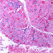
Cardiopulmonary bypass, more commonly known as heart-and-lung bypass, has some unique impacts on the creation and growth of brain cells in the area of a child’s brain called the subventricular zone (SVZ), according to a study in the Annals of Neurology. The SVZ is a critical area for the growth and migration of neurons and nerve cells called neuroblasts, both of which ultimately contribute to the proper development of key brain structures and functions during the early years of life.
The findings, from a study conducted in the Cardiac Surgery Research Laboratory at Children’s National Hospital, provide new insight into the cellular impacts of the cardiopulmonary bypass machine on brain growth and development for newborn infants with congenital heart disease. They will have an important role in the refinement of strategies to help protect the fragile brains of children who require lifesaving cardiac surgery with cardiopulmonary bypass immediately after birth.
Specifically, the research team found that during cardiopulmonary bypass:
- Creation of neurons (neurogenesis) in the neonatal and infant subventricular zone is altered.
- Migration of nerve cells, called neuroblasts, to the frontal lobe is potentially disrupted.
- Changes to the growth and movement of neurons in the SVZ are prolonged.
- Cortical development and expansion is impaired.
- Specific types of neurons found only in the brain and spinal cord, called interneurons, are also affected.
The study uses an innovative pre-clinical model of the developing brain that is more anatomically and physiologically similar to human neonates and infants than those used in prior studies and in most neurological laboratory-based research.
Cardiopulmonary bypass is one of several key factors thought to cause children with congenital heart disease to sometimes demonstrate delays in the development of cognitive and motor skills. These disabilities often persist into adolescence and adulthood and can ultimately represent long-term neurocognitive disabilities. It is also believed that genetic factors, abnormal blood flow to the brain while in utero or low cardiac output after surgical procedures on the heart may contribute to these challenges.
“Unraveling cellular and molecular events during surgery using this preclinical model will allow us to design therapeutic approaches that can be restorative or reparative to the neurogenic potential of the neuronal stem precursor cells found in the subventricular zone of the neonatal or infant brain,” says Nobuyuki Ishibashi. M.D., Foglia-Hills Professor of Pediatric Cardiac Research, director of the Cardiac Surgery Research Laboratory at Children’s National and senior author on the study. “In particular, previous studies in our laboratory have shown improvement in the neurogenic activities of these precursor cells when they are treated with mesenchymal stromal cells (MSCs).”
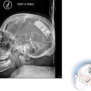
The findings from this study further support the work already underway in the NIH-funded MeDCaP clinical trial for neonates and infants undergoing cardiac surgery using the cardiopulmonary bypass machine. That trial uses the heart and lung machine itself to deliver MSCs directly into the main arteries that carry blood to the brain.