A new imaging device with AI may reduce complications during thyroid surgery
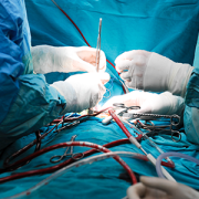
 Surgeons perform approximately 150,000 thyroidectomies in the United States. Post-surgical complications from this procedure frequently occur due to the misidentification or accidental removal of healthy parathyroid glands. On average, 27% of these patients suffer from transient or permanent hypocalcemia, a condition in which the blood has too little calcium, leading to lifelong complications and socioeconomic burden.
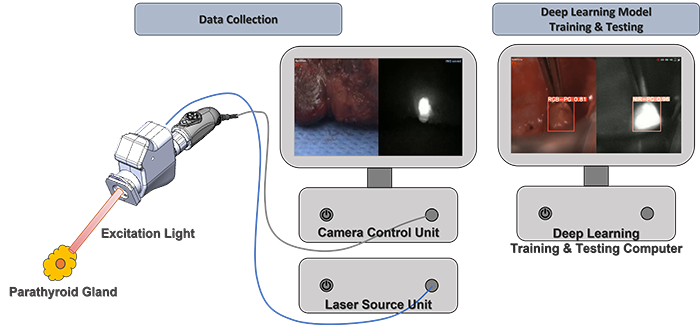
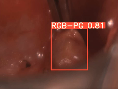
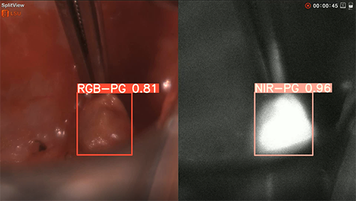
To improve parathyroid detection during surgery, Children’s National Hospital experts developed a prototype equipped with a dual-sensor imaging device and a deep learning algorithm that accurately detects parathyroids, according to a new study published in the Journal of Biophotonics.
“What excited us in this study was that even deep-seated tissues were able to be imaged without light loss, and high resolution imaging was possible due to the unique optical design,” said Richard Jaepyeong Cha, Ph.D., council member of the International Society of Innovative Technologies for Endocrine Surgery and principal investigator for the Sheikh Zayed Institute for Pediatric Surgical Innovation at Children’s National Hospital. “Moreover, in several cases, parathyroid autofluorescence was detected even before the surgeon dissected the parathyroid gland, and while it was covered by fat and/or fascia.”
What’s unique
This is the first study that uses color RGB/NIR paired imaging-based parathyroid detection by incorporating multi-modal (both RGB light and near-infrared autofluorescence, or NIRAF, ground truth imaging) data into parathyroid identification using a deep learning algorithm.
The patient benefit
“We envision that our technology will open a new door for the digital imaging paradigm of dye-free, temporally unlimited, and precise parathyroid detection and preservation,” said Richard. “Successful translation of this technology will potentially reduce the risk of hypoparathyroidism after common thyroid surgery and improve the clinical outcomes.”
The results support the effectiveness of their novel approach despite the small sample size, which can potentially improve specificity in the identification of parathyroid glands during parathyroid and thyroid surgeries.
The hold-up in the field
It is often difficult for surgeons with naked eyes to identify parathyroid glands from thyroid tissue because of the small size, the variable position, and similar appearance to the surrounding tissues.
Since 2011, surgeons have benefited from using NIRAF, a non-invasive optical method for intraoperative real-time localization of parathyroids.
While the NIRAF technology has gained traction among endocrine surgery community, false negatives can occur with current devices that use the NIRAF technology in secondary hyperparathyroidism cases. According to Kim et al., the technology is still suboptimal, and a significant percentage of parathyroid is being missed.
Children’s National Hospital leads the way
Engineers in Children’s National are leading this field through several innovations:
- Non-dye injected, label-free use in real-time in comparison to temporally limited ICG angiography. This technology was featured as the cover article in the journal Lasers in Surgery and Medicine 54(3), 2022.).
- Simultaneous perfusion assessment from four glands at any time during operation.
- Arterial flow detection from pulsatile information in well-perfused PG vasculature.
- Quantified parathyroid detection and classification with prediction values using deep learning technique.
You can read the full study “A co-axial excitation, dual-RGB/NIR paired imaging system toward computer-aided detection (CAD) of parathyroid glands in situ and ex vivo” in the Journal of Biophotonics.