Using a multisystem approach to improve access to autism care in Washington, D.C.

Children with autism face significant barriers to accessing evaluations and intervention services.
An article in the journal Pediatrics reviews the outcomes from a collective, targeted advocacy approach to improving access to autism supports and resources for children and their families in Washington, D.C. The effort was led by Children’s National Hospital and engaged a multidisciplinary team from within the hospital and across a wide range of community sectors.
What this means
Children’s National and DC Autism Parents worked collaboratively with a coalition of organizations from the broader District of Columbia community to address some of the biggest challenges and barriers that prevent autistic children and their families from receiving the resources and support they need in the nation’s capital.
Why it matters
Children with autism face significant barriers to accessing evaluations and intervention services often because of confusing referral processes, lack of centralized coordination across organizations serving children with autism, insurance coverage gaps, multiyear waitlists for diagnostic services and limited provider knowledge about autism. Racism and systemic inequities also persist in autism care across the United States.
Long and growing wait times in autism diagnostic clinics and lack of centralized care coordination for autistic children are prevalent across the District of Columbia, and as a result, many children and families in the region continue to lack access to the support they need.
What’s unique
The study describes multiyear efforts (2017–2022) to improve autism care throughout the District of Columbia using a collective impact framework to unite organizations from different sectors. This approach features the creation of a common agenda (including defining goals and priorities), shared measurement, mutually reinforcing activities, continuous communication and infrastructure support.
Together, the group members sought to address barriers and overcome challenges at multiple levels of the healthcare system at the same time by focusing advocacy in three specific areas:
- Infrastructure-building initiatives/system-level approaches.
- Population- and community-level services to build capacity and connect providers and families to needed resources.
- Direct services that provide innovative, gap-filling supports to children and families as a stopgap until the necessary supports can be more sustainably provided across the board.
Bottom line
While more work is needed to continue expanding the availability of needed services, the findings from this initial effort can inform the next steps in Washington, D.C., and serve as a model for a collective framework approach for autism services in other parts of the United States.
You can read the full study “A Multisystem Approach to Improving Autism Care” in the journal Pediatrics.
More information and resources about these autism initiatives can be found at:


 Children’s National Hospital named

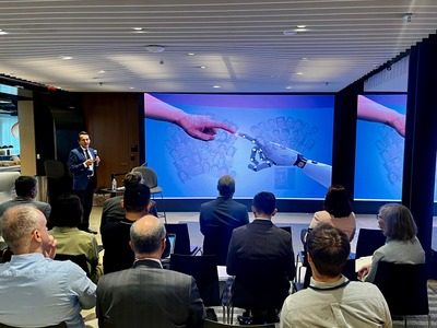 Children’s National Hospital is joining a team of global health researchers to use large language models (LLMs) like ChatGPT to help Kenyan youth learn about their health and adopt lifestyles that may prevent cancer, diabetes and other non-communicable diseases.


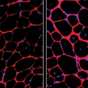
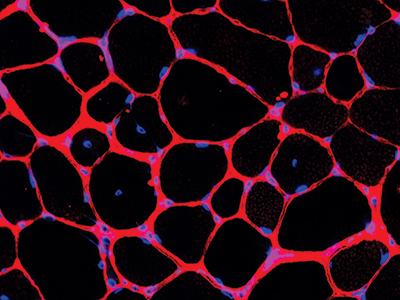
 Children’s National Hospital announced a $96 million investment from an anonymous donor family to transform rare childhood brain tumor research and care. The donation, which strengthens our globally recognized leadership in the field, is one of the largest in the hospital’s history.