ACC.19: A focus on pediatric cardiology
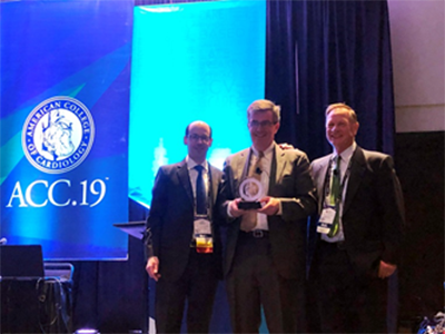
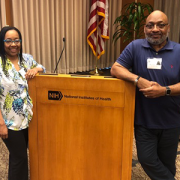
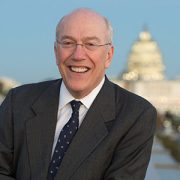
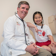
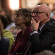
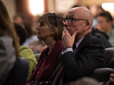
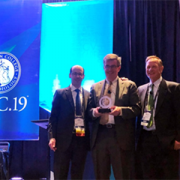
Dr. Gerard Martin, center, accepts an award before delivering the 2019 Dan G. McNamara Keynote lecture at ACC.19.
“Innovation meets tradition,” is how many attendees and journalists described the American College of Cardiology’s 68th Scientific Sessions (ACC.19), which took place March 16-18, 2019 in New Orleans, La.
Gerard Martin, M.D., F.A.A.P., F.A.C.C., F.A.H.A., a pediatric cardiologist and the medical director of Global Services at Children’s National, supported this narrative by referencing both themes in his 2019 Dan G. McNamara keynote lecture, entitled “Improved Outcomes in Congenital Heart Disease through Advocacy and Collaboration.” Dr. Martin highlighted advancements in the field of pediatric cardiology that took place over the past 15 years, while touting modern advancements – such as pulse oximetry screenings for critical congenital heart disease – that were a result of physician-led advocacy and collaboration.
Dr. Martin’s message was to continue to invest in research and technology that leads to medical breakthroughs, but to remember the power of partnerships, such as those formed by the National Pediatric Cardiology Quality Improvement Collaborative. These alliances, which generated shared protocols and infrastructure among health systems, improved interstage mortality rates between surgeries for babies born with hypoplastic left heart syndrome.
A dozen cardiologists and clinicians from the Children’s National Heart Institute also participated in CME panel discussions or delivered poster presentations to support future versions of this template, touching on early-stage innovations and multi-institution research collaborations. The themes among Children’s National Heart Institute faculty, presented to a diverse crowd of 12,000-plus professional attendees representing 108 countries, included:
Personalized guidelines:
- Sarah Clauss, M.D., F.A.C.C., a cardiologist, presented “Unique Pediatric Differences from Adult Cholesterol Guidelines: Lipids and Preventive Cardiology,” before Charles Berul, M.D., division chief of cardiology and co-director of the Children’s National Heart Institute, presented “Unique Pediatric Differences from Adult Guidelines: Arrhythmias in Adults with Congenital Heart Disease,” in a joint symposium with the American Heart Association and the American College of Cardiology.
- Berul, who specializes in electrophysiology, co-chaired a congenital heart disease pathway session, entitled “Rhythm and Blues: Electrophysiology Progress and Controversies in Congenital Heart Disease,” featuring components of pediatric electrophysiology, including heart block, surgical treatment of arrhythmias and sudden death risk.
Early detection:
- Anita Krishnan, M.D., associate director of the echocardiography lab, presented “Identifying Socioeconomic and Geographic Barriers to Prenatal Detection of Hypoplastic Left Heart Syndrome and Transposition of the Great Arteries” as a moderated poster in Fetal Cardiology: Quickening Discoveries.
- Jennifer Romanowicz, M.D., a cardiology fellow, and Russell Cross, M.D., director of cardiac MRI, presented the “Neonatal Supraventricular Tachycardia as a Presentation of Critical Aortic Coarctation” poster in FIT Clinical Decision Making: Congenital Heart Disease 2.
- Pranava Sinha, M.D., a cardiac surgeon, presented the poster “Neuroprotective Effects of Vitamin D Supplementation in Children with Cyanotic Heart Defects: Insights from a Rodent Hypoxia Model” in Congenital Heart Disease: Therapy 2.
Coordinated care:
- Ashraf Harahsheh, M.D., F.A.C.C., F.A.A.P., a cardiologist with a focus on hyperlipidemia and preventive cardiology, co-presented an update about BMI quality improvement (Q1) activity from the American College of Cardiology’s Adult Congenital and Pediatric Quality Network – BMI Q1 leadership panel.
- Niti Dham, M.D., director of the cardio-oncology program, and Deepa Mokshagundam, M.D., cardiology fellow, presented the poster “Cardiac Changes in Pediatric Cancer Survivors” in Heart Failure and Cardiomyopathies: Clinical 3.
- Nancy Klein, B.S.N., R.N., C.P.N., clinical program coordinator of the Washington Adult Congenital Heart program at Children’s National, presented the poster “Improving Completion of Advanced Directives in Adults with Congenital Heart Disease” in Risks and Rewards in Adult Congenital Heart Disease.
Innovation:
- Jai Nahar, M.D., a cardiologist, moderated “Future Hub: Augmented Cardiovascular Practitioner: Giving Doctors and Patients a New Voice.” The session focused on technical aspects of artificial intelligence, such as language processing and conversational artificial intelligence, as well as how applications are used in patient-physician interactions.
- Nahar also participated in a key event on the Heart-to-Heart stage, entitled “Rise of Intelligent Machines: The Potential of Artificial Intelligence in Cardiovascular Care.”
“While I enjoyed the significant representation of Children’s National faculty at the meeting and all of the presentations this year, one research finding that I found particularly compelling was Dr. Krishnan’s poster about geographical disparities in detecting congenital heart disease,” says Dr. Berul. “Her research finds obstetricians providing care to women in the lowest quartile of socioeconomic areas were twice as likely to miss a diagnosis for a critical congenital heart defect during a fetal ultrasound, compared to obstetricians providing care for women in the highest quartiles.”
Dr. Krishnan’s study was the collaborative effort of 21 centers in the United States and Canada, and investigated how socioeconomic and geographic factors affect prenatal detection of hypoplastic left heart syndrome and transposition of the great arteries.
“We studied over 1,800 patients, and chose these diseases because they require early stabilization by a specialized team at a tertiary care center,” says Dr. Krishnan, who led the research in conjunction with the Fetal Heart Society Research Collaborative. “We hope that by understanding what the barriers are, we can reduce disparities in care through education and community-based outreach.”