Cardiology & Heart Surgery

Steady rates of patient satisfaction, reimbursement for cardiac telehealth during COVID-19

a telehealth video visit with a patient family

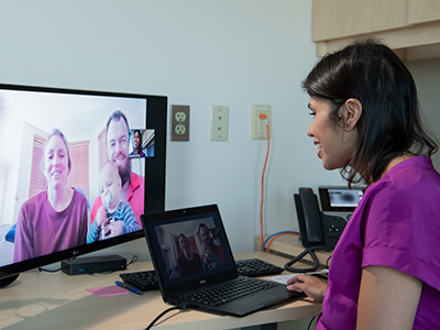
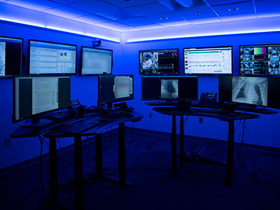
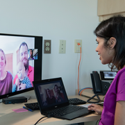
In the first two weeks of COVID-19’s major impact on the U.S., Children’s National Hospital moved most of its subspecialty in-person day-to-day clinics to virtual care. Children’s National Heart Institute was one of the first divisions to offer telehealth visits — in part because the team was an early adopter of telehealth in cardiology for both physician-to-physician consultations and direct-to-patient care, and stood poised to widely implement it.

A poster presentation at the American Heart Association Scientific Sessions 2020 quantified how the rapid transition to direct-to-consumer telehealth services impacted families with children who have congenital heart disease. The findings were presented by first author Kristine Mehrtens, M.S., B.S.N., R.N., C.P.N., clinical manager for the Heart Institute’s Ambulatory Services.

The team found that though in-person cardiology visits decreased during the COVID-19 pandemic, direct-to-patient telehealth visits were able to partially compensate for the sudden drop.

Additionally, payer reimbursement rates for these direct-to-consumer telehealth visits were similar to in-person clinic visits.

”This is exciting as prior to COVID-19  we have seen a lower reimbursement rates for these cardiology direct-to-consumer telehealth visits compared to in-person cardiology clinic visits,” said Ashraf S. Harahsheh, M.D., a pediatric cardiologist at Children’s National Hospital who has utilized direct-to-consumer telehealth visits since 2016 and is a senior author on the new study.

Patient satisfaction scores for care providers, including the likelihood of recommending a care provider from Children’s National Hospital, was the same for telehealth follow-up visits as it was for in-person clinic visits before the pandemic.

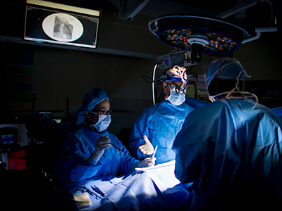
“As a multidisciplinary team, we agreed that diagnostic studies such as echocardiograms were important to include with follow-up visits,” says Mehrtens. “Together we developed a strategy to ensure we could meet the needs of the patients and also safely conduct in-person visits when necessary.”

Why is this important?

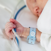
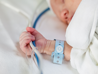
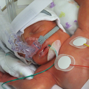
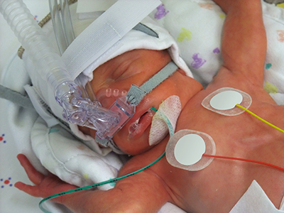
The pandemic and the resulting temporary halt to in-person, non-urgent/emergent visits earlier this year put the most vulnerable people with congenital heart disease at high risk for complications or worsening of their existing heart disease because they are unable to follow the recommended schedule for follow-ups.

The readiness of the Children’s Heart Institute team to quickly move to a telehealth platform successfully bridged the gap between in-person visits for some patients, allowing cardiology surveillance to continue safely.

“I am proud of our team of physicians and advanced care providers,” Harahsheh concludes. “We went from three providers (8%) pre-COVID 19 to 31 (79%) providers offering direct-to-consumer telehealth visits during the pandemic.”

What’s next?

Building on previous, smaller studies of telehealth before the pandemic began, the team will continue to conduct research to assess the safety and efficacy of these telehealth visits over time. The increase in patients who are continuing to see their providers for routine follow-ups via telehealth will allow a larger sample for effective study of this care model.

American Heart Association Scientific Sessions 2020
Impact of Telemedicine on Pediatric Cardiac Center’s Ambulatory Response to the 2019 Novel Coronavirus Disease (covid-19) Pandemic
P1692
9:00am – 10:00am
Fri, Nov 13  (CST)

Read additional news stories about cardiology telehealth:

Cardiac technology advances show promise for kids but only if right-sized

newborn in ICU

“Smaller patients, and those with congenital heart disease, can benefit from minimally-invasive methods of delivering pacemakers and defibrillators without the need for open-chest surgery,” says Charles Berul, M.D.

How to address the growing need for child-sized pacemakers and defibrillators, and finding better surgical techniques to place them, is the topic of an invited session called The Future is Now (or Coming Soon): Updates on New Technologies in Congenital Heart Care at the 2020 American Heart Association Scientific Sessions.

“Smaller patients, and those with congenital heart disease, can benefit from minimally-invasive methods of delivering pacemakers and defibrillators without the need for open-chest surgery,” says Charles Berul, M.D., co-director of the Children’s National Heart Institute and chief of Cardiology at Children’s National Hospital, who presented at the session.

“This unmet need can only be met by innovative pediatric research, geared towards miniaturization technologies for use in the smallest of children,” he says.

His presentation focused on the devices and approaches that have caught the attention of pediatric cardiology, such as pacemakers and subcutaneous defibrillators designed without lead wires, as well as less-invasive surgical approaches that may reduce recovery time for children with congenital heart disease who require these assist devices.

Using them in kids comes with added challenges, however. Often pediatric cardiologists have to be creative in how to make them work for smaller patients, Dr. Berul notes. This reiterates the important point that simply applying an adult technology to a child isn’t the right approach. The subcutaneous defibrillator, for example, is still pretty large for a child’s body. Some studies also show these devices may not be as accurate in children as in adults.

Investigators in the Sheikh Zayed Institute working together with the cardiologists at Children’s National Hospital are focused on product development and commercialization of tools and techniques to allow percutaneous minimally-invasive placement of devices, taking advantage of the newest devices and surgical techniques as they develop.

In his presentation, Dr. Berul stressed that as the technology for adults advances, it creates an opportunity for pediatric cardiology, but only if the devices, and the techniques to place them, are specifically redesigned for pediatric application.

American Heart Association Scientific Sessions 2020
The Future is Now (or Coming Soon): Updates on New Technologies in Congenital Heart Care – On Demand Session
CH.CVS.715
9:00am – 10:00am
Fri, Nov 13  (CST)

Single institution study finds high rates of cardiac complications in MIS-C

coronavirus

At this year’s AHA Scientific Sessions, cardiologists from Children’s National Hospital presented a poster about an interesting finding in children with MIS-C.

During the height of the pandemic, researchers at Children’s National Hospital discovered that as many as one half of children diagnosed with multisystem inflammatory disease in children (MIS-C) at the hospital developed cardiac complications including coronary artery abnormalities, even when diagnosed and treated promptly.

The data was shared as part of a poster presentation at the American Heart Association Scientific Sessions in November 2020. Though analysis was limited to the data from one institution’s confirmed MIS-C cases, the findings are significant enough to warrant further study.

Interestingly, the authors noted that the high rate of cardiac complications far exceeds the rate of similar issues in children with Kawasaki disease — another pediatric inflammatory syndrome that shares many common symptoms with MIS-C. The two are so similar that immunomodulation therapies successfully deployed in children with MIS-C were based on those developed to treat Kawasaki disease.

Knowledge of common cardiac complications in Kawasaki disease also flagged the need for routine echocardiograms in patients with MIS-C, which helped identify the higher rates of cardiac complications seen in the MIS-C patient population.

“This finding, however, is another data point that shows how MIS-C and Kawasaki disease have some specific differences needing further study,” says Ashraf Harahsheh, M.D., a pediatric cardiologist at Children’s National Hospital who studies Kawasaki disease and the first author on the new study.

“Previous clinical advancements made in Kawasaki disease set the stage for our response to MIS-C early on,” he said. ”Now we also need to understand MIS-C as its own syndrome so we can better address what we are seeing in this patient population,” he says.

While most of the cardiac findings resolved during follow up, long-term studies are needed to determine if the cardiac abnormalities are associated with major cardiac events later.

“This work will help inform the community of the importance of diagnosing children with MIS-C promptly and following clinical guidelines for necessary tests and treatments once MIS-C is diagnosed,” Harahsheh concludes.

Next, the research team plans to take a deep dive into patient demographics as well as findings from clinical, laboratory and electrocardiogram data for children who developed cardiac complications with MIS-C. The goal will be to refine treatment algorithms and potentially identify a subgroup of patients who may require different or more intense therapy to prevent cardiac complications.

American Heart Association Scientific Sessions 2020 Poster Session
Cardiac Complications of SARS CoV-2 Associated Multi-System Inflammatory Syndrome in Children (MIS-C)
P1306
9:00am – 10:00am
Fri, Nov 13 (CST)

Speckle tracking echo reveals possible biomarker for SUDEP risk

EEG with electrical activity of abnormal brain

A study published in the journal Epilepsia used speckle tracking echocardiography to detect subtle changes in heart function found in pediatric patients with refractory epilepsy when compared to controls. Children with refractory epilepsy had impaired systolic ventricular strain compared to controls, not correlated to epilepsy history. These differences in ventricular function may be a biomarker that can indicate someone with epilepsy is at higher risk for Sudden Unexpected Death in Epilepsy (SUDEP).

Speckle tracking echocardiography is a non-invasive technique where software automatically identifies and tracks individual “speckles” of the myocardial wall on a routine echocardiogram in order to directly quantify the extent of contraction.

The study’s first authors, John Schreiber, M.D., medical director of Electroencephalography (EEG) and director of the Epilepsy Genetics program, and Lowell Frank, M.D., advanced imaging cardiologist and director of the Cardiology Fellowship Training program, both at Children’s National Hospital, answered some questions about the study findings.

Why is this important work?

Sudden unexpected death in epilepsy (SUDEP) is a rare but devastating consequence of epilepsy. Some of the proposed mechanisms of SUDEP implicate brain stem, cardiac and respiratory pathways.

This study identified alterations in ventricular function that may serve as one potential biomarker for SUDEP risk that can be evaluated non-invasively and regularly.

How will this work benefit patients?

Identification of children or adults with markedly impaired ventricular strain or diastolic function may provide the opportunity to implement a targeted treatment or monitoring strategy to prevent SUDEP.

What did you find that excites you? What are you hoping to discover?

These differences in cardiac strain were true for all patients with refractory epilepsy as a whole, not one particular group. This suggests that refractory convulsive epilepsy itself, rather than other patient-specific factors, produces these changes. Thanks in part to a grant from the Dravet Syndrome Foundation, the team is currently examining a cohort of patients with epilepsy due to pathogenic variants in sodium channel genes, SCN1A and SCN8A, to determine if these patients have greater degrees of impaired cardiac strain. SCN1A and SCN8A are also expressed in the heart, and patients have a considerably higher risk of SUDEP. It will be particularly exciting to examine for differences in specific genetic epilepsies.

How is this work unique?

Strain has been evaluated in many disease states in adult and pediatric populations and may be more sensitive to early myocardial damage than traditional measures of systolic and diastolic function. Children’s National Hospital has been an innovator in using speckle tracking echocardiography and similar techniques to evaluate subtle changes in heart function. This study is a great example of collaboration between The Comprehensive Pediatric Epilepsy Program and the Children’s National Heart Institute that is driving innovative research at Children’s National Hospital.

Boeing gives $5 million to support Research & Innovation Campus

Research & Innovation Campus

Children’s National Hospital announced a $5 million gift from The Boeing Company that will help drive lifesaving pediatric discoveries at the new Children’s National Research & Innovation Campus.

Children’s National Hospital announced a $5 million gift from The Boeing Company that will help drive lifesaving pediatric discoveries at the new Children’s National Research & Innovation Campus. The campus, now under construction, is being developed on nearly 12 acres of the former Walter Reed Army Medical Center. Children’s National will name the main auditorium in recognition of Boeing’s generosity.

“We are deeply grateful to Boeing for their support and commitment to improving the health and well-being of children in our community and around the globe,” said Kurt Newman, M.D., president and CEO of Children’s National “The Boeing Auditorium will help the Children’s National Research & Innovation campus become the destination for discussion about how to best address the next big healthcare challenges facing children and families.”

The one-of-a-kind pediatric hub will bring together public and private partners for unprecedented collaborations. It will accelerate the translation of breakthroughs into new treatments and technologies to benefit kids everywhere.

“Children’s National Hospital’s enduring mission of positively impacting the lives of our youngest community members is especially important today,” said Boeing President and CEO David Calhoun. “We’re honored to join other national and community partners to advance this work through the establishment of their Research & Innovation Campus.”

Children’s National Research & Innovation Campus partners currently include Johnson & Johnson Innovation – JLABS, Virginia Tech, the National Institutes of Health (NIH), Food & Drug Administration (FDA), U.S. Biomedical Advanced Research and Development Authority (BARDA), Cerner, Amazon Web Services, Microsoft, National Organization of Rare Diseases (NORD) and local government.

The 3,200 square-foot Boeing Auditorium will be the focal point of the state-of-the-art conference center on campus. Nationally renowned experts will convene with scientists, medical leaders and diplomats from around the world to foster collaborations that spur progress and disseminate findings.

Boeing’s $5 million commitment deepens its longstanding partnership with Children’s National. The company has donated nearly $2 million to support pediatric care and research at Children’s National through Chance for Life and the hospital’s annual Children’s Ball. During the coronavirus pandemic, Boeing fabricated and donated 2,000 face shields to help keep patients and frontline care providers at Children’s National safe.

Medical device competition announces six winners to share in $250K

Judges award grants for pediatric medical devices that address cardiovascular, NICU, and orthopaedic and spine device innovations.

Yves d’Udekem, M.D., Ph.D., joins Children’s National as Cardiac Surgery Chief

Yves d’Udekem, M.D., Ph.D., a pediatric cardiac surgeon recognized for expertise in the most challenging procedures for newborns and children with complex congenital heart disease, has joined Children’s National Hospital as chief of Cardiac Surgery and co-director of the Children’s National Heart Institute.

“Children’s National has the cases and expertise I was looking for,” he says. “Even better, when you visit it’s clear that the entire Heart Institute team is energetic about working together. They’re constantly seeking better ways to do this work and improve how we care for children and their families.”

Dr. d’Udekem comes to Children’s National from The Royal Children’s Hospital in Melbourne, Australia, an institution that has led the advancement of congenital heart disease care and research, performing more than 500 surgical procedures with cardiopulmonary bypass each year.

He has a broad spectrum of pediatric cardiac surgery expertise, with special emphasis in single ventricle congenital heart defects, when one lower chamber of the heart does not develop. One area of his research portfolio includes clinical research into long-term quality of life for people who had Fontan procedures — a critical surgical approach to adapt blood flow for people born with single ventricle heart disease. He has additional expertise in valve repair, artificial hearts and other cardiac assist devices.

“Sometimes it seems like I’ve been born and put on this earth for that single purpose — I have to bring life to patients with single ventricle hearts or assist devices,” he says. He describes his path to pediatric cardiac surgery as serendipity, “It’s more like pediatric cardiac surgery chose me.”

“It is an honor to welcome Yves d’Udekem to the Children’s National Heart Institute,” says David Wessel, M.D., executive vice president and chief medical officer of Hospital and Specialty Services at Children’s National. “He is a surgeon, physician and leader of the highest caliber. I can say without hesitation that Yves’ leadership of our cardiac surgery team will change the lives of hundreds, if not thousands, of children and adults with congenital heart disease.”

In preparation for Dr. d’Udekem’s arrival, the Children’s National Heart Institute and hospital leadership created a framework plan for success that will build on the legacy of excellence established by Richard Jonas, M.D. Dr. Jonas, a world leader in congenital heart surgery who has made significant clinical and academic advances in the field, will continue his award-winning laboratory-based research at Children’s National on neurodevelopmental outcomes for children with congenital heart disease.

Dr. d’Udekem is Belgian-born with Canadian and Australian citizenship. He received his early training in Belgium and Toronto. As an attending cardiac surgeon in Belgium, he operated on both adults and children with congenital heart disease. Dr. d’Udekem then worked with internationally recognized heart surgeon Marc de Leval, M.D., FRCS, and others in London to train in congenital heart surgery at the Great Ormond Street Hospital for Children. From there, he was recruited to The Royal Children’s Hospital in Melbourne, Australia, where he has been ever since.

Dr. d’Udekem has more than 350 research publications and has obtained more than $7 million in grant funding in the past five years for work to create the first research network of Australian children and adults who have undergone the Fontan procedure.

He started seeing patients at Children’s National in mid-September.

Connections between Kawasaki disease and MIS-C

mother measuring sick child's temperature

A new review article enumerates some key similarities and differences between MIS-C and Kawasaki disease.

Since May 2020, there has been some attention in the general public and the news media to a specific constellation of symptoms seen in children with COVID-19 or who have been exposed to COVID-19. For a time, headlines even called it a “Kawasaki-like” disease. At first glance, both the symptoms and the effective treatments are remarkably similar. However, a new review published in Trends in Cardiovascular Medicine finds that under closer scrutiny, the two conditions have some interesting differences as well.

“At the beginning of this journey, we thought we might be missing actual cases of Kawasaki disease because we identified a few patients who presented late and developed coronary artery abnormalities,” says Ashraf Harahsheh, M.D., senior author of the review article, “Multisystem inflammatory syndrome in children: Is there a linkage to Kawasaki disease?” and a cardiologist at Children’s National Hospital. “But as time passed, children exposed to COVID-19 started to present with a particular constellation of symptoms that actually had some important similarities and distinctions from Kawasaki.”

Similarities between Kawasaki disease and MIS-C

Both disease patterns seem to have a common trigger that provokes the inflammatory cascade reaction in genetically susceptible children, the authors write. However, there is also early evidence that children with each disease have different genetic markers, meaning different populations are genetically susceptible to each disease.

Additionally, the authors found that the massive activation of pro-inflammatory cytokines seen in MIS-C, also known as a “cytokine storm,” overlaps with a similar occurrence seen in Kawasaki disease, adult COVID-19 patients, toxic shock syndrome and some other viral infections.

Primary differences between Kawasaki disease and MIS-C

Overall, when compared to Kawasaki disease, children with MIS-C tend to:

  • Present at an older age
  • Have a more profound form of inflammation
  • Have more gastrointestinal manifestation
  • Show different laboratory findings
  • Have greater risk of left ventricle dysfunction and shock

Further study of both Kawasaki and MIS-C needed

Despite noted differences, the authors are also careful to credit the documented similarities between Kawasaki disease and MIS-C as a key to the quick identification of the new syndrome in children. The study of Kawasaki disease also gave clinicians a valid basis to begin developing diagnostic recommendations and treatment protocols.

The review’s first author Yue-Hin Loke, M.D., who is also a cardiologist at Children’s National, says, “The quick recognition of MIS-C is only possible because of meticulous research conducted by Dr. Tomisaku Kawasaki, who recently passed away on June 5th, 2020. Even though some aspects of both are still shrouded in mystery, the previous research and clinical advancements made in Kawasaki disease set the stage for our immediate response to MIS-C.”

“Previous research provided key information for cardiologists facing this new syndrome, including the necessity of routine echocardiograms to watch for coronary artery abnormalities (CAAs) and for use of  intravenous immunoglobulin (IVIG) to mitigate  the development of CAAs,” says Charles Berul, M.D., chief of Cardiology at Children’s National and a co-author. “Both of these factors have played a key role in reducing the mortality of MIS-C to almost zero.”

The authors note that more research is needed to understand both Kawasaki disease and the specifics of MIS-C, but that what is learned about the mechanisms of one can and should inform study and treatment of the other. And in the meantime, caution and continued surveillance of these patients, especially with respect to coronary artery and myocardial function, will continue to improve the long-term outcomes for both syndromes.

Telehealth and AI reduce cardiac arrest in the cardiac ICU

telemedicine control room

The telehealth command center located a few steps away from the cardiac ICU at Children’s National Hospital.

The cardiac critical care team at Children’s National Hospital has developed an innovative Tele-Cardiac Critical Care model aiming to keep constant watch over the most fragile children with critical heart disease in the cardiac ICU. The system combines traditional remote monitoring and video surveillance with an artificial intelligence algorithm trained to flag early warning signs that a critically ill infant may suffer a serious event like cardiac arrest while recovering from complex cardiac surgery. This second set of eyes helps bedside teams improve patient safety and quality of care.

These high risk post-operative patients are often neonates or small infants born with the most complex and critical congenital heart diseases that require surgery or interventional cardiac catheterization in their first days or weeks of life. At these early stages after crucial cardiac surgery, these patients can decompensate dangerously fast with few outward physical symptoms.

The AI algorithm (T3) monitors miniscule changes in oxygen delivery and identifies any mismatch with a child’s oxygen needs. It also tracks and displays small changes in vital sign trends that could lead to a serious complication. The cardiac ICU command center staff then analyzes additional patient data and alerts the bedside team whenever needed.

The Tele-Cardiac Critical Care program started two years ago. In that time, the program has contributed to a significant decrease in post-operative cardiac arrest for this patient population.

“It’s easy to see how a model  like this could be adapted to other critical care scenarios, including our other intensive care units and even to adult units,” says Ricardo Munoz, M.D., chief of Cardiac Critical Care and executive director of Telehealth. It allows the physicians and nurses to keep constant watch over these fragile patients without requiring a physician to monitor every heartbeat in person for every patient at every hour of the day to maintain optimal outcomes for all of them.”

Dr. Munoz and Alejandro Lopez-Magallon, M.D., medical director of Telehealth and cardiac critical care specialist, presented data from the pilot program at the American Telemedicine Association’s virtual Annual Meeting on June 26, 2020.

U.S. DoD awards $2M for study to protect neurological function after cardiac surgery

doctors operating

A collaboration between clinical and basic science researchers including Drs. Ishibashi, Hashimoto-Torii, Jonas, and Deutsch, seeks to to understand how caspase enzyme activation plays a role in the development of fine and gross motor skills in children who underwent cardiac surgery for CHD repair.

The U.S. Department of Defense has awarded $2 million to Children’s National Hospital to study how a family of protease enzymes known as caspases may contribute to brain cell degeneration when activated by prolonged anesthesia and cardiopulmonary bypass during cardiac surgery for congenital heart disease.

This U.S. Army Medical Research Acquisition Activity Award, Anesthesia Neurotoxicity in Congenital Heart Disease, is led by principal investigator Nobuyuki Ishibashi, M.D., with both clinical and basic science co-investigators including Kazue Hashimoto-Torii, Ph.D., (Neuroscience), Richard Jonas, M.D., (Cardiovascular Surgery) and Nina Deutsch, M.D., (Anesthesiology).

While the specific cellular and molecular mechanisms of how anesthesia and cardiac surgery impact cortical development are poorly understood, both seem to impact brain growth and development in young children. The most common neurologic deficit seen in children after CHD surgical repair is the impairment of fine and gross motor skills.

Both anesthetic agents and inflammation like that seen as a result of cardiopulmonary bypass have also been shown to contribute to the activation of a specific group of enzymes that play an essential role in the routine (programmed) death of cells: caspases. However, recent pre-clinical research shows that these enzymes may also contribute to other alterations to cells beyond cell death, including making changes to other cell structures. In pre-clinical models, these changes cause impairments to fine and gross motor skills – the same neurological deficits seen in children with CHD who have undergone procedures requiring prolonged anesthesia and cardiopulmonary bypass.

The research team hypothesizes that caspases are extensively activated as a result of cardiac surgery and while that activation is rarely causing reduced numbers of neurons, the changes that caspase enzymes trigger in neurons are contributing to neurological deficits seen in children with CHD after surgery.

While the study focuses specifically on the impacts of cardiac surgery for correction of a heart defect, the findings could have major implications for any pediatric surgical procedure requiring prolonged anesthesia and/or cardiopulmonary bypass.

Children’s National ranked a top 10 children’s hospital and No. 1 in newborn care nationally by U.S. News

US News Badges

Children’s National Hospital in Washington, D.C., was ranked No. 7 nationally in the U.S. News & World Report 2020-21 Best Children’s Hospitals annual rankings. This marks the fourth straight year Children’s National has made the list, which ranks the top 10 children’s hospitals nationwide.

In addition, its neonatology program, which provides newborn intensive care, ranked No.1 among all children’s hospitals for the fourth year in a row.

For the tenth straight year, Children’s National also ranked in all 10 specialty services, with seven specialties ranked in the top 10.

“Our number one goal is to provide the best care possible to children. Being recognized by U.S. News as one of the best hospitals reflects the strength that comes from putting children and their families first, and we are truly honored,” says Kurt Newman, M.D., president and CEO of Children’s National Hospital.

“This year, the news is especially meaningful, because our teams — like those at hospitals across the country — faced enormous challenges and worked heroically through a global pandemic to deliver excellent care.”

“Even in the midst of a pandemic, children have healthcare needs ranging from routine vaccinations to life-saving surgery and chemotherapy,” said Ben Harder, managing editor and chief of Health Analysis at U.S. News. “The Best Children’s Hospitals rankings are designed to help parents find quality medical care for a sick child and inform families’ conversations with pediatricians.”

The annual rankings are the most comprehensive source of quality-related information on U.S. pediatric hospitals. The rankings recognize the nation’s top 50 pediatric hospitals based on a scoring system developed by U.S. News. The top 10 scorers are awarded a distinction called the Honor Roll.

The bulk of the score for each specialty service is based on quality and outcomes data. The process includes a survey of relevant specialists across the country, who are asked to list hospitals they believe provide the best care for patients with the most complex conditions.

Below are links to the seven Children’s National specialty services that U.S. News ranked in the top 10 nationally:

The other three specialties ranked among the top 50 were cardiology and heart surgery, gastroenterology and gastro-intestinal surgery, and urology.

R01 grant funds white matter protection study for congenital heart disease

Nobuyuki Ishibashi

Nobuyuki Ishibashi, M.D., is the principal investigator on a $3.2 million NIH R01 to study white matter growth and repair in utero for fetal brains affected by congenital heart disease.

Many of the neurological deficits seen in children with congenital heart disease (CHD) are related to abnormal white matter development early in life caused by reduced oxygen supply to the brain while in utero. Children with immature white matter at birth also commonly sustain additional white matter injuries following cardiac surgery.

The NIH recently awarded a prestigious R01 grant totaling more than $3.2 million to a collaborative project led by the Center for Neuroscience Research, the Sheikh Zayed Institute for Pediatric Surgical Innovation and the Children’s National Heart Institute at Children’s National Hospital as well as MedStar Washington Hospital Center.

The research, titled “White matter protection in the fetus with congenital heart disease,” looks specifically at whether providing a supplemental amount of the naturally occurring tetrahydrobiopterin (BH4) for pregnant women could rescue white matter development of fetuses with congenital heart disease whose brains aren’t receiving enough oxygen – or suffering from hypoxic-ischemic events.

Previous preclinical studies have shown that this lack of oxygen depletes the brain’s natural BH4 level, and the researchers hypothesize that BH4 levels play a critical role in the growth and development of white matter in the fetal brain by triggering key cellular/molecular processes. Specifically, the study will focus on three aims:

  1. Establish in a preclinical model the optimal protective regiment for women pregnant with a fetus who has CHD to receive BH4.
  2. Determine the appropriate approach to deliver BH4 to this population
  3. Leverage genetic tools and biochemical techniques in the laboratory to better understand where and how BH4 levels play a role in the growth (or lack thereof) of oligodendrocytes—the primary cells of white matter.

This laboratory-based work is the first step to determining if the neurodevelopment of babies born with CHD can be preserved or recovered by addressing key brain development that occurs before the baby is even born. Findings related to congenital heart disease may also translate to other populations where white matter development is affected by hypoxia-ischemia, including premature infants.

The project is led by principal investigator Nobuyuki Ishibashi, M.D., with co-investigators Vittorio Gallo, Ph.D., Joseph Scafidi, D.O., and Mary Donofrio, M.D. as well as colleagues at MedStar Washington Hospital Center.

Children’s National Research Institute releases annual report

Vittorio Gallo and Marc Batshaw

Children’s National Research Institute directors Vittorio Gallo, Ph.D., and Mark Batshaw, M.D.

The Children’s National Research Institute recently released its 2019-2020 academic annual report, titled 150 Years Stronger Through Discovery and Care to mark the hospital’s 150th birthday. Not only does the annual report give an overview of the institute’s research and education efforts, but it also gives a peek in to how the institute has mobilized to address the coronavirus pandemic.

“Our inaugural research program in 1947 began with a budget of less than $10,000 for the study of polio — a pressing health problem for Washington’s children at the time and a pandemic that many of us remember from our own childhoods,” says Vittorio Gallo, Ph.D., chief research officer at Children’s National Hospital and scientific director at Children’s National Research Institute. “Today, our research portfolio has grown to more than $75 million, and our 314 research faculty and their staff are dedicated to finding answers to many of the health challenges in childhood.”

Highlights from the Children’s National Research Institute annual report

  • In 2018, Children’s National began construction of its new Research & Innovation Campus (CNRIC) on 12 acres of land transferred by the U.S. Army as part of the decommissioning of the former Walter Reed Army Medical Center campus. In 2020, construction on the CNRIC will be complete, and in 2012, the Children’s National Research Institute will begin to transition to the campus.
  • In late 2019, a team of scientists led by Eric Vilain, M.D., Ph.D., director of the Center for Genetic Medicine Research, traveled to the Democratic Republic of Congo to collect samples from 60 individuals that will form the basis of a new reference genome data set. The researchers hope their project will generate better reference genome data for diverse populations, starting with those of Central African descent.
  • A gift of $5.7 million received by the Center for Translational Research’s director, Lisa Guay-Woodford, M.D., will reinforce close collaboration between research and clinical care to improve the care and treatment of children with polycystic kidney disease and other inherited renal disorders.
  • The Center for Neuroscience Research’s integration into the infrastructure of Children’s National Hospital has created a unique set of opportunities for scientists and clinicians to work together on pressing problems in children’s health.
  • Children’s National and the National Institute of Allergy and Infectious Diseases are tackling pediatric research across three main areas of mutual interest: primary immune deficiencies, food allergies and post-Lyme disease syndrome. Their shared goal is to conduct clinical and translational research that improves what we know about those conditions and how we care for children who have them.
  • An immunotherapy trial has allowed a little boy to be a kid again. In the two years since he received cellular immunotherapy, Matthew has shown no signs of a returning tumor — the longest span of time he’s been tumor-free since age 3.
  • In the past 6 years, the 104 device projects that came through the National Capital Consortium for Pediatric Device Innovation accelerator program raised $148,680,256 in follow-on funding.
  • Even though he’s watched more than 500 aspiring physicians pass through the Children’s National pediatric residency program, program director Dewesh Agrawal, M.D., still gets teary at every graduation.

Understanding and treating the novel coronavirus (COVID-19)

In a short period of time, Children’s National Research Institute has mobilized its scientists to address COVID-19, focusing on understanding the virus and advancing solutions to ameliorate the impact today and for future generations. Children’s National Research Institute Director Mark Batshaw, M.D., highlighted some of these efforts in the annual report:

  • Eric Vilain, M.D., Ph.D., director of the Center for Genetic Medicine Research, is looking at whether or not the microbiome of bacteria in the human nasal tract acts as a defensive shield against COVID-19.
  • Catherine Bollard, M.D., MBChB, director of the Center for Cancer and Immunology Research, and her team are seeing if they can “train” T cells to attack the invading coronavirus.
  • Sarah Mulkey, M.D., Ph.D., an investigator in the Center for Neuroscience Research and the Fetal Medicine Institute, is studying the effects of, and possible interventions for, coronavirus on the developing brain.

You can view the entire Children’s National Research Institute academic annual report online.

Study finds children can become seriously ill with COVID-19

coronavirus

Despite early reports suggesting COVID-19 does not seriously impact children, a new study shows that children who contract COVID-19 can become very ill.

In contrast to the prevailing view that the novel coronavirus known as COVID-19 does not seriously impact children, a new study finds that children who contract the virus can become very ill—many of them critically so, according to physician researchers at Children’s National Hospital. Their results, published in the Journal of Pediatrics and among the first reports from a U.S. institution caring for children and young adults, shows differences in the characteristics of children who recovered at home, were hospitalized, or who required life support measures. These findings highlight the spectrum of illness in children, and could help doctors and parents better predict which pediatric patients are more likely to become severely ill as a consequence of the virus.

In late 2019, researchers identified a new coronavirus, known as SARS-CoV-2, which causes COVID-19. As the disease spread around the world, the vast majority of reports suggested that elderly patients bear the vast majority of the disease burden and that children are at less risk for either infection or severe disease. However, study leader Roberta DeBiasi, M.D., M.S., chief of the Division of Infectious Diseases at Children’s National, states that she and her colleagues began noticing an influx of children coming to the hospital for evaluation of a range of symptoms starting in mid-March 2020, who were tested and determined to be infected with COVID-19. One quarter of these children required hospitalization or life support.

“It was very apparent to us within the first several weeks of the epidemic that this was a very different situation than our colleagues on the West Coast of the US had described as their experience just weeks before,” DeBiasi says. “Right away, we knew that it was important for us to not only care for these sick children, but to examine the factors causing severe disease, and warn others who provide medical care to children.”

To better understand this phenomenon, she and her colleagues examined the medical records of symptomatic children and young adults who sought treatment at Children’s National for COVID-19 between March 15 and April 30, 2020. Each of these 177 children tested positive using a rapid assay to detect SARS-CoV-2 performed at the hospital. The researchers gathered data on each patient, including demographic details such as age and sex; their symptoms; whether they had any underlying medical conditions; and whether these patients were non-hospitalized, hospitalized, or required critical care.

The results of their analysis show that there was about an even split of male and female patients who tested positive for COVID-19 at Children’s National during this time period. About 25% of these patients required hospitalization. Of those hospitalized, about 75% weren’t considered critically ill and about 25% required life support measures. These included supplemental oxygen delivered by intubation and mechanical ventilation, BiPAP, or high-flow nasal cannula – all treatments that support breathing – as well as other support measures such as dialysis, blood pressure support and medications to treat infection as well as inflammation.

Although patients who were hospitalized spanned the entire age range, more than half of them were either under a year old or more than 15 years old. The children and young adults over 15 years of age, Dr. DeBiasi explains, were more likely to require critical care.

About 39% of all COVID-19 patients had underlying medical conditions, including asthma, which has been highlighted as a risk factor for worse outcomes with this infection. However, DeBiasi says, although underlying conditions were more common as a whole in hospitalized patients – present in about two thirds of hospitalized and 80% of critically ill – asthma didn’t increase the risk of hospitalization or critical illness. On the other hand, children with underlying neurological conditions, such as cerebral palsy, microcephaly, or global developmental delay, as well as those with underlying cardiac, hematologic, or oncologic conditions were significantly more likely to require hospitalization.

In addition, although early reports of COVID-19 suggested that fever and respiratory symptoms are hallmarks of this infection, Dr. DeBiasi and her colleagues found that fewer than half of patients had both concurrently. Those with mild, upper respiratory symptoms, such as runny nose, congestion, and cough were less likely to end up hospitalized than those with more severe respiratory symptoms, such as shortness of breath. The frequency of other symptoms including diarrhea, chest pain and loss of sense of smell or taste was similar among hospitalized and non-hospitalized patients.

Dr. DeBiasi notes that although other East Coast hospitals are anecdotally reporting similar upticks in pediatric COVID-19 patients who become seriously ill, it’s currently unclear what factors might account for differences from the less frequent and milder pediatric illness on the West Coast. Some factors might include a higher East Coast population density, differences between the genetic, racial and ethnic makeup of the two populations, or differences between the viral strains circulating in both regions (an Asian strain on the West Coast, and a European strain on the East Coast).

Regardless, she says, the good news is that the more researchers learn about this viral illness, the better prepared parents, medical personnel and hospitals will be to deal with this ongoing threat.

Other researchers from Children’s National who participated in this study include Xiaoyan Song, Ph.D., M.Sc.Meghan Delaney, D.O., M.P.H., Michael Bell, M.D. , Karen Smith, M.D.Jay Pershad, M.D., Emily Ansusinha, Andrea Hahn, M.D., M.S., Rana Hamdy, M.D., M.P.H., MSCE, Nada Harik, M.D.Benjamin Hanisch, M.D.Barbara Jantausch, M.D., Adeline Koay, MBBS, MS.c., Robin Steinhorn, Kurt Newman, M.D. and David Wessel, M.D.

Telehealth connects pediatric heart experts about critical COVID-19 details

telemedicine control room

Telehealth is more than a doctor-to-patient tool during COVID-19. Experts in congenital heart disease meet weekly to share details about how it affects their vulnerable patients.

During the COVID-19 pandemic, telehealth has been crucial in allowing doctors to maintain safe contact with patients who require ongoing medical care without an office visit. Just as important is the role that telehealth is playing to connect care providers with each other to ensure that everyone around the world has the information they need to provide the best care possible for this swift-moving disease.

One good example of this specialist-to-specialist thought leadership connection is the ongoing weekly meeting hosted by the Children’s National Hospital cardiac critical care specialists. Since early in the spread of COVID-19, the Cardiac-ICU team, led by cardiovascular specialists including Ricardo Munoz, M.D., chief of cardiac critical care medicine and executive director of telehealth at Children’s National, have connected pediatric clinicians around the world to discuss how best to care for particularly vulnerable patients with pre-existing heart diseases, and to discuss breaking news in epidemiology of the disease and the effectiveness of various treatment approaches.

The video conference attracts hundreds of physicians and nurses who specialize in pediatric cardiac care from countries all over the world. In the last week of April, the meeting featured a late-breaking session to discuss new pediatric intensive care observations of inflammatory symptoms similar to Kawasaki disease, which were being detected in the United Kingdom, Paris and the United States. While more information is needed about this discovery, the ability of these experts to gather and compare disease phenotypes from country to country facilitates both the additional classification of pediatric-related symptoms and improves how all centers, no matter their location, can prepare to treat children who present locally with these symptoms.

In recent weeks, cardiac physicians and nurses from some of the world’s hardest hit regions, including Italy and Spain, have shared detailed information about their on-the-ground experiences to help colleagues in the U.S. and elsewhere better prepare for new developments.

“This new disease is a moving target, especially when it comes to understanding how it might impact children and adults with existing cardiac disease, particularly those with congenital heart disease,” says Dr. Munoz. “It is extremely important that we learn from each other, especially when we are able to connect with our colleagues in the epicenters of the most serious outbreaks of COVID-19. We are happy to host this important weekly meeting with the goal of helping every specialist keep as many patients with cardiac diseases as safe as possible throughout the global health emergency.”

If you would like to join these weekly telehealth meetings, please send your request to COVIDMultiCICUResponse@childrensnational.org.

New CHD global outcomes set released

The International Consortium for Health Outcomes Measurement (ICHOM) announced the release of a Congenital Heart Disease Standard Set (CHDSS) in late April 2020.

Gerard Martin, M.D., FAAP, FACC, FAHA, cardiologist at Children’s National Hospital, chaired the working group and contributed to the standards’ writing. In ICHOM‘s press release, he noted that, “Having a global set of outcomes that matters most to adult patients and parents of children with congenital heart disease will provide a road map for healthcare professionals and organizations engaged in setting care strategies for this population around the world. I would like to acknowledge the efforts of the Working Group and ICHOM staff for their incredible effort on this project.”

The CHDSS is a minimum core set of standards, comprised of Patient, Parent, and Clinician – Reported Outcome Measures already being collected by most practices in routine clinical care. The CHDSS measures 14 outcomes under the ICHOM framework for comprehensive outcomes measurement. These overarching domains are Overall Health, Social Health, Mental Health, and Physical Health.

Learn more about the CDHSS, the contributors and read the ICHOM press release.

CHD global outcomes set

The CHDSS measures 14 outcomes under the ICHOM overarching domains of Overall Health, Social Health, Mental Health, and Physical Health.

Clinicians and transgender autistic youth create support model

girl talking to doctor

Young people with the co-occurrence of autism and gender diversity and their families partner with clinical researchers to understand care needs and how care providers can meet those needs.

The first ever set of specific recommendations to support transgender autistic young people was co-created by these youth and their families working hand-in-hand with clinical experts. The resulting model offers clinicians a set of concrete ways to provide this unique population the support they need.

The recommendations, A Clinical Program for Transgender and Gender-Diverse Neurodiverse/Autistic Adolescents Developed through Community-Based Participatory Design, were published by the Journal of Clinical Child and Adolescent Psychology on May 4, 2020.

“The idea of patients helping to co-design their own care isn’t new, but including the perspectives of autistic youth in their own care is quite new,” says John Strang, Psy.D., who directs the Gender and Autism Program within the Center for Autism Spectrum Disorders at Children’s National Hospital.

He continues, “And for the many youth who are both transgender and autistic, including their voices and perspectives in their clinical care is critical. Without their input, there is a great risk for misunderstanding their needs – and for marginalization.”

“This was an important process in which to participate, and will hopefully help those — autistic people, trans people, and autistic trans people alike — who often only see themselves represented by cisgender, neurotypical researchers and providers. It is a relief to be a part of creating something like this,” adds Marisa Alexa McCool, a co-researcher who is an autistic transgender woman.

The new publication builds on previously published broad clinical guidelines for providers, now integrating the perspectives of autistic transgender individuals themselves. The program includes specific approaches for supporting young people in their diverse needs, and identifies three key components central to this care:

  1. Helping autistic, gender-diverse young people build community together, which they need and often want, in contrast to clichés about lack of desire for social contact in autism. Many autistic transgender young people prior to entering clinical care have never met another person who is transgender and autistic. The connections that they built with one another through this new clinical care model were critical in helping them develop a positive sense of identity and to know that they are not alone in this world.
  2. Introducing the youth to a broad spectrum of gender diverse and/or neurodiverse role models helps make possibilities for their future more concrete, and builds a sense of hopefulness and pride. Abstract concepts such as gender or future gender can be particularly challenging for autistic youth. The new care model addresses this by providing these youth opportunities to meet and interact with a range of living role models who represent various gender identities as well as neurodiversity experiences. “Being able to see and hear about the diverse journeys of adults who have already navigated gender and/or autism-related diversity has been helpful in making the various options more tangible for gender diverse autistic youth,” says Dr. Strang. “The chance to meet role models with different gender-related experiences – transgender, cisgender, exploring – has helped autistic gender diverse youth to better figure out what is most true for them and what they need from us.”
  3. Supporting the gender expression needs of autistic transgender youth through gender style coaching. Because of autism-related sensory sensitivities and problems with planning and social understanding, autistic transgender young people often have difficulty achieving their desired gender transition. Gender-style coaching can help autistic youth reach their gender-related goals in ways that accommodate and support the young person’s autism-related challenges.

The perspectives included in the new clinical program were from a range of ages and backgrounds, as well as across multiple points in time to make sure that as youths’ own views evolved, their evolving needs were captured as well.

The authors created a specific clinical guide to complement the publication, which is available on the Children’s National website.

“We’re so happy to have been able to partner with self-advocates from the autistic transgender and gender diverse communities, youth who are living this experience, and their families, to co-create a community-driven model that can be used for kids seeking guidance and support,” says Dr. Strang.

“We hear over and over again that what parents and care providers really need are concrete tools to support young people with co-occurring autism and gender diversity, so that’s what we sought to do here,” he concludes. “It’s exciting because, for the first time, we have some simple tools to support these kids. And this is critical, because although the co-occurrence of autism and gender diversity has been of great interest to researchers, nearly all studies to date have focused on how many transgender youth are autistic, instead of how to help and support this poorly understood group.”

Lifesaving heart surgeries for RHD complications in Uganda go on despite COVID-19

Patients and staff at the Uganda Heart Institute

Patients and staff at the Uganda Heart Institute for RHD-related heart surgeries in Uganda, March 2020. These patients were originally scheduled as part of the cancelled medical mission, but UHI cardiovascular surgeon successfully managed these cases without the support of the mission doctors from the U.S.

In early March as countries around the globe began to wrestle with how best to tackle the spread of COVID-19, a group of doctors, nurses, researchers and other medical staff from Children’s National Hospital were wrestling with a distinct set of challenges: What to do about the 10 Ugandan children and adults who were currently scheduled for lifesaving heart surgery (and the countless others who would benefit from the continued training of the local heart surgery team) to correct complications of rheumatic heart disease (RHD) during an impending medical mission in the country.

Rheumatic heart disease impacts over 39 million people globally and causes nearly 300,000 deaths per year. RHD is the result of frequent, untreated streptococcal throat infections in childhood that ultimately cause the body’s immune system to repeatedly damage heart valves. It is completely preventable, yet the majority of the world’s children still live in impoverished and overcrowded conditions that predispose them to RHD. Most patients present with advanced valvular heart disease. For example, in Uganda, an RHD registry includes over 600 children with clinical RHD, of which nearly 40% die within four years and the median survival time from enrollment in the registry is only nine months. For these patients, heart surgery is the only viable solution for long-term survival and normal quality of life.

Patricia: 9-year-old from Gulu

Patricia: 9-year-old from Gulu (northern Uganda), had mitral valve replacement and was doing well on a recent follow-up visit at her home.

The scheduled trip from Washington was part of a nearly 20-year partnership** between doctors, nurses, researchers and other medical staff in the United States, including Craig Sable, M.D., associate chief of cardiology, and and Pranava Sinha, M.D.,pediatric cardiovascular surgeon, at Children’s National Hospital in Washington, D.C., and the Uganda Heart Institute in Kampala, Uganda. The partnership aims to tackle RHD head-on. It provides surgical skill transfer, allows for treatment of more complex patients, and increases sustainable surgical capacity for Uganda’s RHD patients over time. As a result, over the last 15 years more than 1,000 children have received lifesaving heart surgery in Uganda, with the Uganda Heart Institute (UHI) performing one to two heart valve surgeries every two weeks over the last few years.

Jackline: 12-year-old from Gulu

Jackline: 12-year-old from Gulu, had mitral valve repair and aortic valve replacement. Jackline and Patricia were diagnosed through one of our research programs and benefit from our novel telehealth program, which helps connect patients from remote parts of Uganda to specialists at UHI.

COVID-19 was changing the current plan, however. Travel between countries was limited, and the team from the U.S. wouldn’t have been permitted to leave the U.S. and return according to schedule. The trip, and the support teams who were scheduled to arrive to help with the surgeries, were cancelled. The U.S. team members who had already arrived in Uganda were sent home after helping their UHI colleagues set up and prepare for the surgeries as much as possible. Knowing that patients and families were counting on the surgery mission to go forward after waiting for months or years to have surgery for heart valve disease, UHI decided not to cancel the majority of the surgeries. Instead, for the first time, they planned and successfully completed five valve-related cases in a single week – several of them quite complex. The cardiologists and cardiac surgeons from Children’s National who were supposed to be in-country for these procedures were forced to limit their in person assistance to the set-up activities the week prior to surgery and telehealth consult during the procedures.

“It was hard not to be able to stay  and work with the UHI team to help these families,” says Dr. Sable. “But we are so proud of the UHI team for meeting this challenge on their own. We knew they had the skills to perform at this volume and complexity. It’s a proud moment to see the team accomplish this major milestone, and to see the patients they cared for thrive.”

The patients are the most important outcome: The five who had successful open-heart surgery are all doing well, either on their way to recovery or already discharged to their communities, where they will, for the first time in memory, be able to play, exercise and go to school or work.

Longer term, this success demonstrates the UHI medical team’s ability to manage greater surgical capacity even when surgical missions from the U.S. resume. The partnership’s goal is to complete at least 1,000 annual operations (both pediatric and adult), with the majority being performed by the local team. Having this capacity available will mean the difference between life and death for many children and adults who have RHD in Uganda and the surrounding countries.

**This work is supported by the Edwards Life Sciences/Thoracic Surgery Foundation, the Emirates Airline Foundation, Samaritan’s Purse Children’s Heart Project and Gift of Life International.

Experimental model mimics early-stage myogenic deficit in boys with DMD

muscle cells

Muscle regeneration marked by incorporation of muscle stem cell nuclei (green) in the myofibers (red) in dystrophic muscles with low TGFβ level (upper image), but not with high TGFβ level (lower image). Inflammatory and other nuclei are labeled blue.

Boys with Duchenne muscular dystrophy (DMD) experience poor muscle regeneration, but the precise reasons for this remain under investigation. An experimental model of severe DMD that experiences a large spike in transforming growth factor-beta (TGFβ) activity after muscle injury shows that high TGFβ activity suppresses muscle regeneration and promotes fibroadipogenic progenitors (FAPs). This leads to replacement of the damaged muscle fibers by calcified and connective tissue, compromising muscle structure and function. While blocking FAP buildup provides a partial solution, a Children’s National Hospital study team identifies correcting the muscle micro-environment caused by high TGFβ as a ripe therapeutic target.

The team’s study was published online March 26, 2020, in JCI Insight.

DMD is a chronic muscle disease that affects 1 in 6,200 young men in the prime of their lives. The disorder, caused by genetic mutations leading to the inability to produce dystrophin protein, leads to ongoing muscle damage, chronic inflammation and poor regeneration of lost muscle tissue. The patients experience progressive muscle wasting, lose the ability to walk by the time they’re teenagers and die prematurely due to cardiorespiratory failure.

The Children’s National team finds for the first time that as early as preadolescence (3 to 4 weeks of age), their experimental model of severe DMD disease showed clear signs of the type of spontaneous muscle damage, regenerative failure and muscle fiber loss seen in preadolescent boys who have DMD.

“In boys, the challenge due to muscle loss exists from early in their lives, but had not been mimicked previously in experimental models,” says Jyoti K. Jaiswal, MSc, Ph.D., principal investigator in the Center for Genetic Medicine Research at Children’s National, and the study’s co-senior author. “TGFβ is widely associated with muscle fibrosis in DMD, when, in fact, our work shows its role in this disease process is far more significant.”

Research teams have searched for experimental models that replicate the sudden onset of symptoms in boys who have DMD as well as its complex progression.

“Our work not only offers insight into the delicate balance needed for regeneration of skeletal muscle, but it also provides quantitative information about muscle stem cell activity when this balanced is disturbed,” says Terence A. Partridge, Ph.D., principal investigator in the Center for Genetic Medicine Research at Children’s National, and the study’s co-senior author.

This schematic depicts the fate of injured myofibers in healthy or dystrophic muscle

This schematic depicts the fate of injured myofibers in healthy or dystrophic muscle (WT or mdx experimental models) that maintain low TGFβ level, compared with D2-mdx experimental models that experience a large increase in TGFβ level. As the legend shows, various cells are involved in this regenerative response.

“The D2-mdx experimental model is a relevant one to use to investigate the interplay between inflammation and muscle degeneration that is seen in humans with DMD,” adds Davi A.G. Mázala, co-lead study author.  “This model faithfully recapitulates many features of the complex disease process seen in humans.”

Between 3 to 4 weeks of age in the experimental models of severe DMD disease, the level of active TGFβ spiked up to 10-fold compared with models with milder disease. Intramuscular injections of an off-the-shelf drug that inhibits TGFβ signaling tamped down the number of FAPs, improving the muscle environment by lowering TGFβ activity.

“This work lays the foundation for studies that could lead to future therapeutic strategies to improve patients’ outcomes and lessen disease severity,” says James S. Novak, Ph.D., principal investigator in Children’s Center for Genetic Medicine Research, and co-lead study author. “Ultimately, our goal is to improve the ability of patients to continue to maintain muscle mass and regenerate muscle.”

In addition to Mázala, Novak, Jaiswal and Partridge, Children’s National study co-authors include Marshall W. Hogarth; Marie Nearing; Prabhat Adusumalli; Christopher B. Tully; Nayab F. Habib; Heather Gordish-Dressman, M.D.; and Yi-Wen Chen, Ph.D.

Financial support for the research described in this post was provided by the National Institutes of Health under award Nos. T32AR056993, R01AR055686 and U54HD090257; Foundation to Eradicate Duchenne; Muscular Dystrophy Association under award Nos. MDA295203, MDA480160 and MDA 477331; Parent Project Muscular Dystrophy; and Duchenne Parent Project – Netherlands.

Validating a better way to stratify BPD risk in vulnerable newborns

preterm baby

Factoring in the total number of days that extremely preterm infants require supplemental oxygen and tracking this metric for weeks longer than usual improves clinicians’ ability to predict respiratory outcomes according to bronchopulmonary dysplasia severity.

Factoring in the total number of days that extremely preterm infants require supplemental oxygen and tracking this metric for weeks longer than usual improves clinicians’ ability to predict respiratory outcomes according to bronchopulmonary dysplasia (BPD) severity, a research team led by Children’s National Hospital writes in Scientific Reports. What’s more, the researchers defined a brand-new category (level IV) for newborns who receive supplemental oxygen more than 120 days as a reliable way to predict which infants are at the highest risk of returning to the hospital due to respiratory distress after discharge.

About 1 in 10 U.S. infants is born preterm, before 37 weeks gestation, according to the Centers for Disease Control and Prevention. That includes extremely preterm infants who weigh about 1 lb. at birth. These very low birthweight newborns have paper thin skin, frail hearts and lungs that are not yet mature enough to deliver oxygen throughout the body as needed. Thanks to advances in neocritical care, an increasing number of them survive prematurity, and many develop BPD, a chronic lung disease characterized by abnormal development of the lungs and pulmonary vasculature.

“About half of the babies born prematurely will come back to the hospital within the first year of life with a respiratory infection. The key is identifying them and, potentially, preventing complications in this high-risk population,” says Gustavo Nino, M.D., a Children’s National pulmonologist and the study’s lead author.

For decades, the most common way to stratify BPD risk in these vulnerable newborns has been to see if they require supplemental oxygen at 36 weeks corrected gestational age.

“The problem with this classification is it doesn’t take into account the very premature babies who are on oxygen for much longer than other babies. So, we asked the question: Can we continue risk stratification beyond 36 weeks in order to identify a subset of babies who are at much higher risk of complications,” Dr. Nino says.

The longitudinal cohort study enrolled 188 infants born extremely preterm who were admitted to the neonatal intensive care unit (NICU) at Children’s National and tracked their data for at least 12 months after discharge. The team used a multidimensional approach that tracked duration of supplemental oxygen during the newborns’ NICU stay as well as scoring lung imaging as an independent marker of BPD severity. To validate the findings, these U.S.-born newborns were matched with 130 infants who were born preterm and hospitalized at two NICUs located in Bogotá, Colombia.

“Babies who are born very preterm and require oxygen beyond 120 days should have expanded ventilation of the lungs and cardiovascular pulmonary system before going home,” he notes. “We need to identify these newborns and optimize their management before they are discharged.”

And, the babies with level IV BPD risk need a different type of evaluation because the complications they experience – including pulmonary hypertension – place them at the highest risk of developing sleep apnea and severe respiratory infection, especially during the first year of life.

“The earlier we identify them, the better their outcome is likely to be,” Dr. Nino says. “We really need to change the risk stratification so we don’t call them all ‘severe’ and treat them the same when there is a subset of newborns who clearly are at a much higher risk for experiencing respiratory complications after hospital discharge.”

In addition to Dr. Nino, Children’s National study co-authors include Awais Mansoor, Ph.D., staff scientist at the Sheikh Zayed Institute for Pediatric Surgical Innovation (SZI); Geovanny F. Perez, M.D., pediatric pulmonologist; Maria Arroyo, M.D., pulmonologist; Xilei Xu Chen, M.D., postdoctoral fellow; Jered Weinstock, pediatric pulmonary fellow; Kyle Salka, MS, research technician; Mariam Said, M.D., neonatologist, and Marius George Linguraru, DPhil, MA, MSc, SZI principal investigator and senior author. Additional co-authors include Ranniery Acuña-Cordero, Universidad Militar Nueva Granada, Bogotá, Colombia; and Monica P. Sossa-Briceño and Carlos E. Rodríguez-Martínez, both of Universidad Nacional de Colombia.

Funding for research described in this post was provided by the National Institutes of Health (NIH) under award Nos. HL145669, AI130502 and HL141237. In addition, the NIH has awarded Dr. Nino an RO1 grant to continue this research.