Research reveals physiological sex differences in medial amygdala neurons
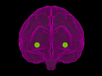
The medial amygdala (MeA) is a region of the brain that modulates innate social and non-social behaviors in several mammals, including humans.
The medial amygdala (MeA) is a region of the brain that modulates innate social and non-social behaviors in several mammals, including humans. Notedly sexually dimorphic, MeA neurons exhibit well-documented sex differences in anatomy, morphology and molecular characteristics. Recently, a pioneer study published in eNeuro from the Children’s National Hospital Center for Neuroscience Research has unveiled new information regarding physiological sex differences in MeA neurons, which, until now, has remained a missing piece in understanding how the MeA codes differently in males and females.
Previous research from Children’s National has shown that two subpopulations of MeA inhibitory output neurons descended from Dbx1 and Foxp2 transcription factors display different responses to innate olfactory cues and in a sex-specific manner. The newest study examines whether these transcription factor defined neurons also possess sex-specific biophysical signatures. The scientists posit that understanding how sex and lineage impact upstream differences at the neuronal level can help illuminate how the MeA processes information and codes for sex-specific behavioral differences.
Using whole-cell patch clamp recording and stepwise current injection, the researchers were able to analyze the intrinsic electrophysiological profiles of the two subclasses of MeA neurons in males and females in a pre-clinical model. Data revealed that the spike frequency of Dbx1-lineage and Foxp2-lineage neurons differed by lineage, sex and stimulus strength. Dbx1-lineage neurons in females discharged more spikes than those in males during high-amplitude current injection, while Foxp2-lineage neurons in females discharged more spikes than those in males during low-amplitude current injection. Across lineage, researchers observed that Dbx1-lineage neurons discharged more spikes than Foxp2-lineage neurons in females, but only at the highest amplitude stimulus, while Dbx1-lineage neurons spiked more than Foxp2-lineage neurons in males during low rather than high-amplitude current injection.
Different spiking patterns are generally indicative of different intrinsic cell properties. However, this study found that the intrinsic properties of the cell – such as membrane potential, resistance, and rheobase – were the same at rest across sex and lineage. The only significant difference was found in capacitance, an electrical measurement that roughly corresponds with cell size. Additionally, the study found that spike frequency adaptation correlated with neuronal lineage and sex, with males having a higher adaptation factor than females and Foxp2-lineage neurons displaying a higher adaptation factor than Dbx1-lineage neurons. In tandem, these results indicated that changes in the intrinsic properties were taking place during stimulation.
The researchers then used waveform phase-plots to visualize phases of the different action potentials and contrived an innovative new method of analyzing these quantitatively instead of solely qualitatively. This allowed them to know that broadly, ion channels that work with repolarization are likely different, and prompted them to focus on the family of ion channels that are known to modify the repolarization phase. From 62 candidate ion channels, the researchers chose 10 to investigate. Experiments ultimately revealed that only one ion channel was found to exhibit statistically significant sex differences in the Foxp2 population. This result indicated that molecular expression of these ion channels are likely driving differences in the physiology of the cells which may be the basis of behavioral expression. Future research topics include how and when sex hormones shape MeA neuronal firing properties and how this relates to network function.
“This is a small piece of contribution to the overall understanding of how the brain as a biological machine codes for different outputs,” says first author Heidi Y. Matos, Ph.D.
By showing sex differences in neural function, this research represents progress in understanding the biological underpinnings of a host of developmental disorders, particularly those diagnosed in different proportions between males and females. Autism spectrum disorders, for example, often have symptoms that manifest through social interaction, and understanding these disorders requires a better understanding of normal MeA physiology.
“In order to get to the why, we have to get to the how of that circuit,” says Dr. Matos.
Just as the brain harnesses the collective power of a diverse range of neurons, the Center for Neuroscience harnesses the aggregate talent of a diverse group of neuroscientists to produce innovative work. This study in particular champions diversity in the sciences, with more than half of the authors coming from underrepresented minorities, including Dr. Matos.
“I think this work is a shining example of the tremendous contributions that are made by neuroscientists from all backgrounds,” says principal investigator Joshua G. Corbin, Ph.D.
“Sex Differences in Biophysical Signatures across Molecularly Defined Medial Amygdala Neuronal Subpopulations” was published in eNeuro. Additional authors include David Hernandez-Pineda, Claire M. Charpentier, Allison Rusk and Kevin S. Jones, Ph.D.