Tracking environmental stress damage in the brain
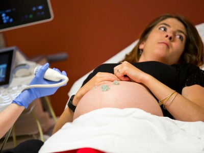
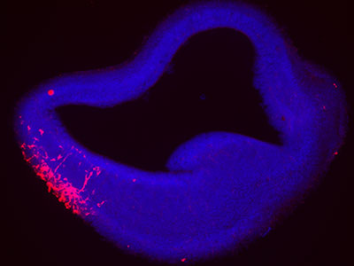 A team led by Children’s National developed a fluorescence reporter system in an experimental model that can single out neurons that have survived prenatal damage but remain vulnerable after birth. What’s knownWhen fetuses are exposed to environmental stressors, such as maternal smoking or alcohol consumption, radiation or too little oxygen, some of these cells can die. A portion of those that survive often have lingering damage and remain more susceptible to further environmental insults than healthy cells; however, researchers haven’t had a way to identify these weakened cells. This lack of knowledge has made it difficult to discover the mechanisms behind pathological brain development thought to arise from these very early environmental exposures, as well as ways to prevent or treat it. What’s newA team led by Kazue Hashimoto-Torii, Ph.D., a principal investigator in the Center for Neuroscience Research at Children’s National Health System, developed a marker that makes a protein known as Heat Shock Factor 1 glow red. This protein is produced in cells that become stressed through exposure to a variety of environmental insults. Gestation is a particularly vulnerable time for rapidly dividing nerve cells in the fetal brain. Tests showed that this marker worked not just on cells in petri dishes but also in an experimental model to detect brain cells that were damaged and remained vulnerable after exposure to a variety of different stressors. Tweaks to the system allowed the researchers to follow the progeny of cells that were affected by the initial stressor and track them as they divided and spread throughout the brain. By identifying which neurons are vulnerable, the study authors say, researchers eventually might be able to develop interventions that could slow or stop damage before symptoms arise. Questions for future researchQ: How do different environmental insults damage brain cells during gestation? Source: Torii, M., S. Masanori, Y.W. Chang, S. Ishii, S.G. Waxman, J.D. Kocsis, P. Rakic and K. Hashimoto-Torii. “Detection of vulnerable neurons damaged by environmental insults in utero.” Published Dec. 22, 2016 by Proceedings of the National Academy of Sciences. doi: 10.1073/pnas.1620641114
|