Tag Archive for: neurology

Daniel J. Licht, M.D.

Q&A with Daniel J. Licht, M.D.: The future of medicine is in light

Daniel J. Licht, M.D.

A pediatric neurologist who specializes in children with congenital heart disease, Dr. Licht initially came to this area of research as he considered ways to ensure children’s brains have adequate oxygen delivery during heart care, preserving neurological health and improving long-term outcomes.

Daniel J. Licht, M.D., joins Children’s National Hospital with a vision: He believes non-invasive devices built using biomedical optics – or instruments using light – can give clinicians invaluable information about how the brain and other organs are functioning.

A pediatric neurologist who specializes in children with congenital heart disease, Dr. Licht initially came to this area of research as he considered ways to ensure children’s brains have adequate oxygen delivery during heart care, preserving neurological health and improving long-term outcomes. He sees countless applications for using the properties of light in pediatric medicine.

Dr. Licht, whose name coincidentally also means “light” in German, is planning to establish a program for biomedical optics at Children’s National, built on the pillars of education, innovation and commercialization. He wants to tap into the resources of the Sheik Zayed Institute of Pediatric Surgical Innovation and expertise across the hospital. He is launching this effort as part of the new Center for Prenatal, Neonatal & Maternal Health Research.

Q: How can light be used diagnostically?

A: I believe that light is truly the future of biomedical devices, especially in children. Light can penetrate human tissues deeply, whether it’s muscle, liver or kidney. For example, you can put a light at the end of an endoscope and someday do virtual biopsies. It’s all a matter of understanding the properties of light, and how to manipulate light to give you the answers that you need. The applications are truly infinite.

Q: What has your initial work in neurology shown?

A: One of the instruments that we have developed can measure cerebral blood flow and quantitatively show the oxygen use of the brain. That’s important because it’s easy to measure oxygen delivery, but it’s hard to balance supply-and-demand without knowing the patient’s unique demand. We now have preclinical data and information from about 500 patients.

In terms of what’s ahead, many therapies today aren’t targeted to the individual, so Johnny’s brain-oxygen demand may not be the same as Sarah’s brain-oxygen demand, even if they both have congenital heart disease. As a patient waits for surgery, we also have found that the brain-oxygen demand increases, but if the demand is not met, this can lead to pre-operative brain injury. This technology could change the whole conversation about the timing of surgery. In addition, we can measure the brain-oxygen demand intraoperatively. We are finding that we can actually define the right perfusion strategy for each patient, rather than making uniform decisions for all patients with a shared diagnosis.

Lastly, beyond the operating room, we can use this technology for countless conditions. It would help with the treatment of almost any disease in the critical care unit, when we are using tools like ECMO (extracorporeal membrane oxygenation, a salvage technique), and we need to monitor a patient’s status. We can also use it to measure intracranial pressure. In very simple terms, if a child with a shunt comes into the emergency room with a headache, we can noninvasively measure the pressure and see how it’s changed without a head CT. We can decide who needs to go to the operating room – and who doesn’t – without radiation.

Q: How did your career bring you to this point?

A: My interest has always been in brain injury and kids with congenital heart disease. Years ago, I started out using MRI because it was the technology that was bright and shiny at the time. I was part of a team that developed an MRI sequence for measuring cerebral blood flow. We made some discoveries that indicated the culprit for brain injury was not the surgeries. Instead, there was something with the babies.

Unfortunately, with MRI, it’s a big, expensive instrument, and you have to take the baby to the machine for a single point-in-time measurement. So I started working with a physicist at the University of Pennsylvania to develop a way to measure the motion of particles, specifically red blood cells, to study cerebral blood flow. We found ways to use light, and this is what I hope to build and commercialize at Children’s National. By the end of my career, I hope to be able to say that we got this into clinical care.

U.S. News Badges

Children’s National Hospital ranked #5 in the nation on U.S. News & World Report’s Best Children’s Hospitals Honor Roll

U.S. News BadgesChildren’s National Hospital in Washington, D.C., was ranked #5 in the nation on the U.S. News & World Report 2023-24 Best Children’s Hospitals annual rankings. This marks the seventh straight year Children’s National has made the Honor Roll list. The Honor Roll is a distinction awarded to only 10 children’s hospitals nationwide.

For the thirteenth straight year, Children’s National also ranked in all 10 specialty services, with eight specialties ranked in the top 10 nationally. In addition, the hospital was ranked best in the Mid-Atlantic for neonatology, cancer, neurology and neurosurgery.

“Even from a team that is now a fixture on the list of the very best children’s hospitals in the nation, these results are phenomenal,” said Kurt Newman, M.D., president and chief executive officer of Children’s National. “It takes a ton of dedication and sacrifice to provide the best care anywhere and I could not be prouder of the team. Their commitment to excellence is in their DNA and will continue long after I retire as CEO later this month.”

“Congratulations to the entire Children’s National team on these truly incredible results. They leave me further humbled by the opportunity to lead this exceptional organization and contribute to its continued success,” said Michelle Riley-Brown, MHA, FACHE, who becomes the new president and CEO of Children’s National on July 1. “I am deeply committed to fostering a culture of collaboration, empowering our talented teams and charting a bold path forward to provide best in class pediatric care. Our focus will always remain on the kids.”

“I am incredibly proud of Kurt and the entire team. These rankings help families know that when they come to Children’s National, they’re receiving the best care available in the country,” said Horacio Rozanski, chair of the board of directors of Children’s National. “I’m confident that the organization’s next leader, Michelle Riley-Brown, will continue to ensure Children’s National is always a destination for excellent care.”

The annual rankings are the most comprehensive source of quality-related information on U.S. pediatric hospitals and recognizes the nation’s top 50 pediatric hospitals based on a scoring system developed by U.S. News.

“For 17 years, U.S. News has provided information to help parents of sick children and their doctors find the best children’s hospital to treat their illness or condition,” said Ben Harder, chief of health analysis and managing editor at U.S. News. “Children’s hospitals that are on the Honor Roll transcend in providing exceptional specialized care.”

The bulk of the score for each specialty service is based on quality and outcomes data. The process includes a survey of relevant specialists across the country, who are asked to list hospitals they believe provide the best care for patients with the most complex conditions.

The eight Children’s National specialty services that U.S. News ranked in the top 10 nationally are:

The other two specialties ranked among the top 50 were cardiology and heart surgery, and urology.

Illustration of brain and brainwaves

Risk factors for pharmacoresistant pediatric epilepsy

Illustration of brain and brainwaves

New study evaluates risk factors for the timing and development of drug-resistant pediatric epilepsy.

Focal cortical dysplasia (FCD) is the most common cause of surgically-treatable epilepsy in children. In a new study published in Neurology, researchers evaluated 143 children with confirmed FCD risk factors for the timing and development of pharmacoresistant epilepsy.

What this means

The current definition of pharmacoresistance requires failure of two appropriately-dosed and selected antiseizure medications before being able to be considered for epilepsy surgery.

“We found that the failure of just one antiseizure medication is associated with an enormous increased incidence and earlier development of pharmacoresistance,” says Nathan Cohen, M.D., neurologist at Children’s National and lead author of the study. “Our data supports the redefinition of pharmacoresistant epilepsy to the failure of just one antiseizure medication in this population, which would potentially allow these patients to benefit from earlier curative surgery.”

Why it matters

The findings showed that in children with FCD the failure of just one antiseizure medication is associated with an enormous risk and earlier incidence of pharmacoresistance. Therefore, the authors advocate for its redefinition in FCD-related epilepsy to the failure of just one antiseizure medication.

“This will allow children to be considered much earlier for potentially curative epilepsy surgery,” adds Dr. Cohen. “We find that the majority of FCD patients develop epilepsy and that the majority of those with epilepsy develop pharmacoresistance.”

In a multivariate analysis, the authors show that the FCD cortical lobar location, pathologic subtype, and age of seizure onset are not important factors in the development of pharmacoresistance.

What’s next

This data supports operational re-definition of pharmacoresistance for surgical planning in FCD-related epilepsy to the failure of one antiseizure medication, and support early, potentially curative surgery to improve outcomes in this patient population.

You can read the full study, Prevalence and Risk Factors for Pharmacoresistance in Children With Focal Cortical Dysplasia–Related Epilepsy, in Neurology.

Digital background depicting innovative technologies in (AI) artificial systems, neural interfaces and internet machine learning technologies

AI algorithm that detects brain abnormalities could help cure epilepsy

Digital background depicting innovative technologies in (AI) artificial systems, neural interfaces and internet machine learning technologies

A new AI algorithm can detect subtle brain abnormalities that cause epileptic seizures.

An artificial intelligence (AI) algorithm that can detect subtle brain abnormalities that cause epileptic seizures has been developed by a UCL-led team of international researchers, including Children’s National Hospital.

To do this, the team quantified features from MRI scans, such as how thick or folded the brain was at nearly 300,000 locations in each case.

They then trained the AI algorithm using examples labelled by expert radiologists as either a healthy brain or one with focal cortical dysplasia (FCD) based on their patterns and features.

The results, published in Brain, showed that in the main cohort of 538 patients, the algorithm was able to detect the FCD in 67% of cases.

“We put an emphasis on creating an AI algorithm that was interpretable and could help doctors make decisions. Showing doctors how the Multicentre Epilepsy Lesion Detection project (MELD) algorithm made its predictions was an essential part of that process,” said Mathilde Ripart, research assistant at UCL and the study’s co-first author.

Around 1% of the population have epilepsy and, of these, 20-30% do not respond to medications.

“We are excited to collaborate with MELD on ways to improve the treatment of pharmacoresistant epilepsy,” said Nathan Cohen, M.D., neurologist at Children’s National Hospital and co-author of the study. “This advanced imaging platform is open source and demonstrates the benefit of team science at the broadest scale.”

In children who have had surgery to control their epilepsy, FCD is the most common cause, and in adults it is the third most common cause.

Additionally, of patients who have epilepsy that have an abnormality in the brain that cannot be found on MRI scans, FCD is the most common cause.

You can read the full UCL press release here.

US News Badges

Children’s National named to U.S. News & World Report’s Best Children’s Hospitals Honor Roll

US News BadgesChildren’s National Hospital in Washington, D.C., was ranked No. 5 nationally in the U.S. News & World Report 2022-23 Best Children’s Hospitals annual rankings. This marks the sixth straight year Children’s National has made the list, which ranks the top 10 children’s hospitals nationwide. In addition, its neonatology program, which provides newborn intensive care, ranked No.1 among all children’s hospitals for the sixth year in a row.

For the twelfth straight year, Children’s National also ranked in all 10 specialty services, with seven specialties ranked in the top 10.

“In any year, it would take an incredible team to earn a number 5 in the nation ranking. This year, our team performed at the very highest levels, all while facing incredible challenges, including the ongoing pandemic, national workforce shortages and enormous stress,” said Kurt Newman, M.D., president and chief executive officer of Children’s National. “I could not be prouder of every member of our organization who maintained a commitment to our mission. Through their resilience, Children’s National continued to provide outstanding care families.”

“Choosing the right hospital for a sick child is a critical decision for many parents,” said Ben Harder, chief of health analysis and managing editor at U.S. News. “The Best Children’s Hospitals rankings spotlight hospitals that excel in specialized care.”

The annual rankings are the most comprehensive source of quality-related information on U.S. pediatric hospitals and recognizes the nation’s top 50 pediatric hospitals based on a scoring system developed by U.S. News.

The bulk of the score for each specialty service is based on quality and outcomes data. The process includes a survey of relevant specialists across the country, who are asked to list hospitals they believe provide the best care for patients with the most complex conditions.

The seven Children’s National specialty services that U.S. News ranked in the top 10 nationally are:

The other three specialties ranked among the top 50 were cardiology and heart surgerygastroenterology and gastro-intestinal surgery, and urology.

illustration of the brain

LIFU successfully delivers targeted therapies past the blood-brain barrier

illustration of the brain

LIFU offers doctors the first opportunity to open the blood-brain barrier and treat the entire malignant brain tumor.

Children’s National Hospital will leverage low-intensity focused ultrasound (LIFU) to deliver therapy directly to a child’s high-grade glioma. The approach offers doctors the first opportunity to open the blood-brain barrier and treat the entire malignant brain tumor.

Children’s National will be the first hospital in the U.S. to treat high-grade pediatric brain tumors with LIFU to disrupt the blood-brain barrier. Crossing it has been a major hurdle for effective therapy. The barrier, a network of blood vessels and tissue, prevents harmful substances from reaching the brain but also stops molecular targeted therapy and immunotherapy from getting into the tumor site and staying there.

“LIFU gives us a way to potentially transiently open up the barrier, so we can deliver novel therapy directly to the tumor and improve the likelihood of survival,” said Roger Packer, M.D., senior vice president of the Center for Neurosciences and Behavioral Medicine at Children’s National. “It is the greatest breakthrough we’ve potentially had in the past 50 years or more for the management of these tumors. We made great strides in our understanding of molecular genetics and the molecular drivers of tumors, but we have not yet translated that knowledge into better therapies; this may be our most effective mechanism to overcome the barrier.”

In 2020, Children’s National was recognized as the first worldwide Center of Excellence by the Focused Ultrasound Foundation.

Focused ultrasound (FUS) is a non-invasive therapeutic technology with the potential to transform the treatment of many medical disorders by using ultrasonic thermal energy to specifically target tissue deep in the body. The technology can treat without incisions or the need of radiation.

How it works

Doctors at Children’s National will be using LIFU in two different types of procedures:

  • 5-ALA: Doctors will give the patient 5-aminolevulinic acid (5-ALA) with the LIFU treatment. 5-ALA enters rapidly dividing cells and is activated by the ultrasound to a state where it kills the dividing cells of the tumor. The surrounding normal brain cells around the tumor are not dividing, so they do not take up the 5-ALA and are left unharmed after ultrasound therapy.
  • Microbubbles: While receiving different doses of LIFU over a one- to two-hour period, the patient is given “microbubbles,” which are widely used in medical imaging and as carriers for targeted drug delivery. These microbubbles bounce around against the walls like seltzer, opening the blood vessels and transiently opening that space.

Both studies are the first in the world for pediatric gliomas of the brain stem, allowing experts to treat patients 4-6 weeks after radiotherapy. The patient then receives medication orally or intravenously as it passes through the bloodstream. It does not go at high levels anywhere within the brain except where the blood-brain-barrier was opened, allowing oral medication or immune therapies to rush into the tumor.

The launch of this program comes a few months after the hospital successfully performed the first-ever high-intensity focused ultrasound surgery on a pediatric patient with neurofibromatosis.

Watch this video to learn more.

model of the brain

Treating newborns with hemimegalencephaly by inducing strokes

model of the brain

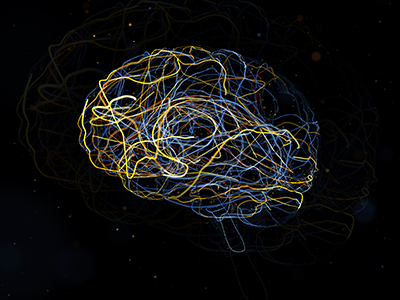
Experts at Children’s National Hospital have pioneered a novel approach using controlled strokes to stop seizures and improve neurodevelopmental outcomes in newborns under three months born with hemimegalencephaly (HME). They now consider it their new standard of care for babies in this age group with HME and refractory epilepsy.

Asking a physician to induce strokes in newborns is asking her to do something contrary to her training. But over the past eight years, experts at Children’s National Hospital have pioneered a novel approach using controlled strokes to stop seizures and improve neurodevelopmental outcomes in newborns under three months born with hemimegalencephaly (HME). They now consider it their new standard of care for babies in this age group with HME and refractory epilepsy.

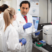
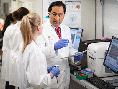
“We have demonstrated the ability to intervene and stop the intractable seizures during a critical time of neurodevelopment in which no other effective medical or surgical option exists. That is extremely rewarding,” said Monica Pearl, M.D., director of the Neurointerventional Radiology Program at Children’s National. Children’s National is the only center in the world currently offering this treatment. A multi-disciplinary team led by Dr. Pearl; Taeun Chang, M.D., director of the Neonatal Neurology and Neonatal Neurocritical Care Program; neurophysiologist and neonatal neurologist Tammy Tsuchida, M.D., Ph.D.; and other experts has now successfully treated seven patients using this minimally-invasive approach.

“We want patients and providers to understand this is a better alternative to a delayed hemispherectomy, the standard of care currently offered to newborns with HME,” said Dr. Chang.

The best treatment for newborns with hemimegalencephaly

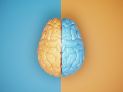
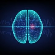
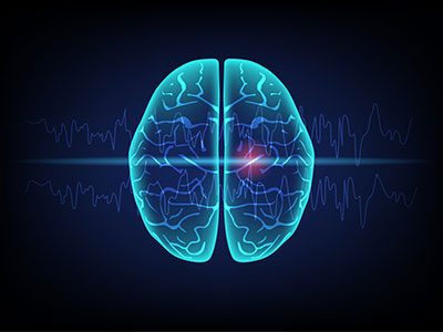
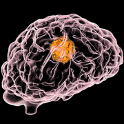
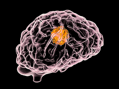
HME, a rare congenital condition occurring in a handful of newborns each year, is characterized by abnormal growth and enlargement of half of the brain which leads to intractable seizures. The seizures often result in severe cognitive delays and hemiparesis. The standard treatment is an anatomic hemispherectomy — surgical removal of the affected half of the brain, allowing the remaining half of the brain to develop and function without constant seizures.

Such a large and complex surgery poses serious risks for infants younger than three months, leaving doctors with the difficult choice to delay surgery until these newborns grow bigger and stronger, even as they are experiencing seizures. These persistent seizures compromise the development of the healthy half of the brain. One study reports as much as a drop of 10 to 20 IQ points with each month’s delay in surgical hemispherectomy.

“I was willing to consider performing these procedures because there is a clear, unmet medical need and these babies are in dire circumstances,” Dr. Pearl said. “Waiting for curative hemispherectomy means more than just lost time; uncontrolled seizures and anti-seizure medications have detrimental effects on the ‘normal,’ unaffected parts of the brain. We needed a better option for these patients.” Dr. Pearl said that complete embolization of the affected hemisphere as both primary and definitive treatment had never been described. They could only find one example in the literature – a paper from 1995 – suggesting embolization as an adjunct to surgery, and nothing suggesting it as a primary modality.

About the care received

Dr. Pearl is one of only a handful of dedicated pediatric neurointerventionalists across the country with neurovascular expertise in people of all ages, in particular neonates and young infants. For these procedures to be performed safely, the neurointerventionalist must be proficient in obtaining femoral arterial access and navigating small caliber cervicocerebral blood vessels that are less than one millimeter in diameter.

Additionally, one needs a neonatal neurocritical care service and NICU that can medically manage large strokes and their potential complications in newborns. Dr. Chang has developed a specialized protocol based on decades of managing strokes and other acute brain injuries in newborns. She created the neonatal neurocritical care service at Children’s National, the only one in the region and the largest in the world.

“Our teams are fortunate in that we each respectively have extensive prior experience in treating and managing neonates and very young infants for various cerebrovascular disorders,” Dr. Pearl said. “We relied on this collective experience to make this hemispheric embolization pathway possible.”

How it happens

To perform the embolizations, Dr. Chang and her team first optimizes control of the seizures using medications. Dr. Pearl places a sheath in the femoral artery using ultrasound guidance – a delicate task in a neonate whose femoral artery diameter is only two to three millimeters. She then navigates a catheter up the aorta and selects the targeted carotid artery using radiographic guidance. What follows is a set of intricate navigations to direct the microcatheter through small blood vessels in the brain, often less than one millimeter.

Using x-ray guidance, Dr. Pearl injects contrast through the microcatheter to visualize the arterial anatomy and advance the microcatheter into position for embolization. She uses glue that hardens when exposed to blood, blocking off the blood supply to the seizure-inducing areas. The process is repeated until the blood supply to the entire affected hemisphere is occluded. Meanwhile, Dr. Chang and her team monitor the brain’s electrical activity using an electroencephalogram (EEG) to watch how the brain responds to each stroke. The surgical epilepsy, neonatal neurocritical care and neonatology teams are all in constant communication throughout the procedure.

Together, they have to contend with the same symptoms patients have immediately following a stroke, most notably brain swelling that can cause bleeding and herniation. The resultant brain swelling is complicated further by the already enlarged hemisphere of the brain. Using neuroprotective strategies learned from treating over a thousand newborns with perinatal brain injury, Dr. Chang and her team and the NICU coordinate to minimize brain swelling and protect the healthy half of the brain by tightly controlling the brain temperature, glucose, sodium levels, and blood pressure. Over the course of a few weeks, Dr. Pearl performs three to four embolization sessions to halt blood supply to the seizing half of the brain.

“The risks of intracranial vasospasm and hemorrhage during embolization are higher in this distinct group of patients compared to other neonates requiring embolization, such as in vein of Galen malformations. These events must be controlled immediately to prevent complications and I know I only have seconds to react,” Dr. Pearl said.

“Here, we have the cultivation of brain-centric neonatal care, a large level IV tertiary NICU with expertise in keeping critically ill babies alive and rare pediatric neurologic subspecialists like Dr. Pearl and myself. All of this is what makes this level of innovation possible,” Dr. Chang said. Now, they wish this minimally invasive approach to be available to all newborns with HME and refractory epilepsy.

“This is not a fluke. This is not a one-time thing. Our team at Children’s National has been perfecting this method for close to a decade,” Dr. Chang said. As for proof, her answer is clear.

illustration of brain tumor

International initiative aims to find rare brain tumor treatments

illustration of brain tumor

Rare brain tumors are not as well characterized due to the paucity of biological and clinical data available.

Certain brain tumors can be hard to diagnose. And as such, that makes it complicated to find a treatment.

In an effort to identify and tailor treatments to patients with rare brain tumors, Children’s National is launching a rare brain tumor initiative. The hospital is collaborating with other hospitals in North America, South America and Europe to compile a registry of children diagnosed with rare brain tumors. The registry will collect biospecimens, clinical and radiological data from patients diagnosed with certain rare brain tumors.

The goal is to find a correlation between the molecular findings and the clinical findings to categorize them. This will help doctors get different prognosticators or different treatment approaches.

Here, Adriana Fonseca Sheridan, M.D., pediatric neuro-oncologist at Children’s National Hospital, tells us more about this international initiative.

What’s been the hold-up in the field?

The recent incorporation of molecular features as part of the diagnostic criteria and classification of brain tumors highlighted a high biological and molecular heterogeneity within previously histologically defined entities. The improvement in our diagnostic capabilities have been incredibly useful to stratify patients into different disease-specific risk groups and tailor therapeutic approaches accordingly in the most common brain tumors. In contrast, rare brain tumors are not as well characterized due to the paucity of biological and clinical data available. Additionally, newly molecularly defined entities lack specific clinical and therapeutic data and represent a major challenge to patients and doctors alike.

How does this work move the field forward?

The overarching objective of the international rare brain tumor registry is to deepen our understanding of the biological underpinnings of rare brain tumors. The registry also seeks to create infrastructure that allows for development of rational and personalized treatment strategies for children with rare entities.

What are you hoping to discover?

We hope to characterize the clinicopathological features and identify risk factors for survival and optimal therapeutic approaches of:

  • CNS sarcomas
  • BCOR-ITD tumors
  • Astroblastoma/MN1 altered tumors
  • Histologically ambiguous/unclassifiable brain tumors

How unique is this work?

Children’s National will spearhead the development of this initiative and lead an effort to prospectively collect biological specimens, radiological and clinical data that allow us to better understand the biologic mechanisms and therapeutic susceptibilities of these rare diseases.

We know that the best way to lead the advancement of the field in rare diseases is through collaboration. Therefore, we will synchronize efforts and collaborate with our European colleagues. They will be running a similar initiative. Our goal is to generate meaningful and robust data that will allow us to better understand how to successfully treat patients with these rare brain tumors across the globe.

mitochondria

Grant funds study of two maternally inherited mitochondrial diseases

mitochondria

The National Institutes of Health awarded George Washington University and Children’s National Hospital a grant to study two maternally inherited mitochondrial diseases.

The National Institutes of Health awarded George Washington University and Children’s National Hospital a grant to study two maternally inherited mitochondrial diseases. Andrea Gropman, M.D., division chief of Neurodevelopmental Pediatrics and Neurogenetics at Children’s National, along with her co-investigator, Anne Chiaramello, M.D., from the George Washington University School of Medicine, will lead the study.

The proposed studies focus on two ultra-rare maternally inherited mitochondrial diseases:

  • Mitochondrial Encephalopathy, Lactic Acidosis and Stroke-like episodes (MELAS); and
  • Leber’s Hereditary Optic Neuropathy-Plus (LHON-Plus).

Both diseases are among those studied by the Rare Diseases Clinical Research Network.

“We are really pleased to be able to change the landscape for MELAS and LHON, two mitochondrial disorders with relentless progression and no treatment,” Dr. Gropman said. “This grant represents the fruition of an eight-year collaboration with my colleague Dr. Chiaramello and we are fortunate to be able to deliver this at Children’s National and serve our patients and community.”

Because patients currently do not have access to effective therapeutic intervention, this results in significant disability, morbidity and premature death. The UG3 phase of the study will focus on translational MELAS and LHON-Plus studies and submission of an IND protocol to the Food and Drug Administration. The UH3 phase will focus on a basket clinical trial with MELAS and LHON-Plus to:

  • Provide proof-of-concept that the basket design can be applied to divergent ultra-rare diseases.
  • Advance the dataset for safety and pharmacokinetics/pharmacodynamics of our lead compound for a larger number of patients than in a conventional clinical trial setting.
  • Gather outcomes and practical information for optimizing the design of future basket clinical trial.

“Dr. Gropman is dedicated to giving children with MELAS the very best care,” said Elizabeth Wells, M.D., vice president of Neuroscience and Behavioral Medicine Center at Children’s National. “This new research funding is exciting and means more patients can benefit from the expertise she has developed at Children’s National.”

brain network illustration

Changing the surgical evaluation of epilepsy

brain network illustrationThe choice between stereoelectroencephalography (SEEG) and subdural evaluation is not mutually exclusive, according to a new opinion piece published in JAMA Neurology.

In their article, Chima Oluigbo, M.D., pediatric epilepsy neurosurgeon, William D. Gaillard, M.D., division chief of Epilepsy and Neurophysiology and Neurology, both at Children’s National Hospital, and Mohamad Z. Koubeissi, M.D., M.A., from The George Washington University Hospital, discuss how the practicing epileptologist requires a profound understanding of the roles of different technologies. It also looks at how to integrate both traditional and emerging paradigms to optimize seizure control. This issue is particularly relevant to choosing the best method of invasive intracranial electroencephalography monitoring in individual cases.

Noting that despite the dramatic increase in SEEG use in recent years, the authors talk about how many patients still benefit from invasive monitoring using subdural grids. Therefore, it is important to define the considerations that should guide decision-making on the choice of SEEG versus subdural monitoring in each patient. The authors expand on their statement explaining that it is critical to define the roles of SEEG vs subdural grid investigation in each patient as subdural grid evaluations are still indicated in specific circumstances.

Additionally combined hybrid deployment of both techniques may be indicated in specific situations. Accommodation should be made to allow customization of the technique chosen to available technical expertise and equipment as well as patient preference.

2021 neurology infographic

2021 at a glance: Neurology and Neurosurgery at Children’s National

2021 neurology infographic

MRI Room

Children’s National uses HIFU to perform first ever non-invasive brain tumor procedure

MRI Room

Children’s National Hospital successfully performed the first-ever high-intensity focused ultrasound (HIFU) procedure on a pediatric patient with neurofibromatosis (NF). This is the youngest patient to undergo HIFU treatment in the world. Image provided by Insightec.

Children’s National Hospital successfully performed the first-ever high-intensity focused ultrasound (HIFU) procedure on a pediatric patient with neurofibromatosis (NF). This is the youngest patient to undergo HIFU treatment in the world. The advancement of children’s medical devices in the U.S. continues to significantly lag behind adult devices. This is why this milestone marks a significant advance in making pediatric surgery more precise and less invasive.

The hospital is offering this treatment to patients under an ongoing research clinical trial. Children’s National is one of the first pediatric hospitals in the nation to use HIFU for neuro-oncology patients. It’s also the first hospital in the world to use it to treat a pediatric patient with NF. NF is a condition that occurs in approximately 1 in 3,500 births and causes tumors to form in the brain, spinal cord and nerves.

“Using HIFU to treat our pediatric patients is a quantum leap towards non-invasive surgery for kids,” said Robert Keating, M.D., division chief of Neurosurgery and co-director of the HIFU program at Children’s National. “It’s exciting because the future is now here and it’s significantly better for our kids, in terms of non-invasive surgery with lower risk of complications and no exposure to radiation.”

Focused ultrasound (FUS) is a non-invasive therapeutic technology with the potential to transform the treatment of many medical disorders by using ultrasonic thermal energy to specifically target tissue deep in the body. The technology can treat without incisions or the need of radiation.

FUS, which has been used for adult clinical trials for many decades, can be delivered through high- or low-intensity focused ultrasound (LIFU). HIFU uses non-invasive therapy that uses focused ultrasound waves to thermally ablate a focal area of tissue. Children’s National will now use HIFU to treat low-grade type tumors located in difficult locations of the brain, such as hypothalamic hamartomas and pilocytic astrocytoma, as well as for movement disorders and epilepsy.

An alternative approach, LIFU uses lower levels of energy to disrupt the blood-brain barrier. Unlike medications, which often have difficulty crossing the blood-brain barrier, LIFU can transiently open the blood-brain barrier to chemotherapy. This may allow more effective treatment of tumors and offer opportunities to treat, for the first time, the entire extent of a malignant brain tumor.

“Having focused ultrasound technology as a tool and conducting clinical trials will allow our neurologists and oncologists to offer a non-invasive treatment option to many patients who suffer from neurological conditions,” said Hasan Syed, M.D., co-director of the HIFU program at Children’s National. “The milestone of performing this first HIFU procedure will lead the way to better understanding of the effect of this technology and provide patients with more options.”

At Children’s National, the HIFU program is being led by Dr. Keating and a multidisciplinary team, including clinicians and investigators from the Sheik Zayed Institute for Pediatric Innovationradiologyoncologysurgery and orthopedics. In an effort to collaborate with the region’s adult hospitals, Children’s National will also treat adult patients on a selective basis who have movement disorders such as essential tremor and Parkinson’s. There is a scarcity of similar resources in the metro region. Many adult patients face one-year wait periods for treatment of their movement disorders, requiring many to travel out of state for treatment.

The LIFU program is scheduled to be operational in 2022. It will likely be the first in the U.S. to treat high-grade pediatric brain tumors with disruption of the blood-brain barrier and provide more effective routes for chemotherapy as well as potential immunotherapy and molecular approaches.

“The use of LIFU with microbubbles to open up the blood-brain barrier is an exciting, potentially game-changing approach for children with these tumors,” said Roger Packer, M.D., senior vice president of the Center for Neurosciences and Behavioral Medicine at Children’s National. “It should safely allow the blood-brain barrier to open and allow delivery of potentially life-saving personalized therapy to the tumor and spare the rest of the brain. It is the most exciting, new development in brain tumor therapy for these malignant midline tumors in the past 50 years.”

Children’s National continues to be a leader in pediatric HIFU use. In 2015, Children’s National doctors became the first in the U.S. to use MR-HIFU to treat pediatric osteoid osteoma – a benign, but painful bone tumor. Successful clinical trial results led to FDA approval in early 2021 for the use of the technology for this treatment. In 2020, the Focused Ultrasound Foundation also designated Children’s National as the first global pediatric Center of Excellence for using this technology to help patients with specific types of childhood tumors.

boy with headache

Kids’ headaches can be disruptive. We need solutions.

Experts leading the Headache Program at Children’s National Hospital recognize how common these disorders are. They also know how disruptive they can be in the day-to-day of children.

Marc DiSabella, D.O., is the director of the program. He is currently leading five pediatric headache trials. In this Q&A, he tells us about the ongoing trials, offering insight into innovative solutions and how he’s carving a new path to improve the quality of life of his patients.

Q: How has your team advised other neurologists on innovative care for patients with headaches that have been refractory to medicines?

A: We receive referrals from outside institutions when they need additional input for diagnostic and management options. We receive patient consult requests from around the country – and sometimes out of the country – to help improve symptoms. In most instances, these headaches tend to be difficult to control and do not respond to available medications. We really try to take a holistic approach to their care, and use treatments in parallel. For example, diagnostic, lifestyle techniques, medications, pain focused cognitive behavioral therapy and physical therapy. We also use complementary medicine as needed, such as acupuncture, injections and infusions.

Q: It is unusual for neurology divisions to run multiple pediatric trials focused on headaches. You are currently leading five that are open. How does this work move the field forward?

A: The medications we offer through our trials allow us to offer treatments that would otherwise not be available to pediatric patients. We do this in hopes of providing them relief while advancing the field. We are hopeful that these new therapies are as effective in pediatrics as they have shown to be in adults. But it is necessary to complete randomized clinical trials to prove this is the case. Historically, pediatric patients in clinical trials investigating painful conditions like migraines have had a disproportionately high placebo response rate. This means even the patients receiving a benign placebo have a high chance of symptom improvement. The newer medications show much better tolerability to the drugs used historically.

Q: What excites you about this work?

A: Pediatric pain disorders are unbelievably gratifying to treat because we take a mysterious disorder that waxes and wanes with no clear reason and give patients back control of their lives. It is extremely frustrating for a patient and their family to know that their day-to-day life can be abruptly derailed by a pain crisis. We work to provide them with several tools they can use daily to take back their lives.

Q: How is this work unique?

A: Our program was created organically over the years through our experiences with our patients. First, we noticed the disruption to patients’ personal and school performance from having untreated pain and recognized the need for pain psychology. Then, we expanded to have physical therapy to recondition patients and perform desensitization. Finally, we recognized our patients need additional medication options not offered through the standard of care. So, we expanded to open our various clinical trials, including those with pharma and internal protocols. As a result, we incorporated the use of Botox injections, for example, and soon will use a novel remote electro-neuromodulatory device.

Dr. Javad Nazarian

Q&A with Dr. Javad Nazarian on his upcoming work on low-grade gliomas

Dr. Javad Nazarian

Supported by the Gilbert Family Foundation, Dr. Nazarian’s return is part of a special research program within the Gilbert Family Neurofibromatosis Institute that focuses on NF1 research.

Javad Nazarian, Ph.D., M.Sc., associate professor of Pediatrics at George Washington University and professor at the University of Zurich, has expanded his research group at Children’s National to focus on Neurofibromatosis type 1 (NF1) transformed low-grade gliomas (LGGs). Dr. Nazarian will apply his expertise from establishing a successful DIPG (diffuse intrinsic pontine glioma) and DMG (diffuse midline glioma) program in Zurich Switzerland and previously at Children’s National.

In addition to his continued research in Zurich, as a principal investigator at the Department of Genomics and Precision Medicine at Children’s National Dr. Nazarian plans on aggregating his knowledge to the new research and work spearheaded at Children’s National. As one of the first research teams to move to the Children’s National Research & Innovation Campus, Dr. Nazarian’s group is excited to use the opportunity to establish cutting-edge and clinically translational platforms.

Supported by the Gilbert Family Foundation, Dr. Nazarian’s return is part of a special research program within the Gilbert Family Neurofibromatosis Institute that focuses on NF1 research. This research includes associated gliomas with a special emphasis on NF1-associated transformed anaplastic LGGs. His team will develop new avenues of research into childhood and young adult NF-associated LGGs with a special emphasis on transformed high-grade gliomas.

Dr. Nazarian is excited for what’s to come and his goals are clear and set. Here, Dr. Nazarian tells us more about his main objectives and what it means for the future of pediatric neuro-oncology care at Children’s National.

  1. What excites you most about being back at Children’s National?

I have received most of my training at Children’s National, so this is home for me. Being one of the nation’s top children’s hospitals gives a unique advantage and ability to advocate for childhood diseases and cancers. It is always exciting to play a part in the vision of Children’s National.

  1. What are some of the lessons learned during your time working in Zurich? And how do you think these will compliment your work at Children’s National?

We developed a focused group with basic research activities intertwined with clinical needs.  The result was the launch of two clinical trials. I also helped in developing the Diffuse Midline Glioma-Adaptive Combinatory Trial (DMG-ACT) working group that spans across the world with over 18-member institutions that will help to design the next generation clinical trials. I will continue leading the research component of these efforts, which will have a positive impact on our research activities at Children’s National.

  1. How does your work focusing on low-grade gliomas formulating into high-grade gliomas expand and place Children’s National as a leader in the field?

Scientifically speaking, transformed LLGs are very intriguing. I became interested in the field because these tumors share molecular signatures similar to high-grade gliomas (HGGs). Our team has done a great job at Children’s National to develop tools – including biorepositories, avatar models, drug screening platforms, focused working groups, etc. – for HGGs. We will apply the same model to transformed LGGs with the goal of developing biology-derived clinical therapeutics for this patient population.

  1. How will this work support families and patients seeking specific neuro-oncology care?

We will develop new and high thruput tools so that we can better study cancer formation or transformation. These tools and platforms will allow us to screen candidate drugs that will be clinically effective. The main focus is to accelerate discovery, push drugs to the clinic, feed information back to the lab from clinical and subsequently design better therapies.

  1. You are one of the first scientists to move to the Children’s National Research & Innovation Campus. What are some of the valuable changes or advancements you hope to see as a result of the move?

The campus will provide high-end facilities, including cutting-edge preclinical space, and allow for team expansion. The close proximity to Virginia Tech will also provide an environment for cross-discipline interactions.

  1. Anything else you think peers in your field should know about you, the field or our program?

The team at Children’s National includes Drs. Roger Packer and Miriam Bornhorst. Both have provided constant clinical support, innovation and clinical translation of our findings. I look forward to working with them.

brain network illustration

Cardiopulmonary bypass may cause significant changes to developing brain and nerve cells

brain network illustration

Cardiopulmonary bypass, more commonly known as heart-and-lung bypass, has some unique impacts on the creation and growth of brain cells in the area of a child’s brain called the subventricular zone (SVZ), according to a study in the Annals of Neurology. The SVZ is a critical area for the growth and migration of neurons and nerve cells called neuroblasts, both of which ultimately contribute to the proper development of key brain structures and functions during the early years of life.

The findings, from a study conducted in the Cardiac Surgery Research Laboratory at Children’s National Hospital, provide new insight into the cellular impacts of the cardiopulmonary bypass machine on brain growth and development for newborn infants with congenital heart disease. They will have an important role in the refinement of strategies to help protect the fragile brains of children who require lifesaving cardiac surgery with cardiopulmonary bypass immediately after birth.

Specifically, the research team found that during cardiopulmonary bypass:

  • Creation of neurons (neurogenesis) in the neonatal and infant subventricular zone is altered.
  • Migration of nerve cells, called neuroblasts, to the frontal lobe is potentially disrupted.
  • Changes to the growth and movement of neurons in the SVZ are prolonged.
  • Cortical development and expansion is impaired.
  • Specific types of neurons found only in the brain and spinal cord, called interneurons, are also affected.

The study uses an innovative pre-clinical model of the developing brain that is more anatomically and physiologically similar to human neonates and infants than those used in prior studies and in most neurological laboratory-based research.

Cardiopulmonary bypass is one of several key factors thought to cause children with congenital heart disease to sometimes demonstrate delays in the development of cognitive and motor skills. These disabilities often persist into adolescence and adulthood and can ultimately represent long-term neurocognitive disabilities. It is also believed that genetic factors, abnormal blood flow to the brain while in utero or low cardiac output after surgical procedures on the heart may contribute to these challenges.

“Unraveling cellular and molecular events during surgery using this preclinical model will allow us to design therapeutic approaches that can be restorative or reparative to the neurogenic potential of the neuronal stem precursor cells found in the subventricular zone of the neonatal or infant brain,” says Nobuyuki Ishibashi. M.D., Foglia-Hills Professor of Pediatric Cardiac Research, director of the Cardiac Surgery Research Laboratory at Children’s National and senior author on the study. “In particular, previous studies in our laboratory have shown improvement in the neurogenic activities of these precursor cells when they are treated with mesenchymal stromal cells (MSCs).”

The findings from this study further support the work already underway in the NIH-funded MeDCaP clinical trial for neonates and infants undergoing cardiac surgery using the cardiopulmonary bypass machine. That trial uses the heart and lung machine itself to deliver MSCs directly into the main arteries that carry blood to the brain.

microglia cells damage the myelin sheath of neuron axons

Katrina Adams, Ph.D., awarded fellowship to help restore functions in MS patients

microglia cells damage the myelin sheath of neuron axons

Multiple sclerosis is a demyelinating disease in which the insulating covers of nerve cells are damaged. Microglia cells (orange) attack the oligodendrocytes that form the insulating myelin sheath around neuron axons, leading to the destruction of the myelin sheath and to the loss of nerve function.

For her contributions to Multiple Sclerosis (MS) research, Katrina Adams, Ph.D., postdoctoral researcher at Children’s National Hospital, received the career transition fellowship from The National Multiple Sclerosis Society. The $600,000 fellowship will support a two-year period of advanced postdoctoral training in MS research and the first three years of research support in a new faculty appointment.

MS symptoms, including vision loss, pain, fatigue and reduced motor coordination, result from the demyelination of neuronal axons that transport critical information across the brain and spinal cord. Demyelination is the loss of myelin protein, which is normally produced by oligodendrocyte cells.

In the healthy brain, oligodendrocytes repair demyelinated areas by replacing damaged or lost myelin in a process called remyelination. Recent evidence has shown that oligodendrocytes display differences in their molecular and functional properties. One source of new oligodendrocytes in the adult brain is neural stem cells, which have been shown to generate oligodendrocytes that contribute to remyelination.

“The goal of this project is to determine whether neural stem cell-derived oligodendrocytes are distinct from other oligodendrocytes, both in the healthy brain and in MS,” said Adams. “I aim to understand the molecular mechanisms that regulate generation of oligodendrocytes from neural stem cells, with the goal of identifying signals that could be targeted in MS patients to promote remyelination.”

Remyelination is very limited in MS patients and current therapies for MS have very little impact on promoting remyelination.

This study will take advantage of the state-of-the-art facilities for single-cell analysis, transcriptomics, microscopy, and animal research in Children’s Research Institute at Children’s National. Adams also added that her postdoctoral mentor, Vittorio Gallo, Ph.D., interim chief academic officer and interim director of the Children’s National Research Institute, and principal investigator for the DC-IDDRC, has renowned expertise in glial biology, animal models of MS and white matter injury.

“This research will be the first to directly compare neural stem cell-derived oligodendrocytes with other resident oligodendrocytes in MS brain samples,” said Adams. “The results of this study will provide critical insight into the role that neural stem cells play in repair of MS demyelinated lesions.”

Adams received her doctorate in molecular biology from the University of California, Los Angeles where she used pluripotent stem cells to study motor neuron development. She currently investigates signaling pathways that regulate neural stem and progenitor cell maintenance and differentiation in the developing postnatal and adult brain, with a focus on the Endothelin-1 pathway. She is interested in understanding how stem and progenitor cells respond to disease or injury, such as in Multiple Sclerosis, with the hope of identifying new therapeutic targets.

doctor looking at brain MRIs

NINDS awards $10 million for pediatric concussion research

doctor looking at brain MRIs

Researchers will use advanced brain imaging and blood tests to explore biological markers—changes in blood pressure, heart rate and pupil reactivity—that could predict which children will develop persistent symptoms after concussion.

The National Institute of Neurological Disorders and Stroke has awarded a $10-million grant to the Four Corners Youth Consortium, a group of academic medical centers studying concussions in school-aged children. Led in part by the Safe Concussion Outcome Recovery and Education (SCORE) program at Children’s National Hospital, the project is named Concussion Assessment, Research and Education for Kids, or CARE4Kids.

Researchers will use advanced brain imaging and blood tests to explore biological markers—changes in blood pressure, heart rate and pupil reactivity—that could predict which children will develop persistent symptoms after concussion. The five-year CARE4Kids study will enroll more than 1,300 children ages 11-18 nationwide.

The five-year study will be led by Gerard Gioia, Ph.D., division chief of Neuropsychology at Children’s National Hospital, Frederick Rivara, M.D., M.P.H., at Seattle Children’s Center for Child Health, Behavior and Development and University of Washington’s Medicine’s Department of Pediatrics, and Dr. Chris Giza at University of California, Los Angeles (UCLA).

“We will be gathering innovative data to help answer the critical question asked by every patient: ‘When can I expect to recover from this concussion?’” said Dr. Gioia. “We have a great team and are excited to have been selected to study this important issue.”

Christopher G. Vaughan, Psy.D., neuropsychologist, and Raquel Langdon, M.D., neurologist, both at Children’s National, will join Dr. Gioia as principal investigators of the study at this site.

Every year, more than 3 million Americans are diagnosed with concussions. Symptoms continue to plague 30 percent of patients three months after injury—adolescents face an even higher risk of delayed recovery. Chronic migraine headaches, learning and memory problems, exercise intolerance, sleep disturbances, anxiety and depressed mood are common.

“Providing individualized symptom-specific treatments for youth with a concussion has been a longstanding aim of the SCORE program,”Dr. Vaughan said. “This project will lead to a better understanding of the specific markers for which children may have a longer recovery. With this knowledge, we can start individualized treatments earlier in the process and ultimately help to reduce the number of children who experienced prolonged effects after concussion.”

The grant was announced on September 9, 2021.

In Washington, D.C., an estimated 240 children ages 11 to 18, will participate in the study.

The study will unfold in two phases. The first part will evaluate children with concussion to identify a set of biomarkers predictive of persistent post-concussion symptoms. To validate the findings, the next stage will confirm that these biomarkers accurately predict prolonged symptoms in a second group of children who have been diagnosed with concussion. The goal is to develop a practical algorithm for use in general clinical practice for doctors and other health professionals caring for pediatric patients.

Institutions currently recruiting patients for the study include Children’s National Hospital, UCLA Mattel Children’s Hospital, Seattle Children’s, the University of Washington, University of Rochester, University of Texas Southwestern Medical Center and Wake Forest School of Medicine. Indiana University, the National Institute of Nursing Research, University of Arkansas, University of Southern California and the data coordinating center at the University of Utah are also involved in the project.

Earlier research conducted by the Four Corners Youth Consortium that led to this project was funded by private donations from Stan and Patti Silver, the UCLA Steve Tisch BrainSPORT Program and the UCLA Easton Clinic for Brain Health; Children’s National Research Institute; as well as from the Satterberg Foundation to Seattle Children’s Research Institute; and an investment from the Sports Institute at UW Medicine.

3d render of brain form

LEND program to support physicians with interdisciplinary training for NDD and ASD

3d render of brain form

In a time with dearth of specialties, LEND will train allied health professionals, parent advocates and self-advocates, provide continuing education and technical assistance, research and consultation while preparing professionals for leadership roles in the provision of health and related care.

A new program at Children’s National Hospital, known as The Leadership Education in Neurodevelopmental and Other Related Disabilities (LEND CN), will provide interdisciplinary training to enhance clinical expertise and leadership skills while reducing the shortage of medical specialists — a hurdle also present nationwide. Participating institutions such as Children’s National Hospital, Howard University and University of the District of Columbia will enhance the care for children and families with neurodevelopmental disorders (NDD), including autism spectrum disorder (ASD).

The program seeks to improve the health of infants, children and adolescents with or at risk for NDD and related disabilities. LEND CN will also prepare future leaders in this space that offer a comprehensive support tailored to a child’s specific condition.

“There are very few opportunities for training a broad multidisciplinary team to work with and provide leadership in the neurodevelopmental and autism space,” said Andrea Gropman, M.D., neurodevelopmental pediatrics and neurogenetics division chief at Children’s National Hospital and principal investigator of the LEND CN program. “This grant funding will allow the LEND CN leadership and curriculum team to develop innovative training and leverage community resources, universities and institutions to provide a broad, diverse and inclusive training.”

The Health Resources and Services Administration (HRSA) awarded the program with $2,200,000. The funding will help develop, implement, evaluate and innovate the curriculum and experiential activities of LEND CN. These efforts will be led by Dr. Gropman and Anne Pradella Inge, Ph.D., clinical director of the Center for Autism Spectrum Disorders at Children’s National Hospital and LEND educational content director.

In a time with dearth of specialties, LEND will train allied health professionals, parent advocates and self-advocates, provide continuing education and technical assistance, research and consultation while preparing professionals for leadership roles in the provision of health and related care.

“We have a broad multidisciplinary team of specialists in developmental pediatrics, neuropsychology, speech and hearing, and other allied health specialists,” Dr. Gropman said, adding that Children’s National is uniquely positioned to participate in this grant opportunity. “This grant is exciting because it allows us to take advantage of the full potential the D.C. area has to offer to establish comprehensive and individualized training.”

Many of the trainees of this program remain local and in the field of developmental disabilities and autism, while many others also have risen to leadership positions. Some who have completed the program return as LEND educators to the next generation of trainees, proving the many doors this program can open for those seeking a career in neurodevelopmental pediatrics and work that intersects with developmental disabilities and their families.

3d illustration of blood cells, plasmodium causing malaria disease

International projects spearheaded by Children’s National Neurology leaders

NIH approves grant for clinical trial on pediatric cerebral malaria in Malawi

3d illustration of blood cells, plasmodium causing malaria disease

Cerebral malaria, when patients lapse into coma after developing a malaria infection, is the most severe neurological complication of infection with Plasmodium falciparum.

The National Institutes of Health (NIH) approved a $5.8 million grant for a Phase I/IIa randomized clinical trial of 6-diazo-5-oxo-L-norleucine (DON), a new medication for pediatric cerebral malaria. Douglas Postels, M.D., neurologist at Children’s National Hospital, will serve as the trial’s principal investigator. The clinical trial will enroll participants in Blantyre, Malawi.

More than 400,000 people die each year from malaria. Cerebral malaria, when patients lapse into coma after developing a malaria infection, is the most severe neurological complication of infection with Plasmodium falciparum. Many children who survive are left suffering from neurological complications because of the disease, leaving some unable to walk, see or go to school. Dr. Postels and others are seeking to initiate this clinical trial with the primary goal to save lives and improve the quality of life for children who survive the disease.

“The purpose of this study is to see if DON is safe in the Malawian population,” Dr. Postels said, noting that adult participants will be enrolled in the first year and children subsequently. “Once the medication has proven to be safe, our intention is to expand this research elsewhere in Africa allowing us to enroll more children and evaluate whether DON decreases the likelihood of death or neurological disability in pediatric cerebral malaria.”

DON was originally tested 50 years ago as an anti-cancer agent but was recently repurposed by the National Institute of Allergy and Infectious Diseases (NIAID) for pediatric cerebral malaria. The current clinical trial is a collaborative project with the NIAID scientists who performed the pre-clinical testing with DON.

“There are currently no adjunctive treatments, used in combination with intravenous anti-malarial medications, that decrease death or disability in pediatric cerebral malaria,” Dr. Postels said. “Our hope is that DON will be the “magic bullet” that helps these critically ill children.”

Improving access to epilepsy care in Ethiopia

Over the next three years, Tesfaye Zelleke, M.D., neurophysiologist at Children’s National Hospital, the Comprehensive Pediatric Epilepsy Program team and the Children’s National Global Health Initiative will create a sustainable program to reduce the epilepsy treatment gap in Ethiopia in collaboration with the Ethiopian Ministry of Health.

In a three-tier approach, the program is looking to help children in the country benefit from the increased access to the treatment and care for epilepsy, the most common neurologic disorder affecting about 1% of the population.

Ethiopia is one of the poorest countries in Africa with very limited access to epilepsy care — there are a handful of pediatric neurologists for a population of over 120 million. Only few referral hospitals have neurology clinics and those clinics are largely concentrated in Addis Ababa, the capital city. Improving access to epilepsy care in resource poor countries like Ethiopia would require utilizing non-neurologist providers, a task-shifting model.

“In the first year, we will focus on creating an epilepsy center of excellence, training of trainers (local non-neurologist providers), create treatment guidelines for epilepsy, and produce health education material for families and public,” said Dr. Zelleke. “In the subsequent years, we plan to expand to other areas outside of Addis Ababa — the Ethiopian capital — and collaborate with epilepsy advocacy groups to continue to increase access to care.”

After the three years, Dr. Zelleke and the team have envisioned working closely with the country’s Ministry of Health to further the impact of the project at a national level.

fetus in utero

Loss of placental hormone linked to brain and social behavior changes

fetus in uteroPreterm birth has been shown to increase the risk of autism spectrum disorders and other developmental problems, particularly in males. The more premature a baby is, the greater the risk of either motor or cognitive deficits. What does the preterm baby lose that is so critical to long-term outcomes?

A new pre-clinical study suggests that one factor may be the loss of a placental hormone that the developing brain would normally see in the second half of pregnancy.

The study is the first to provide direct evidence that loss of a placental hormone alters long-term brain development.

In the study, researchers in the laboratory of Anna Penn, M.D., Ph.D., now at Columbia University Vagelos College of Physicians and Surgeons and previously at Children’s National Hospital in Washington, D.C., found that reducing amounts of a single hormone, called allopregnanolone (ALLO), in the placenta caused brain and behavior changes in male offspring that resemble changes seen in some people with autism spectrum disorder.

The study also found that both brain structure and behavioral changes in the subjects could be prevented with a single injection of ALLO in late pregnancy.

“Our study provides new and intriguing insights into how the loss of placental hormones—which happens in preterm birth or if the placenta stops working well during pregnancy—can lead to long-term structural changes in the brain that increase the risk for autism or other neuropsychiatric disorders,” says lead author Claire-Marie Vacher, Ph.D., assistant professor of neonatal sciences in the Department of Pediatrics at Columbia University’s Vagelos College of Physicians and Surgeons. “What’s encouraging is that these disorders may be preventable if diagnosed and treated early.”

The study was published online August 16 in the journal Nature Neuroscience.

The placenta is an organ that provides the fetus with oxygen and nutrients and removes waste products. It also produces hormones, including high levels of ALLO in late pregnancy that may influence brain development. Penn, now the L. Stanley James Associate Professor of Pediatrics at Columbia University Vagelos College of Physicians and Surgeons and chief of neonatology at Columbia and New York-Presbyterian Morgan Stanley Children’s Hospital, coined the term “neuroplacentology” to describe this new field of research connecting placental function to brain development.

About one in 10 infants is born prematurely (and is thus deprived of normal levels of ALLO and other hormones), and many more pregnancies have poor placental function.

For this study, the researchers created a pre-clinical model in which they were able to selectively decrease the production of ALLO during pregnancy so that some developing pups were exposed to sufficient placental ALLO while others were not. Although male and female fetuses were both subjected to ALLO deficiency, only male subjects showed autism-like behaviors after birth. Working with collaborators in Washington, D.C., France, and Canada, the Penn laboratory analyzed brain development and long-term behavioral outcomes in the offspring.

ALLO reduction led to cerebellum changes, autism-like behaviors

The male subjects that lacked placental ALLO had structural changes in the cerebellum, a brain region that coordinates movement and has been linked to autism, while their littermates did not.

“In particular, we observed thickening of the myelin sheaths, the lipid coating that protects nerve fibers and speeds up neural signaling,” Vacher says. The same type of thickening is also known to occur transiently in the cerebellum of some boys with autism.

The degree of myelin thickening in juvenile male subjects correlated with abnormal behavior, the researchers also found. The more the sheath was thickened (as measured by myelin protein levels), the more the male subjects exhibited autism-like behaviors, such as decreased sociability and repetitive activities.

“Our experimental model demonstrates that losing placental ALLO alters cerebellar development, including white matter development. Cerebellar white matter development occurs primarily after birth, so connecting a change in placental function during pregnancy with lingering impacts on later brain development is a particularly striking result,” says Penn.

“The findings provide a new way to understand poor placental function. Subtle but important changes during pregnancy or after delivery may set in motion neurodevelopmental disorders that children experience later in life.”

Similarities with human tissue

To determine if similar changes occur in infants, the researchers also examined post-mortem cerebellar tissues from preterm and full-term infants who had died soon after birth. Analysis of these human tissues showed similar changes in brain proteins when cerebellum from male babies born preterm were compared to male full-term babies.

“This study is an important first step in understanding how placental hormones may contribute to specific human neurobehavioral outcomes. We look forward to continuing our collaboration with Dr. Penn and her team to help define how cerebellar neurons and glia respond to environmental factors, including placental function, that can compromise the developing brain,” says study co-author Vittorio Gallo, Ph.D., interim chief academic officer at Children’s National Hospital and interim director of the Children’s National Research Institute.

Hormone injection reduced autism symptoms

ALLO’s therapeutic potential was then tested in the preclinical model.

Male offspring of the pre-clinical model given a single injection of ALLO in late pregnancy had fewer autism-like behaviors, the researchers found. Similar results were seen after an injection of muscimol, a drug that enhances the function of GABA receptors—the same receptors that respond to ALLO. Myelin protein levels in the developing cerebellum also normalized with the treatment.

“Identifying when key hormone levels are abnormal, and figuring out how and when to adjust these levels, provides an opportunity to intervene,” Penn says. “Performing additional studies with our pre-clinical model, and measuring hormone levels in moms and babies, may lead to earlier treatment to reduce or prevent long-term cognitive and behavioral impairments in high-risk fetuses and newborns.”

A version of this story appeared on the Columbia University newsroom.

The study is titled “Placental endocrine function shapes cerebellar development and social behavior.” The other contributors: Helene Lacaille (Columbia), Jiaqi J. O’Reilly (Columbia), Jacquelyn Salzbank (Columbia), Dana Bakalar (National Institutes of Health, Bethesda, MD), Sonia Sebaoui (Children’s National Hospital, Washington, DC), Philippe Liere (University Paris Saclay, Le Kremlin‐Bicêtre Cedex, France), Cheryl Clarkson-Paredes (George Washington University, Washington, DC), Toru Sasaki (Children’s National Hospital), Aaron Sathyanesan (Children’s National Hospital), Panagiotis Kratimenos (Children’s National Hospital), Jacob Ellegood (Hospital for Sick Children, Toronto, ON), Jason Lerch (Hospital for Sick Children and University of Oxford, John Radcliffe Hospital, Oxford, UK), Yuka Imamura (Pennsylvania State University College of Medicine, PA), Anastas Popratiloff (George Washington University), Kazue Hashimoto-Torii (Children’s National Hospital and George Washington University), and Michael Schumacher (University Paris Saclay).