Tag Archive for: Mulkey

Sarah Mulkey receives NIH career development grant

Sarah Mulkey

Sarah B. Mulkey, M.D., Ph.D., a fetal-neonatal neurologist in the Division of Fetal and Translational Medicine at Children’s National Health System, has received a KL2 award from the Clinical and Translational Science Institute at Children’s National, which is funded through the National Institutes of Health. This grant, totaling $135,000 over two years, will allow Dr. Mulkey to reserve dedicated research time — apart from her clinical duties — to pursue a research project studying the autonomic nervous system in newborns.

Dr. Mulkey’s project will focus on developing a better understanding of this part of the nervous system — responsible for unconscious control of basic bodily functions, such as heart rate and breathing — in healthy, full-term babies, and how this system integrates with other brain regions responsible for mood and stress responses. Dr. Mulkey and colleagues then will compare these findings to those from babies whose autonomic nervous systems might have abnormal development, such as infants born pre-term or those with congenital heart defects or intrauterine growth restriction. The findings could help researchers develop new interventions to optimize autonomic nervous system development in vulnerable patients and improve long-term neurologic and psychological health in children.

“This award is an incredible opportunity for a young investigator since it provides protected time both for research and career development,” Dr. Mulkey says. “We need more clinicians in pediatric research to improve medical care and outcomes for children. This award makes it possible for me to devote significant time to research in order to contribute to new knowledge about babies throughout my career.”

To that end, NIH’s National Center for Advancing Translational Sciences has created a new LinkedIn page to highlight the innovative work of KL2 scholars.

Drs. DeBiasi and du Plessis

Zika virus, one year later

Drs. DeBiasi and du Plessis

A multidisciplinary team at Children’s National has consulted on 66 Zika-affected pregnancies and births since May 2016.

The first pregnant patient with worries about a possible Zika virus infection arrived at the Children’s National Health System Fetal Medicine Institute on Jan. 26, 2016, shortly after returning from international travel.

Sixteen months ago, the world was just beginning to learn how devastating the mosquito-borne illness could be to fetuses developing in utero. As the epidemic spread, a growing number of sun-splashed regions that harbor mosquitoes that efficiently spread the virus experienced a ballooning number of Zika-affected pregnancies and began to record sobering birth defects.

The Washington, D.C. patient’s concerns were well-founded. Exposure to Zika virus early in her pregnancy led to significant fetal brain abnormalities, and Zika virus lingered in the woman’s bloodstream months after the initial exposure — longer than the Centers for Disease Control and Prevention (CDC) then thought was possible.

The research paper describing the woman’s lengthy Zika infection, published by The New England Journal of Medicine, was selected as one of the most impactful research papers written by Children’s National authors in 2016.

In the intervening months, a multidisciplinary team at Children National has consulted on 66 pregnancies and infants with confirmed or suspected Zika exposure. Thirty-five of the Zika-related evaluations were prenatal, and 31 postnatal evaluations assessed the impact of in utero Zika exposure after the babies were born.

The continuum of Zika-related injuries includes tragedies, such as a 28-year-old pregnant woman who was referred to Children’s National after imaging hinted at microcephaly. Follow-up with sharper magnetic resonance imaging (MRI) identified severe diffuse thinning of the cerebral cortical mantle, evidence of parenchymal cysts in the white matter and multiple contractures of upper and lower extremities with muscular atrophy.

According to a registry of Zika-affected pregnancies maintained by the CDC, one in 10 pregnancies across the United States with laboratory-confirmed Zika virus infection has resulted in birth defects in the fetus or infant.

“More surprising than that percentage is the fact that just 25 percent of infants underwent neuroimaging after birth – despite the CDC’s recommendation that all Zika-exposed infants undergo postnatal imaging,” says Roberta L. DeBiasi, M.D., M.S., chief of the Division of Pediatric Infectious Diseases and co-director of the Congenital Zika Virus Program at Children’s National. “Clinicians should follow the CDC’s guidance to the letter, asking women about possible exposure to Zika and providing multidisciplinary care to babies after birth. Imaging is an essential tool to accurately monitor the growing baby’s brain development.”

Adré du Plessis, M.B.Ch.B., M.P.H., director of the Fetal Medicine Institute and Congenital Zika Virus Program co-leader, explains the challenges: ”When it comes to understanding the long-term consequences for fetuses exposed to the Zika virus, we are still on the steepest part of the learning curve. Identifying those children at risk for adverse outcomes will require a sustained and concerted multidisciplinary effort from conception well beyond childhood.”

In addition to counseling families in the greater Washington, D.C. region, the Children’s research team is collaborating with international colleagues to conduct a clinical trial that has been recruiting Zika-infected women and their babies in Colombia. Pediatric Resident Youssef A. Kousa, D.O., Ph.D., M.S., and Neurologist Sarah B. Mulkey, M.D., Ph.D., will present preliminary findings during Research and Education Week 2017.

In Colombia as well as the District of Columbia, a growing challenge continues to be assessing Zika’s more subtle effects on pregnancies, developing fetuses and infants, says Radiologist Dorothy Bulas, M.D., another member of Children’s multidisciplinary Congenital Zika Virus Program.

The most severe cases from Brazil were characterized by interrupted fetal brain development, smaller-than-normal infant head circumference, brain calcifications, enlarged ventricles, seizures and limbs folded at odd angles. In the United States and many other Zika-affected regions, Zika-affected cases with such severe birth defects are outnumbered by infants who were exposed to Zika in utero but have imaging that appears normal.

In a darkened room, Dr. Bulas pores over magnified images of the brains of Zika-infected babies, looking for subtle differences in structure that may portend future problems.

“There are some questions we have answered in the past year, but a number of questions remain unanswered,” Dr. Bulas says. “For neonates, that whole area needs assessment. As the fetal brain is developing, the Zika virus seems to affect the progenitor cells. They’re getting hit quite early on. While we may not detect brain damage during the prenatal period, it may appear in postnatal images. And mild side effects that may not be as obvious early on still have the potential to be devastating.”

Sarah B. Mulkey

Researchers tackle Zika’s unanswered questions

Youssef A. Kousa

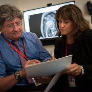
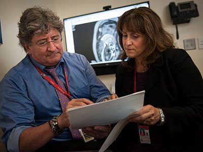
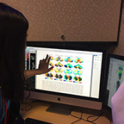
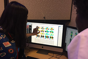
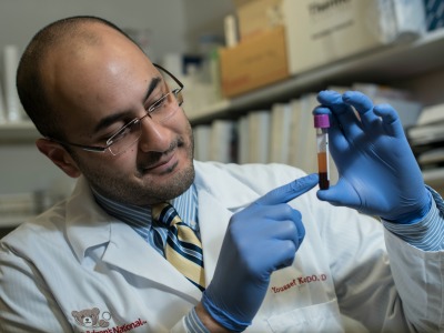
Youssef A. Kousa, D.O., Ph.D., M.S., is examining whether interplays between certain genes make some women more vulnerable to symptomatic Zika infections.

A Maryland woman traveled to the Dominican Republic early in her pregnancy, spending three weeks with family. She felt dizzy and tired and, at first, attributed the lethargy to jet lag. Then, she experienced a rash that lasted about four days. She never saw a bite or slapped a mosquito while in the Dominican Republic but, having heard about the Zika virus, asked to be tested.

Her blood tested positive for Zika.

Why was this pregnant woman infected by Zika while others who live year-round in Zika hot zones remain free of the infectious disease? And why was she among the slim minority of Zika-positive people to show symptoms?

Youssef A. Kousa, D.O., Ph.D., M.S., a pediatric resident in the child neurology track at Children’s National Health System, is working on a research study that will examine whether interplays between certain genes make some women more vulnerable to symptomatic Zika infections during pregnancy, leaving  some fetuses at higher risk of developing microcephaly.

Dr. Kousa will present preliminary findings during Research and Education Week 2017 at Children’s National.

At sites in Puerto Rico, Colombia and Washington D.C., Dr. Kousa and his research collaborators are actively recruiting study participants and drawing blood from women whose Zika infections were confirmed in the first or second trimester of pregnancy. The blood is stored in test tubes with purple caps, a visual cue that the tube contains an additive that binds DNA, preventing it from being cut up. Additional research sites are currently being developed.

When the blood arrives at Children’s National, Dr. Kousa will use a centrifuge located in a sample preparation room to spin the samples at high speed for 11 minutes. The sample emerges from the centrifuge in three discrete layers, separated by weight. The rose-colored section that rises to the top is plasma. Plasma contains tell-tale signs of the immune system’s past battles with viruses and will be analyzed by Roberta L. DeBiasi, M.D., M.S., Chief of the Division of Pediatric Infectious Diseases at Children’s National, and Dr. Kousa’s mentor.

A slender line at the middle indicates white blood cells. The dark red layer is heavier red blood cells that sink to the bottom. This bottom half of the test tube, where the DNA resides, is where Dr. Kousa will perform his genetic research.

For years, Dr. Kousa has worked to identify genetic risk factors that influence which fetuses develop cleft lip and palate. In addition to genetic variances that drive disease, he looks at environmental overlays that can trigger genes to respond in ways that cause pediatric disease. When Zika infections raced across the globe, he says it was important to apply the same genetic analyses to the emerging disease. Genes make proteins that carry out instructions, but viral infection disrupts how genes interact, he says. Cells die. Other cells do not fully mature.

While certain poverty-stricken regions of Brazil have recorded the highest spikes in rates of microcephaly, more is at play than socioeconomics, he says. “It didn’t feel like all of the answers lie in the neighborhood. One woman with a Zika-affected child can live just down the street from a child who is more or less severely affected by Zika.”

As a father, Dr. Kousa is particularly concerned about how Zika stunts growth of the fetal brain at a time when it should expand exponentially. “I have three kids. You see them as they achieve milestones over time. It makes you happy and proud as a parent,” he says.

Sarah B. Mulkey

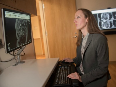
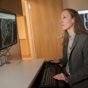
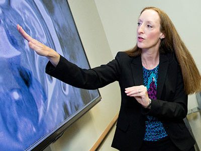
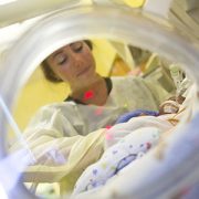
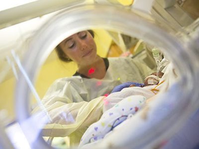
Sarah B. Mulkey, M.D., Ph.D., is studying whether infants exposed to Zika in utero achieve the same developmental milestones as uninfected infants.

While Dr. Kousa concentrates on Zika’s most devastating side effects, his colleague Sarah B. Mulkey, M.D., Ph.D., is exploring more subtle damage Zika can cause to fetuses exposed in utero. In the cohort of Colombian patients that Dr. Mulkey is researching, just 8 percent had abnormal fetal brain magnetic resonance images (MRIs). At first glance, the uncomplicated MRIs appear to be reassuring news for the vast majority of pregnant women.

Dr. Mulkey also will present preliminary findings during Research and Education Week 2017 at Children’s National.

In the fetus, the Zika virus makes a beeline to the developing brain where it replicates with ease and can linger after birth. “We need to be cautious about saying the fetal MRI is ‘normal’ and the infant is going to be ‘normal,’ ” Dr. Mulkey says. “We know with congenital cytomegalovirus that infected infants may not show symptoms at birth yet suffer long-term consequences, such as hearing deficits and vision loss.”

Among Zika-affected pregnancies in Colombia in which late-gestational age fetal MRIs were normal, Dr. Mulkey will use two different evaluation tools at 6 months and 1 year of age to gauge whether the babies accomplish the same milestones as peers. One evaluation tool is a questionnaire that has been validated in Spanish.

At 6 months and 1 year of age, the infants’ motor skills will be assessed, such as their ability to roll over in both directions, sit up, draw their feet toward their waist, stand, take steps independently and purposefully move their hands. Videotapes of the infants performing the motor skills will be scored by Dr. Mulkey and her mentor, Adre du Plessis, M.B.Ch.B., Chief of the Division of Fetal and Transitional Medicine at Children’s National. The Thrasher Research Fund is funding the project, “Neurologic outcomes of apparently normal newborns from Zika virus-positive pregnancies,” as part of its Early Career Award Program.

Both research projects are extensions of a larger multinational study co-led by Drs. du Plessis and DeBiasi that explores the impact of prolonged Zika viremia in pregnant women, fetuses and infants; the feasibility of using fetal MRI to describe the continuum of neurological impacts in Zika-affected pregnancies; and long-term developmental issues experienced by Zika-affected infants.

Sarah B. Mulkey

Puzzling symptoms lead to collaboration

Sarah B. Mulkey, explaining the research

Sarah B. Mulkey, M.D., Ph.D., is lead author of a study that describes a brand-new syndrome that stems from mutations to KCNQ2, a genetic discovery that began with one patient’s unusual symptoms.

Unraveling one of the greatest mysteries of Sarah B. Mulkey’s research career started with a single child.

At the time, Mulkey, M.D., Ph.D., a fetal-neonatal neurologist in the Division of Fetal and Transitional Medicine at Children’s National Health System, was working at the University of Arkansas for Medical Sciences. Rounding one morning at the neonatal intensive care unit (NICU), she met a new patient: A newborn girl with an unusual set of symptoms. The baby was difficult to wake and rarely opened her eyes. Results from her electroencephalogram (EEG), a test of brain waves, showed a pattern typical of a severe brain disorder. She had an extreme startle response, jumping and twitching any time she was disturbed or touched, that was not related to seizures. She also had trouble breathing and required respiratory support.

Dr. Mulkey did not know what to make of her new patient: She was unlike any baby she had ever cared for before. “She didn’t fit anything I knew,” Dr. Mulkey remembers, “so I had to get to the bottom of what made this one child so different.”

Suspecting that her young patient’s symptoms stemmed from a genetic abnormality, Dr. Mulkey ran a targeted gene panel, a blood test that looks for known genetic mutations that might cause seizures or abnormal movements. The test had a hit: One of the baby’s genes, called KCNQ2, had a glitch. But the finding deepened the mystery even further. Other babies with a mutation in this specific gene have a distinctly different set of symptoms, including characteristic seizures that many patients eventually outgrow.

Dr. Mulkey knew that she needed to dig deeper, but she also knew that she could not do it alone. So, she reached out first to Boston Children’s Hospital Neurologist Philip Pearl, M.D., an expert on rare neurometabolic diseases, who in turn put her in touch with Maria Roberto Cilio, M.D., Ph.D., of the University of California, San Francisco and Edward Cooper, M.D., Ph.D., of Baylor College of Medicine. Drs. Cilio, Cooper and Pearl study KCNQ2 gene variants, which are responsible for causing seizures in newborns.

Typically, mutations in this gene cause a “loss of function,” causing the potassium channel to remain too closed to do its essential job properly. But the exact mutation that affected KCNQ2 in Dr. Mulkey’s patient was distinct from others reported in the literature. It must be doing something different, the doctors reasoned.

Indeed, a research colleague of Drs. Cooper, Cilio and Pearl in Italy — Maurizio Taglialatela, M.D., Ph.D., of the University of Naples Federico II and the University of Molise — had recently discovered in cell-based work that this particular mutation appeared to cause a “gain of function,” leaving the potassium channel in the brain too open.

Wondering whether other patients with this same type of mutation had the same unusual constellation of symptoms as hers, Dr. Mulkey and colleagues took advantage of a database that Dr. Cooper had started years earlier in which doctors who cared for patients with KCNQ2 mutations could record information about symptoms, lab tests and other clinical findings. They selected only those patients with the rare genetic mutation shared by her patient and a second rare KCNQ2 mutation also found to cause gain of function — a total of 10 patients out of the hundreds entered into the database. The researchers began contacting the doctors who had cared for these patients and, in some cases, the patients’ parents. They were scattered across the world, including Europe, Australia and the Middle East.

Dr. Mulkey and colleagues sent the doctors and families surveys, asking whether these patients had similar symptoms to her patient when they were newborns: What were their EEG results? How was their respiratory function? Did they have the same unusual startle response?

She is lead author of the study, published online Jan. 31, 2017 in Epilepsia, that revealed a brand-new syndrome that stems from specific mutations to KCNQ2. Unlike the vast majority of others with mutations in this gene, Dr. Mulkey and her international collaborators say, these gain-of-function mutations cause a distinctly different set of problems for patients.

Dr. Mulkey notes that with a growing focus on precision medicine, scientists and doctors are becoming increasingly aware that knowing about the specific mutation matters as much as identifying the defective gene. With the ability to test for more and more mutations, she says, researchers likely will discover more cases like this one: Symptoms that differ from those that usually strike when a gene is mutated because the particular mutation differs from the norm.

Such cases offer important opportunities for researchers to come together to share their collective expertise, she adds. “With such a rare diagnosis,” Dr. Mulkey says, “it’s important for physicians to reach out to others with knowledge in these areas around the world. We can learn much more collectively than by ourselves.”

Neonatal baby

Thrasher to fund Children’s project

Neonatal baby

The Thrasher Research Fund will fund a Children’s National Health System project, “Defining a new parameter for post-hemorrhagic ventricular dilation in premature infants,” as part of its Early Career Award Program, an initiative designed to support the successful training and mentoring of the next generation of pediatric researchers.

The proposal was submitted by Rawad Obeid, M.D., a neonatal neurology clinical research fellow at Children’s National who will serve as the project’s principal investigator. The competition for one-year Thrasher Research Fund awards is highly competitive with just two dozen granted across the nation. Research clinicians at Children’s National received two awards this funding cycle, with another awarded to support a neurologic outcomes study about Zika-affected pregnancies led by Fetal-Neonatal Neurologist Sarah B. Mulkey, M.D., Ph.D.

“Preterm infants born earlier than the 29th gestational week are at high risk for developing cerebral palsy and other brain injuries,” Dr. Obeid says. “Infants with intraventricular hemorrhage (IVH) followed by hydrocephalus (post-hemorrhagic hydrocephalus) face the highest risks of such brain injuries.”

Dr. Obeid hypothesizes that measuring distinct frontal and temporal horn ratio trajectories in extremely premature infants with and without IVH will help to definitively characterize post-hemorrhagic ventricular dilation (PHVD). Right now, experts disagree about the degree of PHVD that should trigger treatment to avoid life-long impairment.

He will be mentored by Anna A. Penn, M.D., Ph.D., Director, Translational Research for Hospital-Based Services & Board of Visitors Cerebral Palsy Prevention Program; Taeun Chang, M.D., Director of the Neonatal Neurology Program within the Division of Neurophysiology, Epilepsy & Critical Care; and Dorothy Bulas, M.D., F.A.C.R., F.A.I.U.M., F.S.R.U., Vice Chief of Academic Affairs.

In the award nomination letter, Dr. Penn noted that in “clinical settings and in the laboratory, I have supervised many trainees, but a trainee like Dr. Obeid is rare. He has pursued his research interests with great commitment. Before coming to Children’s National, he already had multiple job offers, but chose further training to enhance his research skills. While I have worked with many accomplished students, residents and fellows, Dr. Obeid stands out not only for his strong clinical skills, but also for his eagerness to learn and his dedication to both his patients and his research.”


Thrasher Research Fund supports Zika virus neurologic outcomes study

The Thrasher Research Fund will fund a Children’s National project, “Neurologic Outcomes of Apparently Normal Newborns From Zika Virus-Positive Pregnancies,” as part of its Early Career Award Program, an initiative designed to support the successful training and mentoring of the next generation of pediatric researchers.

The project was submitted by Sarah B. Mulkey, M.D., Ph.D., a fetal-neonatal neurologist who is a member of the Congenital Zika Virus Program at Children’s National. During the award period, Dr. Mulkey will be mentored by Adre du Plessis, M.B.Ch.B., director of the Fetal Medicine Institute, and Roberta L. DeBiasi, M.D., M.S., chief of the Division of Pediatric Infectious Diseases. Drs. du Plessis and DeBiasi co-direct the multidisciplinary Zika program, one of the nation’s first.

In the award letter, the fund mentioned Children’s institutional support for Dr. Mulkey, as demonstrated by the mentors’ letter of support, as “an important consideration throughout the funding process.”

The search for precise blood biomarkers of neonatal brain injury

Bloodbiomarkers

PDF Version

What’s Known:
Hypoxic-ischemic encephalopathy (HIE) is characterized by reduced blood and oxygen flow to a baby’s brain around birth and may cause neurologic disability or death. It occurs most commonly after intrauterine asphyxia brought on by such difficulties as circulatory problems, placental abruption, or inflammatory processes. Newborns with HIE may suffer seizures, difficulty feeding, and disturbed control of heart rate and breathing. Cooling therapy, which is the standard of care, offers some protection to the developing brain, but up to 50 percent of HIE-affected infants still have poor outcomes.

What’s New:
Research scientists at Children’s National Health System are involved in a multi-center clinical trial to determine if erythropoietin (EPO), a hormone naturally secreted by the kidneys and commonly used to treat anemia, helps to prevent brain injury in these infants. The trial, called the HEAL Study (High Dose Erythropoietin for Asphyxia and Encephalopathy), is exploring whether EPO, given in addition to hypothermia, further lowers the risk of brain injury in HIE-affected babies. As a part of this study, researchers at Children’s National are leading the investigation to identify biomarkers of brain injury. Biomarkers are telltale chemicals in the blood and are used in tests that evaluate whether patients have suffered a heart attack. While available biomarkers warn when the heart, kidney, or liver is in trouble, there is no blood biomarker that signals ongoing brain injury. Such blood biomarkers could help to determine which infants are responding to treatment as well as to precisely identify which HIE-affected infants are still struggling and require additional treatments, such as EPO, to protect the brain and improve outcomes.

Questions for Future Research: 

  • Does EPO, in tandem with hypothermia, improve long- term neurodevelopmental outcomes in newborns with HIE?
  • Which biomarkers, or panel of biomarkers, best reflect the timing and severity of neonatal brain injury?
  • Can biomarkers direct which types of treatments are best for specific patients and when they should be used?

Source: Plasma Biomarkers of Brain Injury in Neonatal HIE (Hypoxic-Ischemic Encephalopathy).” A.N. Massaro, Y. Wu, T.K. Bammler, A. Mathur, R.C. McKinstry, T. Chang, D.E. Mayock, S. Mulkey, K. Van Meurs, L. Dong, R. Ballard, and S. Juul. Presented during the 2016 Pediatric Academic Societies Annual Meeting, Baltimore, MD. May 3, 2016.