Dominant Fontan approach may be associated with increased liver cirrhosis

The amount of long-term liver cirrhosis in children with single ventricle congenital heart disease who underwent the Fontan procedure may depend on which surgical approach is chosen by the pediatric cardiac surgeon.
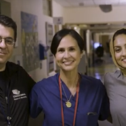
The amount of long-term liver cirrhosis in children with single ventricle congenital heart disease who underwent the Fontan procedure may depend on which surgical approach is chosen by the pediatric cardiac surgeon, according to researchers at Children’s National Hospital who presented their findings this week at the American Association of Thoracic Surgery annual meeting. The full manuscript appears in the Journal of Thoracic and Cardiovascular Surgery.
What this means
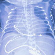
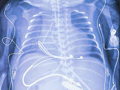
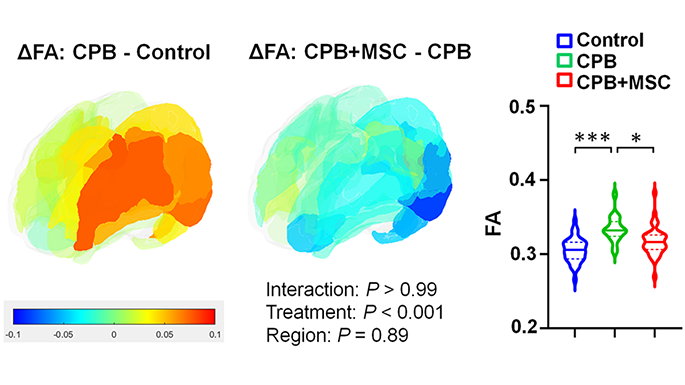
Senior study author Yves d’Udekem, M.D., Ph.D., chief of Cardiac Surgery at Children’s National, says that the vast majority of Fontan procedures in the United States use an extracardiac conduit approach to redirect blood flow to the lungs. However, a retrospective review of 332 patients who underwent the Fontan at Children’s National showed that children who received the extracardiac Fontan may experience liver cirrhosis at a rate of 30% after 15 years compared to the lateral tunnel approach which showed 15-year liver cirrhosis at a significantly lower rate of 4.4%. The lateral tunnel was a well-established method pioneered in Europe by pediatric cardiac surgeon Marc de Leval in the 1980s. This technique lost traction in the field and people started in the 1990s to perform a variation of the technique called the extracardiac Fontan because it was thought that it would be giving more favorable flows and protect the patients against rhythm issues. Thirty years later, these predictions did not reveal themselves to be true.
“Since the 1990s, the vast majority of Fontan procedures in the United States are performed creating an extracardiac conduit rather than the lateral tunnel,” says Dr. d’Udekem. “But what we see when we follow long-term outcomes of these children is a consequence not reported before.”
Children’s National leads the way
Dr. d’Udekem and the research team, including presenter and first author Eiri Kisamori, M.D., a cardiac surgery fellow at Children’s National, are the first to report these findings based on reviews of 15-year outcome data. These retrospective reviews of long-term outcomes are a critical tool to inform and improve clinical approaches with the goal of optimizing the long-term quality of life for children born with these critical congenital conditions.
What’s next
While more research is needed, the authors hypothesize that the size of the conduit for blood flow may be the culprit for higher levels of liver damage. For children who have already received an extracardiac Fontan, Dr. d’Udekem says that widening their existing conduit in a reoperation may successfully improve blood flow to the liver. For future procedures, he notes that in his own practice, he now uses the lateral tunnel approach whenever possible.
Read the study: Alarming rate of liver cirrhosis after the small conduit Extracardiac Fontan. A comparative analysis with the Lateral Tunnel.
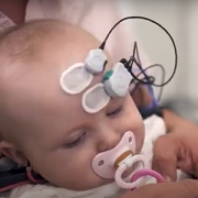


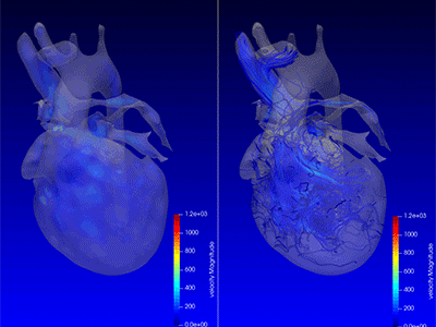
 Advanced MRI visualization techniques to follow blood flow in the hearts of cardiac patients. Gene therapy for pediatric patients with Duchenne muscular dystrophy. 3D-printed casts for treating clubfoot. These were among the most popular articles we published on Innovation District in 2023. Read on for our full list.

 The hospital will advocate for the unique needs of children as part of nationwide network working to accelerate transformative health solutions.
 Children’s National Hospital was awarded nearly $7.5 million in a five-year grant to continue its leadership of an FDA-funded pediatric device consortium. Building upon a decade of previous consortium leadership, the new consortium is Alliance for Pediatric Device Innovation (APDI) and features a new and expanded roster of partners that reflects its added focus on providing pediatric innovators with expert support on evidence generation, including the use of real-world evidence (RWE), for pediatric device development.


 Children’s National Hospital in Washington, D.C., was ranked #5 in the nation on the U.S. News & World Report 2023-24 Best Children’s Hospitals annual rankings. This marks the seventh straight year Children’s National has made the Honor Roll list. The Honor Roll is a distinction awarded to only 10 children’s hospitals nationwide.