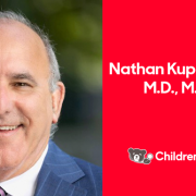
Nathan Kuppermann, M.D., M.P.H., named chief academic officer and chair of Pediatrics

Nathan Kuppermann, M.D., M.P.H.

Dr. Kuppermann will oversee research, education and innovation for the Children’s National Research Institute as well as academic and administrative leadership in the Department of Pediatrics at George Washington University School of Medicine & Health Services.

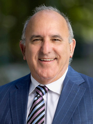
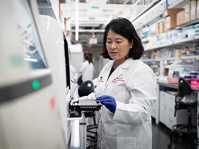
Children’s National Hospital has appointed Nathan Kuppermann, M.D., M.P.H., as its new executive vice president (EVP), chief academic officer (CAO) and chair of Pediatrics. In this role, Dr. Kuppermann will oversee research, education and innovation for the Children’s National Research Institute as well as academic and administrative leadership in the Department of Pediatrics at George Washington University School of Medicine & Health Services. He comes to Children’s National from UC Davis Health and UC Davis School of Medicine in Sacramento, CA, and will start in September.

After a national search, Dr. Kuppermann stood out for his exceptional contributions to clinical and academic research, focusing on pediatric emergency care, and his dedication to mentorship. For the past 18 years he has served as the Bo Tomas Brofeldt endowed chair of the Department of Emergency Medicine and is currently a distinguished professor of Emergency Medicine and Pediatrics, and the associate dean for Global Health at UC Davis Health.

“I was drawn to Children’s National by its nationally recognized work and dedication to innovation and team science,” says Dr. Kuppermann. “I’m eager to contribute to the remarkable work being done in both the research and education space to continue to improve the understanding, prevention and treatment of childhood diseases.”

Dr. Kuppermann is a pediatric emergency medicine physician and clinical epidemiologist, and a leader in emergency medical services for children, particularly in multicenter research. With more than 300 peer-reviewed research publications to his credit, Dr. Kuppermann has contributed extensively to high-impact journals including the New England Journal of Medicine, JAMA, BMJ and the Lancet.

“The Children’s National Research Institute is a key part of our health system’s ecosystem – it’s where we nurture innovation and pursue the most promising research,” says Michelle Riley-Brown, MHA, FACHE, president and CEO of Children’s National. “Dr. Kuppermann’s unwavering commitment to excellence in pediatric healthcare, research and innovation set him apart in a competitive field. I am confident he will advance our efforts in making breakthrough discoveries for kids everywhere.”

Dr. Kuppermann received his undergraduate degree from Stanford University, his medical degree from UC San Francisco School of Medicine and his Master of Public Health degree from the Harvard School of Public Health. He completed a pediatrics residency and chief residency at Harbor-UCLA Medical Center and a fellowship in Pediatric Emergency Medicine at Boston Children’s Hospital.

He has been recognized nationally and internationally for his research and mentorship. He was a Fulbright Distinguished Scholar in the U.K. and in 2010 was elected to the National Academy of Medicine. In 2022, he received the Maureen Andrew Mentor Award from the Society for Pediatric Research.

“Dr. Kuppermann’s leadership will undoubtedly propel the hospital’s efforts in advancing pediatric healthcare innovation, reinforcing Children’s National as a top-ranking institution,” says Horacio Rozanski, chair of the Children’s National Board of Directors. “We look forward to the positive impact he will make to the hospital’s overall mission, as well as its research and academic endeavors.”

Pioneering research center aims to revolutionize prenatal and neonatal health

Catherine Limperopoulos, Ph.D., was drawn to understanding the developing brain, examining how early adverse environments for a mother can impact the baby at birth and extend throughout its entire lifetime. She has widened her lens – and expanded her team – to create the new Center for Prenatal, Neonatal & Maternal Health Research at Children’s National Hospital.

“Despite the obvious connection between mothers and babies, we know that conventional medicine often addresses the two beings separately. We want to change that,” said Dr. Limperopoulos, who also directs the Developing Brain Institute. “Given the current trajectory of medicine toward precision care and advanced imaging, we thought this was the right moment to channel our talent and resources into understanding this delicate and highly dynamic relationship.”

Moving the field forward

Since its establishment in July 2023, the new research center has gained recognition through high-impact scientific publications, featuring noteworthy studies exploring the early phases of human development.

Dr. Limperopoulos has been at the forefront of groundbreaking research, directing attention to the consequences of maternal stress on the unborn baby and the placenta. In addition, under the guidance of Kevin Cook, Ph.D., investigators published a pivotal study on the correlation between pain experienced by premature infants in the Neonatal Intensive Care Unit and the associated risks of autism and developmental delays.

Another area of research has focused on understanding the impact of congenital heart disease (CHD) on prenatal brain development, given the altered blood flow to the brain caused by these conditions during this period of rapid development. Led by Josepheen De Asis-Cruz, M.D., Ph.D., a research team uncovered variations in the functional connectivity of the brains of infants with CHD. In parallel, Nickie Andescavage, M.D., and her team employed advanced imaging techniques to identify potential biomarkers in infants with CHD, holding promise for guiding improved diagnostics and postnatal care. Separately, she is investigating the impact of COVID-19 on fetal brain development.

In the months ahead, the team plans to concentrate its efforts on these areas and several others, including the impact of infectious disease, social determinants of health and protecting developing brains from the negative impacts of maternal stress, pre-eclampsia and other conditions prevalent among expectant mothers.

Assembling a team

Given its robust research plan and opportunities for collaboration, the center pulled together expertise from across the hospital’s faculty and has attracted new talent from across the country, including several prominent faculty members:

  • Katherine L. Wisner, M.S., M.D., has accumulated extensive knowledge on the impact of maternal stress on babies throughout her career, and her deep background in psychiatry made her a natural addition to the center. While Dr. Wisner conducts research into the urgent need to prioritize maternal mental health, she will also be treating mothers as part of the DC Mother-Baby Wellness Initiative — a novel program based at Children’s National that allows mothers to more seamlessly get care for themselves and participate in mother-infant play groups timed to align with their clinical appointments.
  • Catherine J. Stoodley, B.S., M.S., D.Phil., brings extensive research into the role of the cerebellum in cognitive development. Dr. Stoodley uses clinical studies, neuroimaging, neuromodulation and behavioral testing to investigate the functional anatomy of the part of the brain responsible for cognition.
  • Katherine M. Ottolini, M.D., attending neonatologist, is developing NICU THRIVE – a research program studying the effects of tailored nutrition on the developing newborn brain, including the impact of fortifying human milk with protein, fat and carbohydrates. With a grant from the Gerber Foundation, Dr. Ottolini is working to understand how personalized fortification for high-risk babies could help them grow.

Early accolades

The new center brings together award-winning talent. This includes Yao Wu, Ph.D., who recently earned the American Heart Association’s Outstanding Research in Pediatric Cardiology award for her groundbreaking work in CHD, particularly for her research on the role of altered placental function and neurodevelopmental outcomes in toddlers with CHD. Dr. Wu became the third Children’s National faculty member to earn the distinction, joining an honor roll that includes Dr. Limperopoulos and David Wessel, M.D., executive vice president and chief medical officer.

Interim Chief Academic Officer Catherine Bollard, M.D., M.B.Ch.B., said the cross-disciplinary collaboration now underway at the new center has the potential to make a dramatic impact on the field of neonatology and early child development. “This group epitomizes the Team Science approach that we work tirelessly to foster at Children’s National,” Dr. Bollard said. “Given their energetic start, we know these scientists and physicians are poised to tackle some of the toughest questions in maternal-fetal medicine and beyond, which will improve outcomes for our most fragile patients.”

Next-generation genomics testing holds key to undiagnosed rare disease

Before and after pictures of the patient's improved gaitSeth Berger, M.D., Ph.D., felt the pull to dig deeper when he started reading the chart. An 11-year-old boy had an abnormal gait and couldn’t even walk in a straight line down the sidewalk to go trick-or-treating. Yet workups with neurology, orthopedics and an exome analysis of the patient’s genetic code did not provide a diagnosis. He had been getting worse for roughly three years.

With one of the largest clinical genetics departments in the country, Children’s National Hospital receives more than 10,000 visits a year from patients like this middle schooler. Often, they are children and caregivers who are searching for answers and follow-up support for diagnoses of genetic disorders, which impact so few people that only highly trained geneticists and genetic counselors can get to the root of the disorder.

“In genetics, we are finding layers of understanding. A negative clinical test is not always the final answer because the significance of variants can often be missed or misunderstood,” said Dr. Berger, a medical geneticist and principal investigator in the Center for Genetics Medicine Research at Children’s National. “It can take extensive research and a deep knowledge of the limits of certain tests to reach a diagnosis.”

The fine print

On page 4 of the patient’s genetics report, Dr. Berger found a reference to a pair of variants with no known clinical impact. Dr. Berger recognized that the genes referenced could affect proteins that drive potentially treatable neurological outcomes.

Dr. Berger ordered further testing, including biochemical testing of the patient’s blood and a phenylalanine loading challenge, a test that measures how the body metabolizes certain amino acids. With the results, he confirmed a recessive GCH1 deficiency in the patient was causing a condition called DOPA-responsive dystonia, a disorder that causes involuntary muscle contractions, tremors and uncontrolled movements. Laura Schiffman Tochen, M.D., director of the Movement Disorders Program at Children’s National, started the patient on levodopa-carbidopa — a drug combination used to treat Parkinson’s disease and other neurological disorders — and within two hours the boy showed improvement. His gait was almost normal.

Why we’re excited

Dr. Berger presents at conferences on this case and several other medical mysteries that he’s recently solved in his clinical practice and his role at the Pediatric Mendelian Genomics Research Center, a Children’s National program immersed in a federally funded research study to better understand how differences in genetic material can affect human health. As part of his work, he’s joined the GREGoR project (Genomic Research to Elucidate the Genetics of Rare Disease), which hopes to increase the number of genetic disorders where a cause can be identified. The elite genetics consortium includes nationally recognized research centers – the University of California at Irvine, Broad Institute, University of Washington, Baylor University, Stanford University, Invitae and Children’s National – which are working together to harness cutting-edge genomics sequencing capabilities. They hope to enroll thousands in their research, funded by the National Institutes of Health.

“It’s truly stunning what genetic sequencing can find. The outcomes can be life-changing,” said Dr. Berger. “These cases with life-altering diagnoses don’t come along every day, but when they do, they make the hunt to find answers all the more worthwhile.”

Prenatal COVID exposure associated with changes in newborn brain

pregnant woman talking to doctor

The team found differences in the brains of both infants whose mothers were infected with COVID while pregnant, as well as those born to mothers who did not test positive for the virus.

Babies born during the COVID-19 pandemic have differences in the size of certain structures in the brain, compared to infants born before the pandemic, according to a new study led by researchers at Children’s National Hospital.

The team found differences in the brains of both infants whose mothers were infected with COVID while pregnant, as well as those born to mothers who did not test positive for the virus, according to the study published in Cerebral Cortex.

The findings suggest that exposure to the coronavirus and being pregnant during the pandemic could play a role in shaping infant brain development, said Nickie Andescavage, M.D., the first author of the paper and associate chief for the Developing Brain Institute at Children’s National.

The fine print

The study’s authors looked at three groups of infants: 108 born before the pandemic; 47 exposed to COVID before birth; and 55 unexposed infants. In all cases, researchers performed magnetic resonance imaging (MRI) scans of the newborns’ brains during the first few weeks of life. The MRI scans, which are non-invasive and do not expose patients to radiation, provided 3D images of the brain, allowing doctors to calculate the volume of different areas.

Researchers found several differences in the brains of babies exposed to COVID. They had larger volumes of the gray matter that makes up the brain’s outermost layer, compared to the two other groups. In contrast, an inner area of the brain, known as deep gray matter, was smaller than in unexposed babies. These are areas that contain large numbers of neurons that generate and process signals throughout the brain. “Their brains formed differently if they were exposed to COVID,” said Dr. Andescavage, adding that “those exposed to COVID had unique signatures” in the brain.

Doctors also measured the depths of the folds in the babies’ brains – a way to determine how the brain is maturing during early development. Babies born to mothers who had COVID in pregnancy had deeper grooves in the frontal lobe, while babies born during the pandemic – even without being exposed to COVID – had increased folds and grooves throughout the brain, compared to babies born before the pandemic. “There was something about being born during the pandemic that changed how the brain developed,” Dr. Andescavage said.

What’s ahead

The study authors can’t fully explain what caused the differences in brain development in these babies, Dr. Andescavage said. But other studies have linked maternal stress and depression to changes in the newborn brain. In a future study, Dr. Andescavage and her colleagues will examine the relationship between infant brain development and how stress and anxiety during the pandemic may have played a role in early development.

Because the babies in the study were just a few weeks old, researchers don’t know if their altered brain development will affect how they learn or behave. Researchers plan to follow the children until age 6, allowing them to observe whether pandemic-era babies hit key developmental milestones on time, such as walking, talking, holding a crayon and learning the alphabet.

Researchers have been worried about the effect of COVID on the fetus since the beginning of the pandemic. Studies show that babies exposed to COVID in the womb may experience developmental impacts, and research is underway to better understand long-term outcomes.

Although the coronavirus rarely crosses the placenta to infect the fetus directly, there are other ways maternal infection can influence the developing baby. Dr. Andescavage said inflammation is one potential harm to a developing baby. In addition, if a pregnant woman becomes so sick that the levels of oxygen in her blood fall significantly, that can deprive the fetus of oxygen, she added.

In recent decades, studies of large populations have found that maternal infections with influenza and other viruses increased the risk of serious problems in children even years later, including autism, attention deficit hyperactivity disorder and schizophrenia, although the reasons behind the association are not well understood. Technology may allow doctors to answer a number of questions about COVID and the infant brain.

“With advanced imaging and MRI, we’re in a position now to be able to understand how the babies are developing in ways we never previously could,” Dr. Andescavage said. “That will better allow us to identify the exposures that may be harmful, and at what times babies may be especially vulnerable, to better position us to promote maternal wellness. This, in turn, helps infant wellness.”

Evidence review: Maternal mental conditions drive climbing death rate in U.S.

mother kissing newborn baby

More than 80% of maternal deaths in the United States are preventable, particularly the nearly 1 in 4 maternal fatalities that are attributable to mental health disorders.

Painting a sobering picture, a research team led by Children’s National Hospital culled years of data demonstrating that maternal mental illness is an under-recognized contributor to the death of new mothers. They are calling for urgent action to address this public health crisis in the latest edition of JAMA Psychiatry.

Backed by dozens of peer-reviewed studies and health policy sources, the journal’s special communication comes as maternal mortality soars in the United States to as much as three times the rate of other high-income countries.

“The contribution of mental health conditions to the maternal morbidity and mortality crisis that we have in America is not widely recognized,” said Katherine L. Wisner, M.D., associate chief of Perinatal Mental Health and member of the Center for Prenatal, Neonatal & Maternal Health Research at Children’s National. “We need to bring this to the attention of the public and policymakers to demand action to address the mental health crisis that is contributing to the demise of mothers in America.”

The evidence review laid out the risks facing new mothers: More than 80% of maternal deaths in the United States are preventable, particularly the nearly 1 in 4 maternal fatalities that are attributable to mental health disorders. Overdose and other maternal mental health conditions are taking the lives of more than twice as many women as postpartum hemorrhage, the second leading cause of maternal death. For non-Hispanic Black mothers, the mortality rate is a striking 2.6 times higher than non-Hispanic White mothers.

Yet the research team found that recent national efforts to combat maternal mortality have failed to address maternal mental health as “the public health crisis that it represents.” Even methodologies to measure maternal health statistics are inconsistent, which challenges efforts to shape health policy.

In examining 30 recent studies and another 15 historical references, the team – which included Caitlin Murphy, MPA, PNP, research scientist at the Milken School of Public Health at George Washington University, and Megan Thomas, M.D., FACOG, obstetrician at the University of Kansas School of Medicine – found ample data to support the need to elevate maternal mental health as a priority. Some examples:

  • Multiple studies show that the perinatal period puts women at higher risk for new and recurrent psychiatric disorders, with 14.5% of pregnant mothers having a new episode of depression and another 14.5% developing an episode three months after birth.
  • Nationwide, more than 400 maternity healthcare centers closed between 2006 and 2020, creating “maternity care deserts” that left nearly 6 million women with limited or no access to maternity care.
  • Mental health conditions such as suicide or opioid overdose are to blame for nearly 23% of maternal deaths in America, according to reports from three dozen Maternal Morbidity and Mortality Review Committees, which are state-based organizations that review each maternal death within a year of pregnancy. That’s followed by hemorrhage (13.7%), cardiac conditions (12.8%) and infection (9.2%).

Even with these sobering statistics, Dr. Wisner says that only 20 percent of women are screened for depression postpartum. “Given that this is a time that many mothers have contact with healthcare professionals, it’s critically important that all mothers are screened and offered treatment,” she said. “Mental health is fundamental to health — of the mother, the child and the entire family.”

Dr. Wisner is board-certified in general and child psychiatry. Throughout her research career, she has conducted research on maternal-infant interactions and family health. She recently joined the new Center for Prenatal, Neonatal & Maternal Health Research because of its vision to improve outcomes for the entire family by understanding the relationship between mothers and their babies.

“Throughout my career, I have fought hard against these silos that try to lock psychiatry into certain age categories,” Dr. Wisner said. “At Children’s National, we have a huge interest in reunifying the family. We want to ensure that we’re caring for unborn babies, infants and toddlers, while focusing on maternal health and the family in its broader context.”

Driving pediatric breakthroughs through 2023

desktop computer showing the CNRI Annual ReportThe Children’s National Research Institute released its 2022-2023 Academic Annual Report. In the report, a summary of the past academic year highlights the accomplishments of each of the institute’s research centers, provides research funding figures and exalts some of the institute’s biggest milestones.

The stories in the report are a testament to the hard work and dedication of everyone at the Children’s National Research Institute.

We celebrated five decades of leadership and mentorship of Naomi Luban, M.D., and her incredible accomplishments in the W@TCH program, which have been instrumental in shaping the future of pediatric research.

We also celebrated innovation, highlighting our recent FDA award to lead a pediatric device consortium, which recognizes our commitment to developing innovative medical devices that improve the lives of children.

Breakthroughs at the Research & Innovation Campus continued as our researchers worked tirelessly to develop new treatments and therapies that will transform the lives of children and families around the world.

Taking a look at the breakthroughs happening in our now six research centers, we spotlighted the following stories:

  • Reflecting on decades of progress in the blood, marrow and cell therapy programs at Children’s National. Our researchers have made significant strides in this field, and we are proud to be at the forefront of these life-saving treatments.
  • In genetic medicine, we continue to be a beacon of hope for families facing rare and complex conditions. Our researchers are making incredible breakthroughs that are changing the landscape of pediatric medicine.
  • We are also proud to share the $90 million award received from an anonymous donor to support pediatric brain tumor research. The predominant focus of this award is to develop new treatments that will improve outcomes for children with this devastating disease.
  • This year, we opened a new Center that enhances our research capabilities in the field of Prenatal, Neonatal & Maternal Health Research. We are excited about the possibilities this new center will bring and look forward to the discoveries that will emerge from it.
  • In addition, we are driving future pandemic readiness with the NIH funded Pediatric Pandemic Network. Our researchers are using cutting-edge technology and innovative approaches to prepare for the next pandemic and protect children.
  • We are also exploring the potential of artificial intelligence (AI) in pediatric breakthroughs. Our researchers are using machine learning and other AI techniques to develop new treatments and therapies that will transform the lives of children.

Black, Hispanic children at greater risk for complications during hospitalization

Boy lying in a hospital bed, surrounded by medical equipment

The research team found that patients who are Black and Medicaid-insured patients experienced the greatest disparities in postoperative sepsis, a rare complication in which patients suffer from infection that can cause multi-organ failure.

Evaluating more than 5 million pediatric hospital stays nationwide, researchers found children who are Black, Hispanic or insured with Medicaid face a greater risk of health events after surgeries than white patients, according to a new study published in the journal Pediatrics.

“We looked at the data, and we calculated the risks,” said Kavita Parikh, M.D., MSHS, medical director of Quality & Safety Research, research director of the Division of Hospital Medicine and first author on the multi-institute study. “Despite decades of focus on eliminating medical errors, we know that children continue to suffer substantial harms in hospital settings, and our study highlights where children who are Black, Hispanic or insured with Medicaid are at the greatest risk.”

The big picture

The study analyzed data from more than 5.2 million hospitalizations collected by the 2019 Kids’ Inpatient Database, a national repository of data on hospital stays. It includes a 10% sample of newborns and an 80% sample of other pediatric discharges from 4,000 U.S. hospitals. More than 80% of patients were younger than 1 year of age.

The research team found that patients who are Black and Medicaid-insured patients experienced the greatest disparities in postoperative sepsis, a rare complication in which patients suffer from infection that can cause multi-organ failure. Patients who are Hispanic experienced the greatest disparity in postoperative respiratory failure, a complication that can limit breathing and ventilation.

Plausible factors cited include structural racism in the U.S. healthcare system, clinician bias, insufficient cultural responsiveness, communication barriers and limited access to high-quality healthcare.

What’s ahead

The study – “Disparities in Racial, Ethnic, and Payor Groups for Pediatric Safety Events in U.S. Hospitals” – is foundational in understanding what is happening among pediatric patients. Dr. Parikh said that researchers now must conduct further studies into these alarming disparities and qualitative work to understand drivers, with the action-oriented goal of developing interventions to improve patient safety in the hospital for all children.

“We brought together leaders in pediatric medicine, health policy and public health to analyze this data, and we are committed to taking the next steps to improve outcomes for pediatric patients,” Dr. Parikh said. “It will take more patient-centered work and research, resources and multifaceted strategies to resolve these worrying disparities for our pediatric patients nationwide.”

Children’s National performs first ever HIFU procedure on patient with cerebral palsy

Doctors performing bilateral high intensity focused ultrasound (HIFU) pallidotomy on a patient with dyskinetic cerebral palsy.

HIFU is a non-invasive therapy that utilizes focused ultrasound waves to thermally ablate a focal area of tissue.

In January, a team of multidisciplinary doctors performed the first case in the world of using bilateral high intensity focused ultrasound (HIFU) pallidotomy on Jesus, a 22-year-old patient with dyskinetic cerebral palsy.

The procedure is part of a clinical trial led by Chima Oluigbo, M.D., pediatric neurosurgeon at Children’s National Hospital.

“The primary objective of the study is to evaluate the safety of ExAblate Transcranial MRgFUS as a tool for creating bilateral or unilateral lesions in the globus pallidus (GPi) in patients with treatment-refractory secondary dystonia due to dyskinetic cerebral palsy,” Dr. Oluigbo explained. “The secondary purpose is to assess the impact of HIFU pallidotomy on dyskinetic cerebral palsy movement disorder in pediatric and young adult patients.”

In addition, the impact of bilateral pallidotomy on motor development, pain perception, speech, memory, attention and cognition in these patients will be assessed.

“We hope that the trial will help us find results that lead to treatments that can reduce the rigidity and stiffness which occurs in cerebral palsy so we can help these children who do not have any effective treatment,” Dr. Oluigbo added.

“This new, first of its kind, non-invasive therapeutic approach – without even a skin incision – will open the door to offering hope for a number of kids with movement disorders who have failed conventional therapy,” said Robert Keating, M.D., chief of neurosurgery at Children’s National. “We are at the beginning of a new era for treating functional disorders in the pediatric patient.”

How it works

HIFU is a non-invasive therapy that utilizes focused ultrasound waves to thermally ablate a focal area of tissue. In the past, Children’s National successfully used HIFU to treat low-grade type tumors located in difficult locations of the brain, such as hypothalamic hamartomas and pilocytic astrocytoma, as well as for epilepsy and other movement disorders.

This most recent procedure was another successful milestone for the hospital, discharging Jesus the following day without any complications.

The team comprised neurosurgeons, MRI techs, anesthesiologists and radiologists, to name a few.

Originally, Jesus came to Children’s National in 2006 when he started working with the Physical Medicine and Rehabilitation team to help him with his muscle hypertonia management as well as equipment, orthoses and therapy concerns.

“As he continued to grow, his muscle hypertonia became more pronounced and caused difficulty with his care, positioning and comfort,” said Olga Morozova, M.D., pediatric rehabilitation specialist at Children’s National. “We have tried multiple oral medications however he has had significant side effects from the majority of the medications.”

Dr. Morozova and Julie Will, M.S.N., F.N.P., the nurse practitioner that worked with Jesus, referred him to Dr. Oluigbo after they learned about HIFU being an option to treat Jesus using a non-invasive approach.

Moving the field forward

This clinical trial highlights the expanding indications for focused ultrasound.

“We are excited about the potential for these innovative treatment strategies in neurosurgery to transform the lives of pediatric patients who suffer from challenging diseases, such as brain tumors, epilepsy, and movement disorders,” said Hasan Syed, M.D., co-director of the Focused Ultrasound Program at Children’s National. “We are redefining what is possible in neurosurgery.”

From low-intensity focused ultrasound (LIFU) treatments for our young DIPG patients to now the groundbreaking research on HIFU for pediatric movement disorders, the dedication to cutting-edge techniques highlights the team’s commitment to patients and transforming pediatric neurosurgical care.

Novel AI platform matches cardiologists in detecting rheumatic heart disease

Artificial intelligence (AI) has the potential to detect rheumatic heart disease (RHD) with the same accuracy as a cardiologist, according to new research demonstrating how sophisticated deep learning technology can be applied to this disease of inequity. The work could prevent hundreds of thousands of unnecessary deaths around the world annually.

Developed at Children’s National Hospital and detailed in the latest edition of the Journal of the American Heart Association, the new AI system combines the power of novel ultrasound probes with portable electronic devices installed with algorithms capable of diagnosing RHD on echocardiogram. Distributing these devices could allow healthcare workers, without specialized medical degrees, to carry technology that could detect RHD in regions where it remains endemic.

RHD is caused by the body’s reaction to repeated Strep A bacterial infections and can cause permanent heart damage. If detected early, the condition is treatable with penicillin, a widely available antibiotic. In the United States and other high-income nations, RHD has been almost entirely eradicated. However, in low- and middle-income countries, it impacts the lives of 40 million people, causing nearly 400,000 deaths a year.

“This technology has the potential to extend the reach of a cardiologist to anywhere in the world,” said Kelsey Brown, M.D., a cardiology fellow at Children’s National and co-lead author on the manuscript with Staff Scientist Pooneh Roshanitabrizi, Ph.D. “In one minute, anyone trained to use our system can screen a child to find out if their heart is demonstrating signs of RHD. This will lead them to more specialized care and a simple antibiotic to prevent this degenerative disease from critically damaging their hearts.”

The big picture

AI system that can detect RHD

The new AI system combines the power of novel ultrasound probes with portable electronic devices installed with algorithms capable of diagnosing RHD on echocardiogram.

Millions of citizens in impoverished countries have limited access to specialized care. Yet the gold standard for diagnosing RHD requires a highly trained cardiologist to read an echocardiogram — a non-invasive and widely distributed ultrasound imaging technology. Without access to a cardiologist, the condition may remain undetected and lead to complications, including advanced cardiac disease and even death.

According to the new research, the AI algorithm developed at Children’s National identified mitral regurgitation in up to 90% of children with RHD. This tell-tale sign of the disease causes the mitral valve flaps to close improperly, leading to backward blood flow in the heart.

Beginning in March, Craig Sable, M.D., interim division chief of Cardiology, and his partners on the project will implement a pilot program in Uganda incorporating AI into the echo screening process of children being checked for RHD. The team believes that a handheld ultrasound probe, a tablet and a laptop — installed with the sophisticated, new algorithm — could make all the difference in diagnosing these children early enough to change outcomes.

“One of the most effective ways to prevent rheumatic heart disease is to find the patients that are affected in the very early stages, give them monthly penicillin for pennies a day and prevent them from becoming one of the 400,000 people a year who die from this disease,” Dr. Sable said. “Once this technology is built and distributed at a scale to address the need, we are optimistic that it holds great promise to bring highly accurate care to economically disadvantaged countries and help eradicate RHD around the world.”

Children’s National Hospital leads the way

To devise the best approach, two Children’s National experts in AI — Dr. Roshanitabrizi and Marius George Linguraru, D.Phil., M.A., M.Sc., the Connor Family Professor in Research and Innovation and principal investigator in the Sheikh Zayed Institute for Pediatric Surgical Innovation — tested a variety of modalities in machine learning, which mimics human intelligence, and deep learning, which goes beyond the human capacity to learn. They combined the power of both approaches to optimize the novel algorithm, which is trained to interpret ultrasound images of the heart to detect RHD.

Already, the AI algorithm has analyzed 39 features of hearts with RHD that cardiologists cannot detect or measure with the naked eye. For example, cardiologists know that the heart’s size matters when diagnosing RHD. Current guidelines lay out diagnostic criteria using two weight categories — above or below 66 pounds — as a surrogate measure for the heart’s size. Yet the size of a child’s heart can vary widely in those two groupings.

“Our algorithm can see and make adjustments for the heart’s size as a continuously fluid variable,” Dr. Roshanitabrizi said. “In the hands of healthcare workers, we expect the technology to amplify human capabilities to make calculations far more quickly and precisely than the human eye and brain, saving countless lives.”

Among other challenges, the team had to design new ways to teach the AI to handle the inherent clinical differences found in ultrasound images, along with the complexities of evaluating color Doppler echocardiograms, which historically have required specialized human skill to evaluate.

“There is a true art to interpreting this kind of information, but we now know how to teach a machine to learn faster and possibly better than the human eye and brain,” Dr. Linguraru said. “Although we have been using this diagnostic and treatment approach since World War II, we haven’t been able to share this competency globally with low- and middle-income countries, where there are far fewer cardiologists. With the power of AI, we expect that we can, which will improve equity in medicine around the world.”

Shaping the future of pediatric endocrinology

Youn Hee Jee

“Select patients will have the opportunity to participate in research studies focused on cutting-edge genomic investigations into their growth disorders,” says Youn Hee Jee, M.D., M.Med., endocrinologist.

“We’re dedicated to unraveling the mysteries that families have long sought answers to,” says Andrew Dauber, M.M.Sc., M.D., chief of Endocrinology at Children’s National Hospital. “There are numerous endocrine and genetic conditions with the potential to impact a child’s growth. That’s why we’ve assembled a team of leading endocrinologists and geneticists to create a new Growth Specialty Clinic and address these issues with a fresh perspective.”

This team, combined with the expertise of the hospital’s translational scientists, is making significant progress in identifying the causes of a variety of growth disorders and developing innovative treatments. And at the core of this work, Dr. Dauber says, is a recognition of the unique impact endocrine disorders have on each individual child.

What’s unique

Leveraging the expertise of Medical Geneticists Natasha Shur, M.D., and Deepika Burkardt, D.O., from the Children’s National Rare Disease Institute – the largest clinical genetics program in the United States – the growth clinic taps into substantial knowledge in the genetics of growth.

Dr. Shur emphasizes the commitment to providing answers for these families. “This collaborative effort goes beyond diagnosis; it opens doors to potential treatment options.”

The Growth Specialty Clinic is for children with severe undiagnosed growth disorders that are suspected to have a genetic etiology and children with rare genetic diagnoses who would benefit from the expertise of practitioners more familiar with those disorders. It is also closely linked to the Center for Genetic Medicine Research.

“Select patients will have the opportunity to participate in research studies focused on cutting-edge genomic investigations into their growth disorders,” says Youn Hee Jee, M.D., M.Med., endocrinologist.

In one case, Dr. Jee identified a new genetic cause of an overgrowth syndrome. Rare genetic conditions known as generalized overgrowth syndromes manifest as excessive body growth during fetal life and/or childhood, frequently resulting in tall stature. She is investigating the mechanisms that promote healthy bone growth.

Additionally, Dr. Jee identified a new genetic cause of short stature. Her research showed that the identified genetic cause impairs the recycling of essential proteins for growth, expanding our knowledge of human growth.

Moving the field forward

“We’re taking innovative approaches to treatment by leveraging our insights into the genetic origin of each patient’s growth disorder,” says Dr. Dauber.

In the brief time since the clinic’s launch, several new diagnoses and treatment pathways have already been offered. In one single-patient study, researchers were able to successfully overcome the patient’s growth hormone resistance using a targeted approach, and the patient has shown significant catch-up growth after one year of treatment.

Children’s National is also at the forefront of other groundbreaking research, launching novel clinical trials that are advancing the field of endocrinology:

  • Vosoritide clinical trial: Children’s National has the first clinical trial in the world testing Vosoritide in children with certain genetic causes of short stature. Researchers have enrolled approximately 50 subjects with exciting preliminary results for patients with Noonan syndrome, Aggrecan gene mutations and NPR2 gene mutations. All 24 hypochondroplasia patients have completed the 18-month trial. Dr. Dauber intends to present results at the 2024 American College of Medical Genetics meeting in Toronto.
  • Hypochondroplasia study: Children’s National is the first site to launch BioMarin’s new natural history study for children with hypochondroplasia which will also be a lead into their future Phase 3 trial.

Read more about our advances in Diabetes & Endocrinology.

AHA’s Outstanding Research Award: Three generations of pediatric cardiac excellence

Drs. Catherine Limperopoulos, Yao Wu and David Wessel

Catherine Limperopoulos, Ph.D., Yao Wu, Ph.D., and David Wessel, M.D.

Children’s National Hospital is celebrating a remarkable milestone as three of its faculty members have been honored over 15 years with the American Heart Association’s Outstanding Research in Pediatric Cardiology Award. Yao Wu, Ph.D., became the latest researcher to earn the accolade for her groundbreaking work into congenital heart disease (CHD).

A research faculty member with the newly established Center for Prenatal, Neonatal & Maternal Health Research, Dr. Wu received the award specifically for her studies on the role of altered placental function, measured by advanced in utero imaging, and neurodevelopmental outcomes in toddlers with CHD.

Honored at the association’s annual meeting in Philadelphia, Dr. Wu returned to Children’s National to warm congratulations from her colleagues who had previously won the award: David Wessel, M.D., executive vice president and chief medical officer, and Catherine Limperopoulos, Ph.D., director of the new center.

“I am thrilled to pass the baton to one of our own,” Dr. Limperopoulos said. “Dr. Wu’s recognition speaks to the outstanding and innovative research happening at Children’s National among junior faculty who are focusing on advancing our understanding of congenital heart disease and its long-term neurodevelopmental outcomes.”

Why we’re excited

The prestigious award represents more than individual accomplishments; it symbolizes three generations of mentorship and collaboration at the hospital. In 2007, Dr. Wessel joined Children’s National to enhance the care of newborns across specialty services by expanding programs and research, with a focus on critically ill newborns with heart disease. He recruited and mentored Dr. Limperopoulos in 2010, who became one of his research partners and creator of the hospital’s Center for Prenatal, Neonatal & Maternal Health Research. Dr. Limperopoulos, in turn, recruited and mentored Dr. Wu, providing her with the tools to conduct advanced imaging on in-utero brains and placentas, as well as the development of children with CHD.

“Each one of us is in different phases of our careers, yet we are connected by our deep interest in advancing cardiac care for critically ill newborns,” Dr. Wessel said. “In this collaborative environment, we learn from each other to improve entire lifetimes for our patients.”

Dr. Wu said she believes in sharing scientific developments for the advancement of the entire medical community. “It was an honor to be chosen to join this esteemed club, which has a relentless focus on improving health outcomes,” she said.

Children’s National leads the way

The award winners shared five collaborations published in leading journals to contribute to the ongoing dialogue in the field and the innovative work happening at Children’s National:

Painful NICU procedures change neurological development in preterm babies

baby in the NICU

Premature infants exposed to pain while in the Neonatal Intensive Care Unit (NICU) are at greater risk for motor delays, language deficits and autism, even in the absence of structural brain injuries, according to findings from the new Center for Prenatal, Neonatal & Maternal Health Research at Children’s National Hospital.

Premature infants exposed to pain while in the Neonatal Intensive Care Unit (NICU) are at greater risk for motor delays, language deficits and autism, even in the absence of structural brain injuries, according to findings from the new Center for Prenatal, Neonatal & Maternal Health Research at Children’s National Hospital.

The research sheds light on the potential outcomes of routine medical interventions – such as heel pricks, venipunctures and IV placements – and correlates these skin breaks to changes in neurological connectivity in the preterm infants’ brains. Published in BMC Medicine, the work provides valuable insights about the far-reaching impact of early medical care.

“We know that premature babies are often exposed to repeated medical interventions, light, sound and other stimuli that they would not experience in utero, and we wanted to better understand the long-term effect,” said Kevin Cook, Ph.D., research faculty at the new center and an expert in fetal and neonatal neurology. “Through this study, we can see that early and repeated exposure to pain appears to alter brain development and put children at risk for poor neurodevelopmental outcomes.”

The big picture

Globally, nearly 1 in 10 babies is born preterm, and the Children’s National team was particularly interested in the experience of those born “very” and “extremely” preterm, which is considered any delivery earlier than 32 and 28 weeks of gestation, respectively. While rates of prematurity have been relatively stable, survival rates of these babies have increased remarkably in recent decades, thanks to improved interventions and therapies for preterm infants. Yet neurodevelopmental challenges among these children persist, with noteworthy risks of autism and other neurological deficits.

At Children’s National, researchers are working to understand the mechanism behind those challenges. Given that the late second trimester and the third trimester are critical periods for brain development, the team wanted to study the effects of exposing babies to the world outside the womb early.

The fine print

Dr. Cook and his colleagues collected resting-state functional MRI (fMRI) scans from 148 infants born at least four weeks prematurely, along with 99 infants born full term. The fMRI scans, uniquely suited for studying the resting state of the brain in non-responsive infants, revealed significant hyperconnectivity within the cerebellum, which coordinates muscle activity, and the limbic and paralimbic regions, which govern emotions, motivation and cognitive functions.

Notably, the hyperconnectivity correlated with the number of skin break procedures, including heel pricks, venipunctures and IV placements. When the children returned for developmental evaluations at 18 months, the skin breaks were strongly associated with an increased risk of autism and lower motor and language scores. The toddlers identified at risk for autism had an average of 118 skin breaks, which is significantly more than the average of 65 skin breaks in those who were not at risk.

What’s ahead

Catherine Limperopoulos, Ph.D., director of the Center for Prenatal, Neonatal & Maternal Health Research, said the findings have important implications for understanding how painful NICU procedures can impact long-term outcomes and how physicians conceptualize the risks of care given to preterm babies. She and her team at the center recommend further research into managing pain in premature babies, especially given the limits of current options and the known risk of opioids.

“With this foundational study, we should consider ways to improve pain management for preterm infants and methods to better weigh the interventions used on these incredibly vulnerable patients,” Dr. Limperopoulos said. “Saving their lives is certainly the priority, and the quality of that life should also be forefront of our minds.”

Children’s National at the American Epilepsy Society Annual Meeting

illustration of neurons with electrical impulsesSeveral experts from Children’s National Hospital will be sharing their knowledge at the upcoming American Epilepsy Society Annual Meeting in Orlando, December 1-5. Here’s a sample of what you can expect.

  • Chima Oluigbo, M.D., a pediatric neurosurgeon, will be on panel with other surgeons discussing different surgical techniques and approaches related to epilepsy surgery followed by hands-on practice at teaching stations. He will focus on extra-temporal epilepsy scenarios and will be presenting on Nuances of Temporal Lobe Surgery in the Pediatric Population at the Neurosurgery Symposium highlighting Surgical Controversies in Temporal Lobe Epilepsies.
  • Ersida Buraniqi, M.D., a child neurologist, will be part of a special interest group on critical care and discuss advances in electroencephalography (EEG) and multimodal neuro-monitoring for seizures in the intensive care unit (ICU). Dr. Buranigui will be doing a special presentation on EEG features to predict electrographic seizures and mortality in the pediatric intensive care unit (PICU).
  • Dana Harrar, M.D., director of Pediatric Stroke Program and co-director of Critical Care Neurology, is presenting at an invitation-only resident EEG course, providing an interactive structured curriculum on pediatric and adult EEG. Dr. Harrar will be focusing on doing an ICU-EEG nomenclature overview.
  • Madison Berl, Ph.D., director of Neuropathy Research and of the Intellectual and Developmental Disabilities Research Center Program, will be presenting during the AES Annual Course. The topic “It’s About Time” will focus on the critical importance the timing in epilepsy care plays in patient outcome. Dr. Berl will be presenting on neuropsych outcomes.
  • Leigh Sepeta, Ph.D., director of Inpatient Neuropsychology, is the vice-chair of the special interest group on neuropsychology. Additionally, Freya Prentice, M.Sc., will be doing a presentation during this session on functional mapping of the cognitive memory circuit in pediatric epilepsy.
Date Time Presenter(s) Title
12/2/23 8:00 am Chima Oluigbo, M.D., FRCSC, FAANS Skills Workshop | Epilepsy Surgery Workshop: Techniques and Clinical Scenarios
12/2/23 5:30 pm Chima Oluigbo, M.D., FRCSC, FAANS SIG | Epilepsy Surgery: Homunculus Revisited: Managing Central Lobe Epilepsies
12/2/23 5:30 pm Ersida Buraniqi, M.D. SIG | Critical Care: Advances in EEG and Multimodal Neuro-monitoring for Seizures in the ICU
12/2/23 7:00am Dana Harrar M.D. Resident EEG Course
12/3/23 9:00 am Chima Oluigbo, M.D., FRCSC, FAANS Neurosurgery Symposium | Surgical Controversies in Temporal Lobe Epilepsies
12/3/23 8:45 am Madison Berl, Ph.D. Annual Course | It’s About Time: Timing in Epilepsy Evaluation and Treatment
12/4/23 7:00 am Leigh Sepeta, M.D. SIG | Neuropsychology: Mapping Cognition in Epilepsy: From the Lab to the Clinic
12/4/23 7:00 am Freya Prentice, M.D. SIG | Neuropsychology: Mapping Cognition in Epilepsy: From the Lab to the Clinic
12/5/23 7:00 am Dana Harrar M.D. SIG | Epilepsy Education: Epilepsy Education Throughout the Training Pipeline


Perinatal Mood and Anxiety Lab to launch at Children’s National

mother with newborn baby

The hospital has been working for years on improving screenings and support for perinatal mood and anxiety disorders.

Physician researchers at Children’s National Hospital secured a $1.8 million grant from the Agency for Healthcare Research and Quality (AHRQ) that will fund a Perinatal Mood and Anxiety Patient Safety Lab. Neonatologists, pediatric emergency medicine physicians, psychologists, computer scientists and the Perinatal Mood and Anxiety Disorder team from Children’s National will partner with systems engineers at Virginia Tech and Human Factors experts at MedStar Patient Safety Institute to set up a learning lab. The lab will improve mental health screening, referral and treatment of parents and caregivers at the hospital.

The need

“After multi-month admission to our NICU, 45% of parents screen positive for depression. I can’t think of any other disorder or disease that screens positive at 45%. This can’t be ignored,” says Lamia Soghier, M.D. M.Ed., M.B.A., neonatologist and medical director of the Neonatal Intensive Care Unit (NICU) at Children’s National. “Our goal is to provide safe, comprehensive, point-of-care access to mental health services for caregivers of infants treated at our hospital. I can’t think of a better team on the cutting edge that’s qualified to tackle this issue.”

The big picture

The new grant will tackle three major aims:

  • Optimize screening, referral and treatment for postpartum depression in the NICU and the Pediatric Emergency Department (ED).
  • Design and develop a novel software dashboard for real-time tracking of the screening, referral and treatment stages for eligible mothers.
  • Implement new solutions and evaluate latent safety threats related to missed screening, referral or treatment in current and future systems.

Researchers from the Center for Prenatal, Neonatal & Maternal Health Research and population health experts from the Child Health Advocacy Institute at Children’s National will also support this work.

Leading the way

“Children’s National is truly an innovator in this space,” says Dr. Soghier. “There are very few pediatric hospitals working with families to screen for mental health in the NICU, and fewer tackling the problem in the ED. Our team is dedicated to paving this path.”

The hospital has been working for years on improving screenings and support for perinatal mood and anxiety disorders, which was originally made possible by an investment from the A. James & Alice B. Clark Foundation to Children’s National aimed at providing families with greater access to mental health care and community resources. This new AHRQ grant will support the trajectory and goals of this work.

The microscopic world of cell healing: A window into future therapies

illustration of laser damaging the plasma membrane from Advanced Science cover
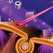
Unraveling how cells mend after injury serves as a key to unlocking potential therapies. Recent findings from the Center for Genetic Medicine Research at Children’s National Hospital offered surprising insights into the cell’s healing mechanisms by illuminating the intricate cellular responses to various types of injuries.

The study, featured on the back cover of the latest issue of Advanced Science, found that cells respond in distinct ways depending on the type of injury, such as a traumatic muscle tear that creates a large injury or tiny holes in the cell membrane caused by pathogenic proteins. Daniel Bittel, DPT, Ph.D., a research postdoctoral fellow at the Center for Genetic Medicine Research, said that cells are routinely injured from even everyday activities, such as walking up a flight of stairs.

“Injuries often involve damage to the plasma membrane,” Bittel said. “We wanted to investigate how healing happens at the subcellular level to better understand diseases and develop targeted therapies. We were especially curious about muscle cells because, interestingly, healthy ones get stronger the more that they are injured.”

The fine print

Using the center’s unique, custom-built microscope, the research team zoomed in on the process of cellular healing to watch how cells activate repair after injuries. Using a laser to damage the plasma membrane, they mimicked mechanically induced trauma. They also used a pathogen-derived protein to create nanoscale pinprick injuries in a cell’s plasma membrane that resemble those that are seen after strenuous muscle exertion.

Then, they watched as cells went to work within seconds, engaging healing mechanisms tailored to the type of injury. In the case of a cell facing numerous pinpricks along the cell membrane, it immediately deployed the endocytic pathway used by the cells to eat and drink. This process helped remove the injurious agents and the tiny holes they made. However, with a larger mechanical injury, the cells demonstrated patience, allowing the plasma membrane to seal before clearing up the damage by the same endocytic pathway.

 The big picture

The paper is part of an ongoing body of research on cell injury that will inform future investigations into a wide range of pediatric health issues including muscular dystrophies, injuries to neurons, orthopedic injuries from sports and other mechanical damage to tissues.

Jyoti Jaiswal, M.Sc., Ph.D., senior investigator at the Center for Genetic Medicine Research, said this work is foundational in the development of new therapies. “Knowing where the problem lies will help us figure out what therapy will work best and target the therapy to address the specific deficit,” he said. “This work will pave the way to help tailor therapies and tackle diseases more effectively.”

NexTGen team assembles to delve into progress on CAR T-cell therapies

The international NexTGen team assembled at the Children’s National Research & Innovation Campus for their annual meeting to share progress made in their first full year of work on the $25-million Cancer Grand Challenge, focused on creating a CAR T-cell therapy for pediatric solid tumors.

“It was invigorating to bring the whole team together from our eight institutions in the U.S., U.K. and France, as we uncover opportunities in our research and share the headway that we have made,” said Catherine Bollard, M.D., M.B.Ch.B., director of the Center for Cancer and Immunology Research and interim chief academic officer at Children’s National Hospital. “Breakthroughs happen when Team Science collaborates, and that is exactly what is happening here with the NexTGen team.”

Why we’re excited

Over the course of two days, more than 85 team members met to discuss the six work packages that are coming together, with the ambitious goal of making CAR T-cell therapies the standard of care for solid tumors within the next decade:

  • Discovery of new targets
  • The tumor microenvironment
  • Component engineering
  • Integration and modeling
  • Clinical studies
  • Data integration

Each work package includes a patient advocate – individuals with a personal connection to cancer as a family member or survivor – who offers their invaluable perspectives on the research and treatment process. Many attended the meeting, sitting alongside the oncologists, immunologists, mathematicians, molecular biologists and other leading experts.

The big picture

The Cancer Grand Challenges are funded by grants from the National Cancer Institute, Cancer Research U.K. and the Mark Foundation for Cancer Research. Their goal is to drive progress against cancer by empowering global leaders in the research community to take on tough challenges and think differently.

“They call it a ‘grand challenge’ for a reason,” Dr. Bollard said. “It’s going to take the effort and expertise of all these individuals to make a new therapy a reality. I have confidence that we can do it.”

Cell & Gene Therapy in the DMV: Experts collaborate for cures

Leaders in medicine, academia, industry and state and local government came together for the first annual Cell and Gene Therapy in the DMV Symposium, hosted at the Children’s National Research & Innovation Campus. The mission: Connect the local scientific community – bursting with expertise and collaboration potential – to develop these cutting-edge therapies for cancers, sickle cell disease and immune-mediated disorders.

The daylong event drew over 100 experts from a range of organizations in the D.C, Maryland and Virginia region, sometimes called the DMV: Children’s National Hospital, the Food and Drug Administration, the National Institute of Standards and Technology, the National Institutes of Health, the General Accounting Office, Virginia Tech, MaxCyte, AstraZeneca, Kite Pharma, Montgomery College, the Maryland State Department of Commerce and more. Together, they unraveled a host of topics including the regulatory environment, workforce development and training, research standards and the promise of these therapies.

“Our Cell & Gene Therapy Symposium brings together our current collaborators and future partners in the D.C., Maryland and Virginia space, which is an incredibly rich area. We see tremendous opportunity and breakthroughs in our future,” said Catherine Bollard, M.D., M.B.Ch.B., interim chief academic officer and chief of Pediatrics at Children’s National Hospital. “Many different diseases rely on the immune system to either be ramped up or to be controlled, and we can seize on these biological processes. Cell and gene therapies are at the heart of where medicine is going.”

The big picture

For decades, oncologists largely have turned to the same menu of treatments to fight cancer, including surgery, chemotherapy and radiation. Cell and gene therapies offer the promise of training the immune system to fight diseases with fewer side effects and potentially higher success rates. Early work has shown progress in liquid cancers, like leukemia, raising the possibility that the therapies could be used on solid tumors and other disorders, such as lupus and sickle cell disease. However, many disciplines must come together to yield discoveries.

“Nobel Prize-winning work doesn’t necessarily translate into available therapies for patients. It takes a whole community like this to make it happen,” said Cenk Sumen, chief scientific officer at MaxCyte Inc., an international cell engineering company based in Rockville, Md. “It has been exciting to see this diverse group of stakeholders come together, which is probably unmatched anywhere on the planet.”

Why we’re excited

Symposium host Patrick Hanley, Ph.D., chief and director of the Cellular Therapy Program at Children’s National, said the goal was to cement the region as the No. 1 location for this highly technical research and development. He believes Children’s National can offer essential elements to this success, given its clinical and research expertise, workforce training opportunities and geographic proximity to the scientific leadership of the federal government. “What makes us unique is our proximity to all the players who can help create new treatment options for patients. We truly are the biomedical capital of the world,” he said.

Michael Friedlander, vice president for health sciences at Virginia Tech, notes that the earliest stages of invention will emanate from academic labs including those at Virginia Tech and Children’s National. “You have basic scientists who are doing fundamental research on properties and procedures that will lead to the new therapies of tomorrow,” he said. “We are putting in place the fundamental pieces to advance children’s health in all dimensions.”

What’s ahead

One challenge is developing a workforce to help prepare cell therapies for patients, following precise standards to ensure the therapy works as designed. Children’s National does this training, as do others in the region. Lori Kelman, Ph.D., M.B.A., biotechnology coordinator and professor at Montgomery College, said that the area is full of people who want to help people and who like science.

“The thing that people might not know is that you don’t need a Ph.D. to work in cell and gene therapy,” she said. “There are opportunities at all levels, including the entry level, which is where a great career often starts.”

Children’s National selected as member of ARPA-H Investor Catalyst Hub spoke network

ARPA-H logoThe hospital will advocate for the unique needs of children as part of nationwide network working to accelerate transformative health solutions.

Children’s National Hospital was selected as a spoke for the Investor Catalyst Hub, a regional hub of ARPANET-H, a nationwide health innovation network launched by the Advanced Research Projects Agency for Health (ARPA-H).

The Investor Catalyst Hub seeks to accelerate the commercialization of groundbreaking and accessible biomedical solutions. It uses an innovative hub-and-spoke model designed to reach a wide range of nonprofit organizations and Minority-Serving Institutions, with the aim of delivering scalable healthcare outcomes for all Americans.

“The needs of children often differ significantly from those of adults. This partnership reflects our commitment to advancing pediatric healthcare through innovation and making sure we’re addressing those needs effectively,” said Kolaleh Eskandanian, Ph.D., M.B.A., vice president and chief innovation officer at Children’s National. “Leveraging the strength of this hub-and-spoke model, we anticipate delivering transformative solutions to enhance the health and well-being of the patients and families we serve.”

Children’s National joins a dynamic nationwide network of organizations aligned to ARPA-H’s overarching mission to improve health outcomes through the following research focus areas: health science futures, proactive health, scalable solutions and resilient systems. Investor Catalyst Hub spokes represent a broad spectrum of expertise, geographic diversity and community perspectives.

“Our spoke network embodies a rich and representative range of perspectives and expertise,” said Mark Marino, vice president of Growth Strategy and Development for VentureWell and project director for the Investor Catalyst Hub. “Our spokes comprise a richly diverse network that will be instrumental in ensuring that equitable health solutions reach communities across every state and tribal nation.”

As an Investor Catalyst Hub spoke, Children’s National gains access to potential funding and flexible contracting for faster award execution compared to traditional government contracts. Spoke membership also offers opportunities to provide input on ARPA-H challenge areas and priorities, along with access to valuable networking opportunities and a robust resource library.

Children’s National awarded nearly $7.5 million by FDA to lead pediatric device innovation consortium

Alliance for Pediatric Device Innovation consortium membersChildren’s National Hospital was awarded nearly $7.5 million in a five-year grant to continue its leadership of an FDA-funded pediatric device consortium. Building upon a decade of previous consortium leadership, the new consortium is Alliance for Pediatric Device Innovation (APDI) and features a new and expanded roster of partners that reflects its added focus on providing pediatric innovators with expert support on evidence generation, including the use of real-world evidence (RWE), for pediatric device development.

Collaborating for success

With the goal of helping more pediatric medical devices complete the journey to commercialization, APDI is led by Children’s National, with Kolaleh Eskandanian, Ph.D., M.B.A., vice president and chief innovation officer, serving as program director and principal investigator, and Julia Finkel, M.D., pediatric anesthesiologist and director of Pain Medicine Research and Development in the Sheikh Zayed Institute for Pediatric Surgical Innovation, serving as principal investigator.

Consortium members include Johns Hopkins University, CIMIT at Mass General Brigham, Tufts Medical Center, Medstar Health Research Institute and MedTech Color. Publicly traded OrthoPediatrics Corp., which exclusively focuses on advancing pediatric orthopedics, is serving as APDI’s strategic advisor and role model for device innovators whose primary focus is children.

Why we’re excited

Consortium initiatives got underway quickly with the announcement of a special MedTech Color edition of the “Make Your Medical Device Pitch for Kids!”competition that focuses on African American and Hispanic innovators. Interested innovators can find details and apply at MedTech Color Pitch Competition. The competition was announced at the recent MedTech Color networking breakfast on Oct. 10,2023 at The MedTech Conference powered by AdvaMed.

“We all benefit from greater equity and inclusion among pediatric MedTech founders, decision-makers, investigators and developers in more effectively addressing the needs of the entire pediatric population,” said Eskandanian. “We need the expertise and insights of innovators from diverse backgrounds, and we want to provide these talented individuals with more opportunities to present their work and share their perspectives on pediatric device development.”

Additional details

APDI is one of five FDA-funded consortia created to provide a platform of services, expertise and funding to help pediatric innovators bring medical devices to the market that specifically address the needs of children.


Babies with congenital heart disease display disrupted brain function before birth

pregnant woman looking at sonogram

In their study, the team at Children’s National Hospital found that specific brain regions become especially vulnerable to injury around 24 weeks of pregnancy when developing babies begin to have high energy demands and rapid neurovascular changes.

For the first time, researchers have found that babies born with congenital heart disease (CHD) have alterations to the emerging functional connectivity of their brains in utero. The changes are related to the subtype of their CHD and their oxygen status before they have lifesaving surgery to treat their cardiac malformation, according to new findings published in the American Heart Association’s Circulation Research.

In their study, the team at Children’s National Hospital found that specific brain regions become especially vulnerable to injury around 24 weeks of pregnancy when developing babies begin to have high energy demands and rapid neurovascular changes. That leaves certain parts of the brain, including the brainstem, more susceptible to injury from cardiac complications and poor circulation.

“We used a special type of magnetic resonance imaging to safely study the brains of these unborn babies, and we found that they have weakened connectivity in the deep grey structures, which are responsible for sensation, movement, alertness and other core functions,” said Josepheen De Asis-Cruz, M.D., Ph.D., assistant professor at the Developing Brain Institute at Children’s National and an author of the study. “This offers an important clue in utero to the type of care the babies will eventually need when they are born.”

The big picture

In the past decade, the survival rate for fetuses with CHD has greatly improved. About 80% of cases – even some of the most high-risk heart defects – can be successfully treated or palliated with surgery and survive. Yet Dr. Cruz said researchers are finding that the rates of poor neurodevelopmental outcomes are about the same. That’s why she and her colleagues are looking at what precisely may be injuring the brains of these newborns prior to surgery, offering a possible roadmap to interventions.

The fine print

The research team studied 107 healthy, low-risk pregnancies and 75 pregnancies known to be complicated by CHD. They used functional connectivity magnetic resonance imaging (fcMRI) to examine the emerging connections of the brains of unborn babies, given fcMRI’s unique ability to query the brain in a resting state when a patient is unable to respond to tasks. They also studied the oxygen saturation levels of the babies after they were born and then mapped all of this information to the type of CHD that they were diagnosed with.

“Our findings indicate that the compromised connectivity in the brains of CHD patients before delivery is linked to hypoxia after birth,” Cruz said. “There were important differences in the low- and high-risk CHD groups. Babies born with transposition of the great arteries or hypoplastic left heart syndrome – two of the most high-risk diagnoses – have notable changes in their brain function, which could someday be used as biomarkers to guide their care.”

What’s ahead

Researchers at Children’s National are working together, using a variety of modalities, to move toward precision imaging in utero to help predict a child’s neurodevelopmental outcomes. The ultimate goal: better interventions sooner.

“This work is foundational. As we fine-tune more techniques to identify babies at risk, we can understand how environmental, genetic and epigenetic factors impact brain development and guide care decisions,” said Catherine Limperopoulos, Ph.D., director of the Center for Prenatal, Neonatal & Maternal Health Research and a senior author of the paper. “We can imagine a day where we can offer pregnant mothers highly detailed and personalized information about their unborn baby, and individualized interventions that lead to healthier lifetimes.”