M.D. in your pocket: New platform allows rare disease patients to carry medical advice everywhere

When someone has a rare disease, a trip to the emergency room can be a daunting experience: Patients and their caregivers must share the particulars of their illness or injury, with the added burden of downloading a non-specialist on the details of a rare diagnosis that may change treatment decisions.

Innovators at Children’s National Hospital and Vanderbilt University Medical Center, supported by Takeda, are trying to simplify that experience using a new web-based platform called the Rare Disease Clinical Activity Protocols, or Rare-CAP. This revolutionary collection of medical information allows patients to carry the latest research-based guidance about their rare disorders in their phones, providing a simple QR code that can open a trove of considerations for any medical provider to evaluate as they work through treatment options for someone with an underlying rare disease.

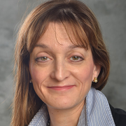
“No one should worry about what happens when they need medical help, especially patients with rare diseases,” said Debra Regier, M.D., division chief of Genetics and Metabolism at Children’s National and Rare-CAP’s lead medical advisor. “We built this new tool because I have watched as my patient-families have wound up in an emergency room — after all, kids get sprains or fractures — but they don’t have the expertise of a rare disease specialist with them. My hope is that they’re going to pull out their phones and access Rare-CAP, which will explain their rare disease to a new provider who can provide more thoughtful and meaningful care.”

The big picture

A rare disease is defined as any disorder that affects less than 200,000 people in the United States. Some 30 million Americans are believed to be living with one of the 7,000 known rare disorders tracked by the National Organization of Rare Diseases (NORD). Led by Dr. Regier, the Rare Disease Institute at Children’s National is one of 40 NORD centers for excellence in the country that provide care, guidance and leadership for the wide array of disorders that make up the rare disease community.

While a key goal of Rare-CAP is to bolster patient self-advocacy, the platform will also allow medical providers to proactively search for protocols on rare diseases when they know they need specialized advice from experts at Children’s National, a network of tertiary care centers and patient organizations.

As a leading values-based, R&D-driven biopharmaceutical company, Takeda has committed $3.85 million to the project to help activate meaningful change and empower a brighter future for rare disease communities, providing a unique understanding of the struggle that patients and caregivers face when they need care.

“Our team, alongside the medical and rare disease community, saw the need for a single portal to collect standardized care protocols, and we are thrilled to see this innovative tool come to life,” said Tom Koutsavlis, M.D., head of U.S. Medical Affairs at Takeda. “People with rare diseases and their caregivers need faster access to authoritative medical information that providers anywhere can act on, this will lead to improving the standard of care, accelerating time to diagnosis and breaking down barriers to increase equitable access.”

The patient benefit

The creators of Rare-CAP imagined its use in a wide range of settings, including emergency rooms, surgical suites, dental offices, urgent care offices and school clinics. The platform will eventually profile thousands of rare diseases and lay out the implications for care, while also creating a dynamic conversation among users who can offer updates based on real-world experience and changes in medical guidance.

“Our patients are unique, and so is this tool,” Dr. Regier said. “As we roll out Rare-CAP, we believe it is just the beginning of the conversation to expand the platform and see its power for the patient and provider grow, with each entry and each new rare disease that’s added to the conversation.”

Andrea L. Gropman, M.D., FAAP, FACMG, FANA, named as the Margaret O’Malley Professor of Genetic Medicine

Andrea L. GropmanChildren’s National Hospital named Andrea L. Gropman, M.D., FAAP, FACMG, FANA, as the Margaret O’Malley Professor of Genetic Medicine at Children’s National Hospital.

Dr. Gropman serves as Chief of the Division of Neurogenetics and Developmental Pediatrics at Children’s National Hospital. She is also a Professor of Pediatrics and Professor of Neurology at George Washington School of Medicine and Health Sciences.

About the award

Dr. Gropman joins a distinguished group of Children’s National physicians and scientists who hold an endowed chair. The Margaret O’Malley Professor of Genetic Medicine is one of 47 endowed chairs at Children’s National.

Professorships support groundbreaking work on behalf of children and their families and foster new discoveries and innovations in pediatric medicine. These appointments carry prestige and honor that reflect the recipient’s achievements and donor’s forethought to advance and sustain knowledge.

Dr. Gropman’s research focuses on neuroimaging, inborn errors of metabolism such as urea cycle disorders and mitochondrial disorders, and neurogenetics. She is the principal investigator of the Urea Cycle Disorders Consortium (UCDC) and the UCDC imaging consortium. She is the deputy clinical director of the Mito EpiGen Program.

Thomas and Mary Alice O’Malley, through their vision and generosity, are ensuring that Dr. Gropman and future holders of this professorship will launch bold, new initiatives to rapidly advance the field of pediatric genetic medicine, elevate our leadership and improve the lifetimes of children with genetic diseases.

About the donors

Tom and Mary Alice O’Malley have partnered with Children’s National to improve the lives of patients with urea cycles disorders for more than two decades. In 2003, their transformational philanthropy helped launch the Urea Cycle Disorders Consortium. This pioneering network grew to include 16-sites worldwide. It garnered 20 years of funding from the NIH’s Rare Diseases Clinical Research Network — the only center to sustain continuous funding over this period. This consortium’s research has yielded multiple effective treatment strategies, including government approval of three lifesaving therapies.

“The O’Malley family’s steadfast generosity helped us grow into the robust community of investigators and families we are today,” says Dr. Gropman. “They transformed care for UCD patients everywhere.”

Researchers publish first-ever atlas of cancerous mutations in histones

Leading genetic researchers at Children’s National Hospital have published the first pan-cancer atlas of key mutations that can drive molecular changes leading to tumors, creating a roadmap that could lead to new treatments for brain tumors and other cancers.

The research – published in npj Genomic Medicine – presents the first-of-its-kind atlas of histone mutations across pediatric, adolescent/young adult and adult cancers. The novel genetic work offers a framework allowing specific cancers to be redefined in the context of changes in histones, which are essential proteins that provide the structural support for chromosomes.

The big picture

“One of the major challenges that we face every day with pediatric, aggressive tumors, including pediatric high-grade gliomas, is that these tumors grow fast. Doctors often have to give patients 9 to 12 months from diagnosis,” said Javed Nazarian, Ph.D., scientific director of the Brain Tumor Institute at Children’s National and principal investigator at the Center for Genetic Medicine Research. “Children’s National has put together a team of clinicians that are truly devoted to finding a therapy for pediatric high-grade gliomas and aggressive pediatric brain tumors. Our dedicated team empowers translational research, from bench to bedside and reverse translation.”

In 2023, the American Cancer Society estimates that 9,910 children under age 15 will be diagnosed with cancer, making it the second leading cause of death among children. Because of treatment advances, 85% will survive, but many will be left with lifelong disabilities from their treatment. Nazarian and his team believe that identifying the underlying molecular alterations leading to cancers will be essential to finding new therapies that extend life expectancies and preserve quality of life.

The fine print

Histones are essential cellular structures, which prevent DNA from getting tangled. Nazarian and other researchers are investigating whether errors in histones could lead to cancers, including high-grade gliomas and other particularly sinister tumors that can strike young children. By mapping the mutations of the histone-encoding genes, Nazarian and his team believe they can find the drivers of tumors in many pediatric and adult cancers. In studying more than 12,000 tumors for the pan-cancer atlas, they cataloged patient ages, survival outcomes and tumor locations to reveal important trends among different cancers.

Overall, the team found that 11% of tumors had somatic histone mutations, with the highest rates observed among chondrosarcoma, a type of bone cancer (67%); pediatric high-grade glioma, a type of cancer that attacks glial cells in the brain and spinal cord (>60%); and lymphoma, a category of cancers in the lymph system (>30%).

“I think one implication of our study is that we are looking at the epigenomic changes of these mutations in a new light,” Nazarian said. “These mutations are not just specific to a particular tumor type, but they are indeed across a large spectrum of cancer types, and they come in different flavors that could potentially show a new avenue for treatments.”

New Becker muscular dystrophy drug on the horizon

photo of muscle collagen

Muscular dystrophies are a collection of genetic disorders that affect boys and cause progressive loss of muscle strength and disability throughout childhood. They impact the protein dystrophin, and other proteins associated with it, which helps strengthen muscles and protect them from injury.

A new corticosteroid – vamorolone – improves symptoms of Becker muscular dystrophy (BMD) with fewer side effects than the off-label treatments currently offered to patients, according to new research from Children’s National Hospital published in iScience.

Currently, there are no drugs approved to treat BMD, an inherited disorder that causes progressive muscle weakness. In preclinical models, daily treatment with vamorolone improved muscle strength and structure with results comparable to prednisolone, which is sometimes prescribed to patients with BMD. Unlike prednisolone, vamorolone is not known to stunt growth, weaken bone and cause negative behaviors.

“Patients with muscular dystrophy can fall anywhere on the spectrum from asymptomatic to facing life-threatening cardiac complications,” said Christopher Heier, Ph.D., principal investigator at the Center for Genetic Medicine Research at Children’s National. “We are excited to have our eye on a drug that may help manage the disease progression, without all the harmful side effects of the steroids currently being offered.”

The big picture

Muscular dystrophies are a collection of genetic disorders that affect boys and cause progressive loss of muscle strength and disability throughout childhood. They impact the protein dystrophin, and other proteins associated with it, which helps strengthen muscles and protect them from injury.

The FDA has approved four drugs to help mitigate the impact of Duchenne muscular dystrophy (DMD), the most severe and most common form of the disease, with dozens more drugs in the research pipeline for that disease subtype. In some cases, these drugs convert DMD into BMD, which is less severe but still greatly affects the quality of life. As a result, the number of patients living with BMD is growing, yet only two drugs are being studied to treat the Becker form of the disease.

Why we’re excited

The Food and Drug Administration is nearing approval for vamorolone to treat DMD. Researchers including Nikki McCormack, Ph.D., a postdoctoral fellow at Children’s National, found it has an added characteristic that makes it particularly helpful to BMD. “Excitingly, by reducing inflammatory signaling in the muscle, we find vamorolone can actually help to correct the underlying dystrophin protein deficiency in BMD through a newly discovered RNA-targeting mechanism.”

Investigators at Children’s National have been interested in expanding vamorolone’s possible use to BMD. Their work builds upon research finished late last year, when they created the first preclinical model to study drugs that could treat BMD. The model provides tremendous hope for those suffering from BMD around the world.

“By creating a pre-clinical model to test possible treatments, we are creating hope for boys living with this life-changing disorder,” said Alyson Fiorillo, Ph.D., principal investigator at the Center for Genetic Medicine Research at Children’s National. “This model, and the drugs it will lead to, will revolutionize how we treat those children living with this disorder.”

New research: Genes that drive testicular cancer identified

In the largest sequencing study to date on testicular cancer, researchers at Children’s National Hospital have identified genes that contribute to testicular germ cell tumors (TGCT), the most common cancer among young, white men.

The findings, published in European Urology, provide direction for future screening and treatment of this disease, which can strike during the teen years and often runs in families. While treatable when identified early, testicular cancer leads to infertility, mental health issues and sometimes death, making its identification crucial for young adults.

“Testicular cancer is really a young person’s disease,” said Louisa Pyle, M.D., Ph.D. , a pediatrician, medical geneticist and research geneticist at the Children’s National Rare Disease Institute. “Most folks who have testicular cancer are between the ages of 15 and 45. Even though testicular cancer is relatively rare in the cancer world, it results in the greatest number of years lost among all adult cancers.”

What we hope to discover

Dr. Pyle led a research team that included experts at the National Cancer Institute and the University of Pennsylvania to study families with multiple members diagnosed with testicular cancer. They used whole exome sequencing to identify variants in many genes that predisposed patients to TGCT. Their work suggests that multiple variants – inherited together – increased the risk for the disease and provides potential routes for drugs that could be used for prevention and treatment.

“We found many genes that help us understand how testicular cancer happens,” Dr. Pyle said. “Our hope is that we can use that to try to come up with better treatments or better ways to preserve fertility for people with testicular cancer or gonadal differences.”

The patient benefit

Testicular cancer most often strikes men of European ancestry. It is also more common among intersex patients and those with differences in sex development, which is a clinical and research focus for Dr. Pyle. Medically, these are children who have a change in the biological characteristics of sex, including their chromosomes, hormones, gonads or physical body parts.

By studying a more common version of testicular cancer, the team learned about the underlying genetics in a way that will benefit intersex patients.

“One of the things we do in medicine is study a common version of the rare thing,” Dr. Pyle said.  “Through this research, we learned that the same genes that cause intersex traits in some patients are also changed in subtle ways for people with testicular cancer. This is a way to study something that could improve care for those kids, by studying a group that has greater numbers.”

Research campus joins Global Network of Innovation Districts

Children’s National Research & Innovation Campus

At the RIC’s 12-acre campus in Northwest Washington, D.C., experts from Children’s National work alongside public and private partners in industry, universities, federal agencies, start-up companies and academic medical centers to find solutions to some of science’s most vexing challenges.

The Children’s National Research & Innovation Campus (RIC) has become the first science ecosystem dedicated to pediatric health to join a network of over three dozen innovation districts worldwide that integrate research space with sustainable communities to create models for urban work and living.

Known as the Global Network of Innovation Districts (GNID), the community was conceived to unlock the design of campuses like the RIC to create collaborations among highly trained professionals. At the RIC’s 12-acre campus in Northwest Washington, D.C., experts from Children’s National work alongside public and private partners in industry, universities, federal agencies, start-up companies and academic medical centers to find solutions to some of science’s most vexing challenges. The campus is surrounded by mass transit, open spaces, retail and housing, and it’s built on deep historic roots in the city as the former home of the Walter Reed Army Medical Center.

Kerstin Hildebrandt, vice president of research administration at the Children’s National Research Institute, said the team at the Research & Innovation Campus is excited to maximize its potential by joining this global network of economic drivers that are enhancing their communities and cities.

“We look forward to sharing our best practices, and we want to learn about how our national and international colleagues are tackling complex issues,” she said. “For example, we can learn how others are leveraging their assets to improve their communities and their response to health crises, climate change and other significant challenges.”

The GNID was launched in March of last year by The Global Institute on Innovation Districts (GIID), an international nonprofit focused on the advancement of innovation districts. With an initial group of 23 districts. GIID is now expanding the network to include approximately 20 additional districts that extend across Europe, North America, Latin America, Australia and Asia.

GIID’s Founder Julie Wagner said innovation districts have become a worldwide phenomenon. She said their leaders are recognizing that working and collaborating with their peers — from Melbourne to Medellin — is a powerful strategy to help these complex geographies leverage their assets in new ways.

“We are finding that innovation districts are willing to execute impactful strategies after holding highly curated exchanges with their peers,” Wagner said. “These are the places armed to solve some of the world’s most vexing challenges. From where I sit, we all need to give them as many tools as possible to help them get there.”

New deep learning system helps scientists edit RNA

RNA molecule

The Children’s National team built DeepCas13 on a newer and less studied CRISPR platform, called CRISPR-Cas13d, which instead focuses on RNA.

Children’s National Hospital scientists have created a revolutionary machine-learning system that predicts the effects of changing ribonucleic acid (RNA) molecules using a gene-editing tool built on CRISPR technology.

Called DeepCas13, the system is among the world’s first deep-learning frameworks to recognize the challenges of editing RNA – and then applying data science and machine learning to solve the intricate problems that stem from modifying biological code. Details of the DeepCas13 system were published recently in Nature Communications.

Born from an international collaboration, DeepCas13 could provide the backbone for treatments for diseases based on errors in RNA, including debilitating neurodegenerative diseases such as Huntington’s disease and muscular dystrophy.

“I am an optimistic person, so I expect to have treatments within five to 10 years,” said Wei Li, Ph.D., principal investigator at the Center for Genetic Medicine Research at Children’s National. “Of course, there are going to be lots of obstacles. If we have a very good system, like DeepCas13, with very good performance that can generate treatments, the next problem is how we deliver the system to the right tissue in the patients.”

The big picture

Most research in this space has focused on a version of CRISPR – or Clustered Regularly Interspaced Short Palindromic Repeats – that edits DNA, called CRISPR-Cas9. The Children’s National team built DeepCas13 on a newer and less studied CRISPR platform, called CRISPR-Cas13d, which instead focuses on RNA. In doing so, researchers are opening the door to treating a host of disorders of RNA, given its biological role in coding, decoding, regulating and supporting gene expression.

DeepCas13 combines hundreds of thousands of data points with considerable computing power to help scientists target errant pieces of RNA, while minimizing any off-target changes that could damage the health of cells.

“We only want to target the RNA molecule that is causing diseases, and we don’t want the system to edit normal RNA,” said Xiaolong Cheng, Ph.D., a member of the Li lab and the first author of the study. “With DeepCas13, we can design highly efficient, and highly specific, rules.”

What’s ahead

The FDA has approved one method for delivering RNA treatments to cells, using a virus known as AAV or adeno-associated virus. So far, the gene therapy method has had limited applications. But Li and other researchers see the potential for life-changing treatments in the coming years, built on DeepCas13 and other advances.

The system was developed with partners from around the world, including the University of Illinois Urbana-Champaign and Northeastern University in Shenyang, China. It is open source and available for free to researchers looking for targets to treat RNA-related diseases.

Li says this international partnership is leading the way: “We tested our DeepCas13 model over other methods, and we confirmed that our method has the highest prediction accuracy.”

DeepCas13 was funded by grants from NIH and the Children’s National Research Institute.

New model to treat Becker Muscular Dystrophy

Researchers at Children’s National Hospital have developed a pre-clinical model to test drugs and therapies for Becker Muscular Dystrophy (BMD), a debilitating neuromuscular disease that is growing in numbers and lacks treatment options.

Their work – recently published in the Journal of Cachexia, Sarcopenia and Muscle – provides scientists with a much-needed method to identify, develop and de-risk drugs for patients with BMD.

“The impact of having a model to test pharmaceutical options cannot be overstated,” said Alyson Fiorillo, Ph.D., principal investigator at the Center for Genetic Medicine Research at Children’s National. “We have patients coming up to us at conferences offering muscle biopsies – on the spot – because they are so excited and relieved that treatments can be investigated.”

Caused by mutations in a gene that produces a protein called dystrophin, Becker is part of a collection of disorders known as muscular dystrophies that cause a progressive loss of muscle strength and increasing disability, starting in childhood. The FDA has approved four drugs to help mitigate the impact of the most common and severe subtype, Duchenne Muscular Dystrophy (DMD). In some cases, these drugs convert the Duchenne form of the disease into Becker, which is less severe but still greatly affects quality of life.

As a result, the population of BMD patients is growing, but patients lack treatments for this incredibly impactful disorder. Currently, the FDA has not approved any drugs for BMD. Only two drugs are in clinical trials, compared to 30 trials underway for DMD.

To address this, Children’s National researchers used CRISPR gene editing to create the first preclinical model of X-linked BMD, called the bmx model. This novel advancement will help researchers better understand BMD and eventually create the first drugs for BMD patients.

“Patients with Becker need therapeutic treatments, and we are excited to start working with the model to someday provide options,” said Christopher Heier, Ph.D., principal investigator at the Center for Genetic Medicine Research and co-author of the study. “Most patients with Becker eventually develop cardiomyopathy, and roughly half die from it. This model is the first step on a path to change that and other heartbreaking outcomes from this genetic disorder.”


CRISPR gene editing identifies possible drug targets for HIV

HIV virus

Working with researchers at Johns Hopkins University, the Children’s National team used CRISPR gene technology to test drug targets that find and attack latent HIV, paving the way for drug treatments that may someday completely cure the virus.

Researchers at Children’s National Hospital have identified several new drug targets that may enhance the elimination of latent HIV in patients, a major bottleneck to the full treatment of the virus, according to new findings published in Science Translational Medicine.

Working with researchers at Johns Hopkins University, the Children’s National team used CRISPR gene technology to test drug targets that find and attack latent HIV, paving the way for drug treatments that may someday completely cure the virus. Currently, anti-retroviral therapies (ARTs) can only slow its progress.

Why we’re excited

“In less than one month, we were able to use CRISPR to test 20,000 gene candidates in one single experiment. It was an amazing application of the technology,” said Wei Li, Ph.D., a co-author of the study and assistant professor at the Center for Genetic Medicine Research at Children’s National. “The CRISPR technology provides a global, unbiased approach to understanding molecular aspects of HIV-1 infection, including the ways that HIV-1 enters cells and replicates. This research could someday revolutionize how we treat the virus pharmaceutically.”

The big picture

More than 30 million people worldwide live with HIV-1, the most common form of the virus that can eventually lead to AIDS. But no single agent can entirely eliminate HIV-1 in these patients.

Researchers have sought ways to attack this elusiveness and turned to the CRISPR gene-editing tool, which can locate specific bits of DNA inside a cell. They trained CRISPR screens on the HIV-1 genome to identify critical factors that allow or prevent the virus from lying latent. In the latter case, these pieces of DNA will be the ideal targets of a drug that will push the virus out of the latent stage so it can be targeted by therapies.

What’s ahead

The findings of the Children’s National and Johns Hopkins scientists point to novel drug therapies and validation systems that could someday eradicate HIV.

Bicna Song, a postdoctoral researcher in Li’s laboratory at the Center for Genetic Medicine, said that reversing HIV-1 latency will allow for the killing of infected cells and give researchers opportunities to actually cure patients with HIV.

“So far, no single latency-reversing agent – alone or in combination with another drug – has been shown to effectively reduce the latent reservoir size in persons living with HIV-1,” said Song, who contributed to the study. “With this work, we are meeting the urgent need to identify factors that can lead to new drug targets.”

Robert Freishtat, M.D., M.P.H., named as Connor Family Professor in Research and Innovation

Robert J. Freishtat

“The Connor Family Professorship will allow my team to act rapidly upon potential transformative discoveries for children’s health” said Dr. Freishtat. “There is no greater honor than to carry the Connor family name as we follow in Dr. Edward Connor’s footsteps to drive breakthroughs that will benefit all children. I am eternally grateful for their support.”

Children’s National Hospital named Robert Freishtat, M.D., M.P.H., as the first Connor Family Professor in Research and Innovation at Children’s National Hospital.

Dr. Freishtat serves as Chief Biotechnology Officer and Senior Investigator, Center for Genetic Medicine Research in the Children’s National Research Institute. He is also a Professor with Tenure in Pediatrics, Emergency Medicine, Genomics and Precision Medicine at The George Washington University School of Medicine and Health Sciences.

About the award

Dr. Freishtat joins a distinguished group of 42 Children’s National physicians and scientists who hold an endowed chair. Professorships at Children’s National support groundbreaking work on behalf of children and their families and foster new discoveries and innovations in pediatric medicine. These appointments carry prestige and honor that reflect the recipient’s achievements and donor’s forethought to advance and sustain knowledge.

Dr. Freishtat is an internationally recognized translational researcher. He is the principal investigator for multiple international collaboratives studying intercellular communication in organ injury/repair. He has authored or co-authored more than 140 articles and book chapters in the fields of lung injury, asthma, obesity, exosomes and emergency medicine.

In 2020, Dr. Freishtat founded the Office of Biotechnology at Children’s National to fast-track novel ideas and forge industry partnerships so solutions can reach patients sooner.

“The Connor Family Professorship will allow my team to act rapidly upon potential transformative discoveries for children’s health” said Dr. Freishtat. “There is no greater honor than to carry the Connor family name as we follow in Dr. Edward Connor’s footsteps to drive breakthroughs that will benefit all children. I am eternally grateful for their support.”

The Connor family, through their vision and generosity, are ensuring that Dr. Freishtat and future holders of this professorship will launch bold, new initiatives to rapidly advance the field of pediatric research and innovation, elevate our leadership and improve the lifetimes of children.

About the donors

Dr. and Mrs. Connor are longtime donors and members of the Children’s National community. Dr. Connor previously served as Director of the Office of Innovation Development and a member of the executive team at the Clinical and Translational Science Institute. His institutional involvement continues through service, formerly as a board member for the Children’s National Research Institute and more recently as a member of the Research, Education, and Innovation Advisory Board. Mrs. Connor, a clinical microbiologist and educator, has worked throughout her career creating a legacy of young people in science.

“We strongly believe in the power of academic entrepreneurship to improve the health and wellbeing of children. This endowment is our way of supporting Children’s National’s work in research and innovation and recognizing Dr. Freishtat’s leadership as an outstanding physician-scientist and role model in clinical and translational pediatrics.”


Researcher to decipher how viruses affect the developing brain with nearly $1M NIH award

zika virus

Zika virus in blood with red blood cells, a virus which causes Zika fever found in Brazil and other tropical countries.

The National Institutes of Health (NIH) awarded Children’s National Hospital nearly $1M of research support toward uncovering the specific cellular response that happens inside a developing brain once it is infected with a virus, including how the neuron gets infected, and how it dies or survives. The research is expected to gather critical information that can inform prenatal neuro-precision therapies to prevent or attenuate the effects of endemic and pandemic viruses in the future.

“We need to use all of the information we have from ongoing and past pandemics to prevent tomorrow’s public health crisis,” said Youssef Kousa, MS, D.O., Ph.D., neonatal critical care neurologist and physician-scientist at Children’s National. “There is still here a whole lot to learn and discover. We could eventually — and this is the vision that’s inspiring us — prevent neurodevelopmental disorders before a baby is born by understanding more about the interaction between the virus, mother, fetus, and environment, among other factors.”

Different viruses, including HIV, CMV, Zika and rubella, injure the developing brain in very similar ways. This line of work was fostered first by the clinical research team led by Adre du Plessis, M.B.Ch.B., and Sarah Mulkey, M.D., supported by Catherine Limperopoulos, Ph.D., chief and director of the Developing Brain Institute at Children’s National.

The clinical research findings then led to the NIH support, which then inspired more basic science research. Fast forward to now, Kousa will study how Zika affects the human brain and extrapolate what is learned and discovered for a broader understanding of neurovirology.

The research program is supported by senior scientists and advisors, including Tarik Haydar, Ph.D., and Eric Vilain, M.D., Ph.D., both at Children’s National and Avindra Nath, M.D., at NIH, as well as other leading researchers at various U.S. centers.

“This is a team effort;” added Kousa, “I’m thankful to have a group of pioneering and seasoned researchers engaged with me throughout this process to provide invaluable guidance.”

Many viruses can harm the developing brain when they replicate in the absence of host defenses, including the gene regulatory networks responsible for the neuronal response. As a result, viral infections can lead to brain injury and neurodevelopmental delays and disorders such as intellectual disability, seizures that are difficult to treat, and vision or hearing loss.

The big picture

Youssef Kousa

Youssef Kousa, MS, D.O., Ph.D., neonatal critical care neurologist and physician-scientist at Children’s National.

The translational research supported by NIH with this award synergistically complements nationally recognized clinical research programs and ongoing prospective cohort studies at Children’s National to identify the full spectrum of neurodevelopmental clinical outcomes after prenatal Zika and other viral infections led by Dr. Mulkey and Roberta DeBiasi, M.D., M.S..

The award also builds upon strengths at the Children’s National Research & Innovation Campus, which is in proximity to federal science agencies. Children’s National experts from the Center for Genetic Medicine Research, known for pediatric genomic and precision medicine, joined forces with the Center of Neuroscience Research and the NIH-NINDS intramural research program to focus on examining prenatal and childhood neurological disorders.

Kousa received this competitive career development award from the National Institute of Neurological Disorders and Stroke of the National Institutes of Health under Award Number K08NS119882. The research content is solely the responsibility of the authors and does not necessarily represent the official views of the National Institutes of Health.

The hold-up in the field

Many neurodevelopmental disorders are caused by endemic viruses, such as CMV, and by viral pandemics, including rubella as seen in the 1960s and Zika since 2015. By studying Zika and other prenatal viral infections, Kousa and team hope to gain deeper biological understanding of the viral effects toward developing therapies for anticipating, treating and preventing virally induced prenatal brain injury in the long-term future.

To date, little is known about how viruses affect developing neurons and, as a result, prenatal brain injury is not yet treatable. To bridge the gap towards prenatal neuro-precision therapies, the research explores how genes regulate neuronal developmental and viral clearance by innovatively integrating three systems:

  • Cerebral organoids, which illuminate how a neuron reacts to a viral infection
  • Pre-clinical models that link prenatal brain injury to postnatal neurodevelopmental outcomes
  • Populational genomics to identify human genetic risk or protective factors for prenatal brain injury

Given the scope and complexity of this issue, the international Zika Genetics Consortium, founded in 2015 by Kousa and a team of leading investigators across the world, provides critical samples and resources for the third arm of the research by performing comprehensive genomic analyses using sequencing data collected from diverse human populations throughout Central and South America, which are not as heavily sequenced as Western populations. Through partnerships with the Centers for Disease Control and Prevention and NIH, the consortium’s database and biorepository houses thousands of patient records and biospecimens for research studies to better understand how viruses affect the developing human brain.

“It is inspiring to imagine that, in the longer term, we could recognize early on the level of brain-injury risk faced by a developing fetus and have the tools to mitigate ensuing complications,” said Kousa. “What is driving this research is the vision that one day, brain injury could be prevented from happening before a baby is born.”

Grant funds study of two maternally inherited mitochondrial diseases

mitochondria

The National Institutes of Health awarded George Washington University and Children’s National Hospital a grant to study two maternally inherited mitochondrial diseases.

The National Institutes of Health awarded George Washington University and Children’s National Hospital a grant to study two maternally inherited mitochondrial diseases. Andrea Gropman, M.D., division chief of Neurodevelopmental Pediatrics and Neurogenetics at Children’s National, along with her co-investigator, Anne Chiaramello, M.D., from the George Washington University School of Medicine, will lead the study.

The proposed studies focus on two ultra-rare maternally inherited mitochondrial diseases:

  • Mitochondrial Encephalopathy, Lactic Acidosis and Stroke-like episodes (MELAS); and
  • Leber’s Hereditary Optic Neuropathy-Plus (LHON-Plus).

Both diseases are among those studied by the Rare Diseases Clinical Research Network.

“We are really pleased to be able to change the landscape for MELAS and LHON, two mitochondrial disorders with relentless progression and no treatment,” Dr. Gropman said. “This grant represents the fruition of an eight-year collaboration with my colleague Dr. Chiaramello and we are fortunate to be able to deliver this at Children’s National and serve our patients and community.”

Because patients currently do not have access to effective therapeutic intervention, this results in significant disability, morbidity and premature death. The UG3 phase of the study will focus on translational MELAS and LHON-Plus studies and submission of an IND protocol to the Food and Drug Administration. The UH3 phase will focus on a basket clinical trial with MELAS and LHON-Plus to:

  • Provide proof-of-concept that the basket design can be applied to divergent ultra-rare diseases.
  • Advance the dataset for safety and pharmacokinetics/pharmacodynamics of our lead compound for a larger number of patients than in a conventional clinical trial setting.
  • Gather outcomes and practical information for optimizing the design of future basket clinical trial.

“Dr. Gropman is dedicated to giving children with MELAS the very best care,” said Elizabeth Wells, M.D., vice president of Neuroscience and Behavioral Medicine Center at Children’s National. “This new research funding is exciting and means more patients can benefit from the expertise she has developed at Children’s National.”

Two new papers advance aggrecan deficiency research

aggrecan protein

Aggrecan (ACAN) is a large protein found in joint cartilage and growth plates.

Andrew Dauber, M.D., M.M.Sc., division chief of Endocrinology at Children’s National Hospital, and colleagues recently published two papers that describe the phenotypic spectrum of aggrecan deficiency and look at treating the condition with human growth hormone.

Aggrecan (ACAN) is a large protein found in joint cartilage and growth plates. It allows joints to move smoothly and without pain. Aggrecan deficiency — due to heterozygous mutations in the ACAN gene — causes dominantly inherited short stature and, in many patients, early-onset osteoarthritis and degenerative disc disease.

Clinical phenotype of patients with aggrecan deficiency

In 2017, Dr. Dauber led an international consortium of researchers that published a manuscript describing the phenotypic spectrum of 103 individuals – 70 adults and 33 children, including 57 females and 46 males – from 20 families with ACAN mutations. In the study, Dr. Dauber and his colleagues established that short stature and accelerated bone age is common among people with ACAN mutations.

In a new study published in the American Journal of Medical Genetics Part A, Dr. Dauber and colleagues further characterize the phenotypic spectrum of aggrecan deficiency, with an emphasis on musculoskeletal health.

Twenty-two individuals from nine families were enrolled in the study. Recorded histories and examinations focused on joint health, gait analysis, joint specific patient reported outcomes and imaging.

“We performed a detailed analysis of the musculoskeletal manifestations in patients with mutations in the aggrecan gene,” says Dr. Dauber. “We found that patients with mutations in this gene had significant short stature which worsened with age. There was a high prevalence of joint complaints and arthritis in adults, and we were able to detect pre-symptomatic joint damage in children using knee MRIs.”

Treating short stature in aggrecan-deficient patients with human growth hormone

Until now, it was unknown how to treat children with aggrecan deficiency. “Providing growth hormone therapy to children with ACAN gene mutations is relatively new in the field of pediatric endocrinology,” explains Dr. Dauber. “Previously, the assumption was that this was just short stature.”

In a new study, published in The Journal of Clinical Endocrinology and Metabolism, Dr. Dauber and colleagues reported the results of a trial that evaluated the efficacy and safety of recombinant human growth hormone (rhGH) therapy on linear growth in children with ACAN deficiency.

“This is the first prospective trial of growth hormone therapy in patients with mutations in the aggrecan gene,” says Dr. Dauber. “Mutations in the gene are the cause for short stature in approximately 2%  of individuals with idiopathic short stature.”

The open-label, single-arm, prospective study enrolled ten treatment-naïve patients with a confirmed heterozygous mutation in ACAN. Participants were treated with rhGH (50 µg/kg/day) over 1 year. Main outcomes measured were height velocity and change in height standard deviation score.

The authors found that growth hormone led to short term improvements in growth rate over the course of the year. The treated patients had their growth rate increase from 5.2 centimeters per year to 8.3 centimeters per year while on therapy.

In 2019, the researchers received the 2019 Human Growth Award at the Pediatric Endocrine Society’s Annual Meeting for an abstract related to this work, entitled “Clinical Characterization and Trial of Growth Hormone in Patients with Aggrecan Deficiency: 6 Month Data.”

Algorithm for antibiotic use benefits patient care in cystic fibrosis

boy using a nebulizer

Despite national consensus guidelines for antibiotic treatment for pulmonary exacerbations in people with CF, prior research and systematic reviews have repeatedly demonstrated a longstanding lack of sufficient evidence for empiric antibiotic therapy recommendations, leading to a significant variation in how antibiotics are prescribed across CF care centers in the United States.

In a recent quality improvement project, researchers from Children’s National Hospital found that antimicrobial stewardship initiatives are beneficial in standardizing care and fostering positive working relationships between cystic fibrosis (CF) pulmonologists, infectious disease physicians and pharmacists.

Antimicrobial stewardship is a systematic effort to change prescribing attitudes that can provide benefit for people with CF. The objective of this effort was to decrease unwarranted use of broad-spectrum antibiotics for children and adolescents with CF. Through initiation of the empiric antibiotic algorithm, researchers found that the proportion of pulmonary exacerbations with antibiotic use consistent with the algorithm increased from 46.2% to 79.5%.

“This work will provide people with CF the expectation of a more consistent approach in their care, as well as the benefits of care from a multidisciplinary team of experts,” said Andrea Hahn, M.D., an infectious diseases specialist at Children’s National and co-author. “Implementing antimicrobial stewardship in the context of care to persons with CF provides benefit in this complex patient population.”

Despite national consensus guidelines for antibiotic treatment for pulmonary exacerbations in people with CF, prior research and systematic reviews have repeatedly demonstrated a longstanding lack of sufficient evidence for empiric antibiotic therapy recommendations, leading to a significant variation in how antibiotics are prescribed across CF care centers in the United States.

The researchers found that implementing antimicrobial stewardship when caring for people with CF provides benefit in this complex patient population.

“We would encourage other CF centers to explore their own trends in practice to determine whether a similar intervention may be both feasible and beneficial in the treatment of pulmonary exacerbations in persons with CF,” said Dr. Hahn.

Continuing medical education through online games

Med Games LogoAndrew Dauber, M.D., MMSc., chief of Endocrinology at Children’s National Hospital, participated as the faculty chair of a Med Games CME educational online activity, to provide continuing medical education for physicians. This game-based activity is intended to meet the educational needs of endocrinologists, endocrine fellows, primary care physicians, physician assistants, nurse practitioners and health care professionals who diagnose and manage children with growth hormone deficiency (CGHD). This CME educational program is provided by Med Learning Group and supported by an educational grant from Novo Nordisk Inc.

Learn more about the program and test your knowledge: ‘Accurate Diagnosis and Effective Management of Children with Growth Hormone Deficiency: What Can You Do to Improve Patient Outcomes in Your Clinical Practice?’

Gene therapy offers potential long-term treatment for limb-girdle muscular dystrophy 2B

Microscopic visual of a diseased muscle section

Microscopic visual of a diseased muscle section. Credit: Daniel Bittel.

Children’s National Hospital experts developed a new pre-clinical gene therapy for a rare disorder, known as limb-girdle muscular dystrophy (LGMD) 2B, that addresses the primary cellular deficit associated with this disease. Using a single injection of a low dose gene therapy vector, researchers restored the ability of injured muscle fibers to repair in a way that reduced muscle degeneration and enhanced the functioning of the diseased muscle. The treatment was safe, attenuated fibro-fatty muscle degeneration, and restored myofiber size and muscle strength, according to the study published in the Journal of Clinical Investigation.

With an incidence of less than 1 in 100,000, LGMD2B is a rare disorder caused by a genetic mutation in a large gene called dysferlin. This faulty gene leads to muscle weakness in the arms, legs, shoulder and pelvic girdle. Affected children and adults face trouble walking, climbing stairs and getting out of chairs. Individuals typically lose the ability to walk within years after the onset of symptoms, and often need assistance with everyday tasks such as showering, dressing and transferring.

This study described a new approach that avoids the need for packaging a large gene, like dysferlin, or giving a large vector dose to target the muscles, which are bottlenecks faced in ongoing gene therapy efforts aimed at muscular dystrophies.

“Currently, patients with LGMD2B have no gene or drug-based therapies available to them, and we are amongst the few centers developing therapeutic approaches for this disease,” said Jyoti K. Jaiswal, M.Sc. Ph.D., senior investigator of the Center for Genetic Medicine Research at Children’s National. “We are working to further enhance the efficacy of this approach and perform a longer-term safety and efficacy study to enable the clinical translation of this therapy.”

The genetic defect in dysferlin that is associated with LGMD2B causes the encoded protein to be truncated or degraded. This hinders the muscle fiber’s ability to heal, which is required for healthy muscles. In recessive genetic disorders, like LGMD2B, common pre-clinical gene therapy approaches usually target the mutated gene in the muscle, making them capable of producing the missing proteins.

“The large size of the gene mutated in this disease, and impediments in body-wide delivery of gene therapy vectors to reach all the muscles, pose significant challenges for developing gene therapies to treat this disease,” said Jaiswal.

To overcome these challenges, the researchers found another way to slow down the disease’s progression. The authors built upon their previous discovery that acid sphingomyelinase (hASM) protein is required to repair injured muscle cells. In this current work, the research team administered a single in vivo dose of an Adeno-associated virus (AAV) vector that produces a secreted version of hASM in the liver, which then was delivered to the muscles via blood circulation at a level determined to be efficacious in repairing LGMD2B patient’s injured muscle cells.

“Increased muscle degeneration necessitates greater muscle regeneration, and we found that improved repair of dysferlin-deficient myofibers by hASM-AAV reduces the need for regeneration, causing a 2-fold decrease in the number of regenerated myofibers,” said Daniel Bittel, D.P.T., PhD., research postdoctoral fellow of the Center for Genetic Medicine Research at Children’s National and a lead author of this study.

Sreetama Sen Chandra, Ph.D., who was a research postdoctoral fellow at Children’s National at the time of this study and served as co-lead author, also added that “these findings are also of interest to patients with Niemann-Pick disease type A since the pre-clinical model for this disease also manifests poor sarcolemma repair.”

Children’s National researchers of the Center for Genetic Medicine Research and the Rare Disease Institute (RDI) are constantly pursuing high-impact opportunities in pediatric genomic and precision medicine. Both centers combine its strengths with public and private partners, including industry, universities, federal agencies, start-up companies and academic medical centers. They also serve as an international referral site for rare disorders.

Gene therapy Schematic

Gene therapy Schematic. Credit: Daniel Bittel.

Integrating clinical parameters with lung imaging to predict respiratory outcomes in premature babies

computer circuit board

The team will develop an objective framework to predict the risk and assess the severity of respiratory disease in premature babies using non-invasive low-radiation X-ray imaging biomarkers and clinical parameters from the patient bedside.

Children’s National Hospital received a $1.7M award from the National Institutes of Health (NIH) National Heart, Lung, and Blood Institute (NHLBI) to develop computational tools that integrate continuous clinical parameters with lung imaging to predict respiratory outcomes for babies born severely premature in newborn intensive care unit (NICU) settings.

The multi-disciplinary team of internationally recognized experts in quantitative imaging, machine learning and neonatal respiratory research believes they can improve clinical practice. To get there, they will develop an objective framework to predict the risk and assess the severity of respiratory disease in premature babies using non-invasive low-radiation X-ray imaging biomarkers and clinical parameters from the patient bedside.

“This computational tool will assist clinicians in making critical decisions about the course of therapy and other necessary follow-ups,” said Gustavo Nino, M.D., M.S.H.S., D’A.B.S.M., principal investigator in the Center for Genetic Medicine at Children’s National. “An objective informed decision about the severity of lung disease in prematurity will result in fewer rehospitalizations, better long-term outcomes and life-saving benefits.”

Prematurity is the largest single cause of death in children under five in the world. Lower respiratory tract infections (LRTI) are the top cause of hospitalization and mortality in premature infants. Clinical tools to predict the risk and assess the severity of LRTI in premature babies are needed to allow early interventions that can decrease the high morbidity and mortality in this patient group.

“Our new technology will provide clinicians an accurate, fast and comprehensive summary of the respiratory status of premature babies,” said Dr. Nino. “The data analysis along with the software technology will help determine if a premature baby seen in the NICU can be safely discharged or will require further monitoring and treatment.”

Predictive analytics could help in many ways. For example, there are instances where newborns in the NICU are on the right path with no risks in the future, but there are babies who will come back with severe infections.

“In the first scenario, if we can predict earlier that they’re fine, this could reduce the number of chest X-rays and extra tests, so we assess that this child can be safely sent home,” said Marius George Linguraru, D.Phil., M.A., M.Sc., principal investigator in the Sheikh Zayed Institute for Pediatric Surgical Innovation at Children’s National. “On the other hand, for kids that may come back to the hospital in the near future, we could predict earlier that they are not that well by looking at images and other continuous measurements such as supplemental oxygen.”

This approach, in essence, is a collection of continuous data from the NICU, which is very complex itself because it needs to be collected every day and fed into a machine learning model that digests the data to identify risk patterns for the health of the lung.

“If we find that there is still a risk, it does not necessarily mean that the child has to stay in the NICU any longer, but they might continue treatment, and we will have to define how this integrates into the clinical management of these patients,” said Linguraru. “If there is something in the data that we can put our finger on, we will know which kids require timely attention, hopefully reducing future adverse situations with potential comorbidities and financial burdens.”

American Heart Association grant funds study of vascular complications in ADPKD

cystic kidney disease

Ashima Gulati, M.D., Ph.D., pediatric nephrologist at Children’s National Hospital recently was awarded a grant from the American Heart Association. Dr. Gulati’s work will aim to identify the genetic determinants of vascular complications in autosomal dominant polycystic kidney disease (ADPKD).

Cerebrovascular complications such as vascular aneurysms and anomalies are an important cause of morbidity in ADPKD that need to be studied. The goal of Dr. Gulati’s research is to contribute to knowledge towards using molecular medicine to inform individual genetic risk of clinically significant vascular complications in ADPKD.

This work addresses a clinically significant vascular complication in ADPKD, the most common inherited form of kidney failure world-wide.

Q&A with Hans Pohl, M.D., on the future of pediatric urology

Hans Pohl

Hans Pohl, Division Chief, Urology

The Urology team at Children’s National Hospital is led by Hans Pohl, M.D., and includes seven other fellowship-trained pediatric urologists and three nurse practitioners. Dr. Pohl has experience in treating patients with spina bifida and bladder exstrophy, in addition to the other more common diagnoses and in using laparoscopy to perform surgery through minimally invasive techniques.

Here, Dr. Pohl tells us more about the department he is leading and what it means for the future of pediatric urology patients at Children’s National.

What excites you most about current research in the field of urology?

The most cutting-edge research these days pertains to understanding the complex and diverse role played by bacteria in the urinary tract. We previously thought the urinary tract is sterile but that turns out to not be the case. Just like the friendly bacteria living in our bodies, there are bacteria that live within our urinary tracts. It is thought that when the normal function of the urinary tract is disrupted, the balance of healthy to unhealthy bacteria is disrupted. Our faculty at the Children’s National Urology Division are looking at urinary tract infection (UTI) from various aspects. Michael Hsieh, M.D., is investigating the role of bacteria in causing inflammation; Daniel Casella, M.D., has investigated how a drug called varenicline might reduce inflammation in infected kidneys; and Briony Varda, M.D., will be investigating the socioeconomic factors affecting how people living with spina bifida manage their urinary tracts and UTIs. By looking at UTIs at all levels from bacteria factors to host factors to treatment we will understand better how to reduce the impact of a very common problem on human beings.

What are some of the most valuable changes or advancements for the program you hope to see in the next couple of years?

I believe that our next step in program evolution will focus on improving the quality of life of children with urination abnormalities and UTIs. Everyone has experienced that their child has urinary symptoms at some point during childhood, some also have UTIs. Pediatric urologists have learned that normal lower urinary tract function is critically important in reducing a child’s risk for a UTI. Even if a child does not have UTIs, there can be significant social stigma amongst a child’s peers when loss of urine control happens.

What makes the Urology Division at Children’s National unique from other programs in the country?

We have grown considerably over the past several years, adding to our faculty surgeons with complimentary skills outside of the operating room. For instance, we have pediatric urologists who are also basic scientists, translational scientists, systems scientists, a clinical informaticist and minimally invasive surgeons. These varied qualities create a dynamic group of people who bring diverse perspectives to treating patients’ problems and generating creative solutions. We believe that our democratic process of complex care management where all surgeons can openly think about how to optimize patient management is unique. Patients don’t get one opinion from their surgeon, they get ten.

A look at the clinical utility of anti-Mullerian hormone

Blood sample tube for anti-Müllerian hormone

Anti-Mullerian hormone (AMH), also known as Mullerian inhibiting substance (MIS), is a hormone produced exclusively in the gonads. It was originally described in the context of sexual differentiation in the male fetus but has gained prominence now as a marker of ovarian reserve and fertility in females.

In a mini-review published in The Journal of Clinical Endocrinology and Metabolism, Roopa Kanakatti Shankar, M.D., pediatric endocrinologist at Children’s National Hospital and an associate professor of pediatrics at the George Washington University School of Medicine and Health Sciences, and co-authors offer an updated synopsis on AMH and its clinical utility in pediatric patients.

The authors performed a systematic search for studies related to the physiology of AMH, normative data and clinical role in pediatrics. After reviewing 70 clinical studies and systematic reviews, they conclude that, “AMH has widespread clinical diagnostic utility in pediatrics but interpretation is often challenging and should be undertaken in the context of not only age and sex but also developmental and pubertal stage of the child.”

Other authors from Children’s National Hospital include Andrew Dauber, M.D., MMSc, Tazim Dowlut-Mcelroy, M.D., and Veronica Gomez-Lobo, M.D.

Read the full review in The Journal of Clinical Endocrinology and Metabolism.