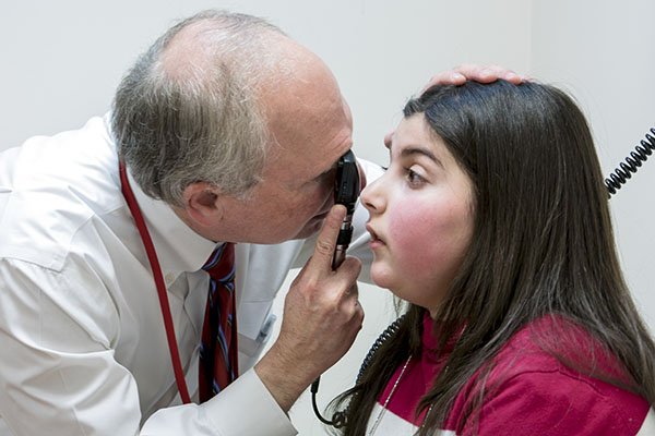
How technology can predict vision loss in neurofibromatosis patients
For the first time, scientists have been able to definitively connect tumor volume and vision loss for children with neurofibromatosis type 1 (NF1). The first study to use quantitative imaging technology to accurately assess the total volume of individual optic nerve glioma (OPG) in NF1 was published in the November 4, 2016 issue of Neurology.
NF1 is a genetic condition that occurs in one in 3,500 births. Children with NF1 develop tumors in multiple locations across the nervous system. About 20 percent of children with NF1 will develop optic pathway gliomas, or tumors that occur in the visual system. Half of those with OPG will have irreversible vision loss, which occurs at a very young age, usually before age 3.
“Neuroradiologists typically assess these tumors through a measurement of the tumor’s radii using magnetic resonance images (MRI) of the patient,” said Marius George Linguraru, D.Phil., M.A., M.S., Principal Investigator in the Sheikh Zayed Institute for Pediatric Surgical Innovation at Children’s National Health System, who is senior author on the study.
“These measurements aren’t detailed enough to serve as a good indicator of whether an OPG will cause vision loss for a child. Through automated computerized analysis, however, we’ve taken the MRI data and systematically analyzed the size and shape, as well as documented changes over time, all in 3-D, to pinpoint the volume of each tumor.”
A look inside the study
The study included children with NF1-related OPGs who are currently cared for at the Gilbert Family Neurofibromatosis Institute at Children’s National. Investigators compared the MRI analysis to the patients’ retinal nerve fiber layer (RNFL), a measure of the health of the visual system. The analysis showed a quantifiable negative relationship between increasing tumor volume within the structures of the anterior visual pathway (the optic nerve, chiasm, and tract) and decreasing thickness of the RNFL, indicating damage to the visual system and vision loss.
“Measuring the tumors in a precise, systematic manner, along with knowing how they grow, is the first step in recognizing which children are at highest risk for vision loss and to potentially identifying them before they suffer any visual symptoms,” added Dr. Linguraru. “If we know which children will probably lose vision, we can treat earlier, and perhaps improve how patients respond to treatment.”
A multicenter collaborative study to validate the findings will begin in 2017.