Children’s National participants share their expertise at PAS meeting
![]()
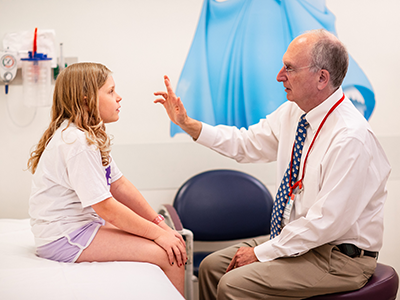
The 2021 Pediatric Academic Societies (PAS) Virtual meeting hosted live-streamed events, on-demand sessions with live Q+A, a virtual exhibit hall, poster presentations and networking events that attracted pediatricians and healthcare providers worldwide. Among the physician-scientists, there were over 20 Children’s National Hospital-affiliated participants at this year’s meeting, adding to the conversation of pediatric research in specialty and sub-specialty areas.
Children’s National experts covered a range of topics, including heart disease, neurology, abnormal glycemia in newborns and antibiotic use in hospitalized children.
The “Neurological Implications of Abnormal Glycemia in Neonatal Encephalopathy and Prematurity” was a hot topic symposium presented by a panel of experts, including Sudeepta Basu, M.B.B.S., M.S., neonatologist at Children’s National.
The experts addressed the importance of recognizing early blood glucose disturbances in newborns with encephalopathy following birth asphyxia and its likely impact on brain injury and long-term outcomes. Although whole body cooling for newborns with encephalopathy after birth asphyxia is now standard of care in most advanced centers like Children’s National, many newborns still die or have neurological impairments. Dr. Basu emphasized on the need of continued advances in newer therapies and optimizing intensive care support for these vulnerable newborns immediately after birth. Dr. Basu’s presentation focused on the association of not only low blood glucose (hypoglycemia) but also high blood glucose (hyperglycemia) with abnormal motor, visual and intellectual outcomes in surviving newborns.
“Recognizing the problem is the first step for further advancement,” Dr. Basu said. “The scientific community needs to recognize the importance of early glucose status as an early marker for disease severity and risk of brain injury.” To sum up, Dr. Basu drew attention to recent newborn resuscitation guidelines from the International Liaison Committee on Resuscitation (ILCOR), which recommends close monitoring of blood glucose levels and optimizing supportive care to maintain it within normal range. Dedicated clinical trials are the need of the hour to guide what are “normal” glucose levels in newborns with encephalopathy and what treatment options are most beneficial.
Rana F. Hamdy, M.D., M.P.H., M.S.C.E., director of the Children’s National Antimicrobial Stewardship Program, delved into the increased number of children receiving care for acute conditions – like acute respiratory tract infections – from urgent care centers and direct-to-consumer (DTC) telemedicine companies during her session “Implementing Antibiotic Stewardship in Telemedicine and Urgent Care Settings.”
Telemedicine, in this case, refers to DTC telemedicine companies—not to be confused with the telemedicine established with primary care providers, like the services provided by Children’s National.
There has been little research focused on promoting good antibiotic stewardship in urgent care settings that tend to overprescribe antibiotics compared to a primary care setting. In addition to her work focusing on improving antimicrobial use within Children’s National, Dr. Hamdy has led collaborative quality improvement work nationally in both the pediatric urgent care and DTC telemedicine settings.
“What we’ve learned from our work with the DTC telemedicine setting is that leadership commitment coming from the company is a necessary core element,” Dr. Hamdy said. “There may be unique opportunities in the telemedicine setting to employ the home-grown computer systems for antimicrobial stewardship interventions, for example, incorporating clinical decision support or feedback reports into the electronic health record systems or displaying a commitment letter in the virtual waiting room.”
In the urgent care setting, Dr. Hamdy’s team recruited approximately 150 pediatric urgent care providers to participate in the national quality improvement initiative. Communication training modules for pediatric urgent care providers with scripted language for target infectious conditions — acute otitis media, pharyngitis and otitis media with effusion — were among the successful intervention approaches that led to improved appropriate antibiotic prescribing practices, according to her team’s findings.
“Understanding the prescribing practices in the urgent care setting is important to knowing where and how to focus on target conditions and to be able to support with education and resources,” Dr. Hamdy said. “And understanding the perceived barriers to judicious antibiotic prescribing can help to identify the highest yield interventions.”
This also reflects the approach taken by the outpatient antibiotic stewardship team at the Children’s National Goldberg Center, led by Ariella Slovin, M.D., primary care pediatrics provider at Children’s National Hospital. Dr. Slovin’s oral abstract entitled “Antibiotic Prescribing Via Telemedicine in the Time of COVID-19,” examined the effect that a shift to telemedicine due to the COVID-19 pandemic had on antibiotic use for acute respiratory tract infections. Overall, her team found a decrease in the proportion of acute respiratory tract infections prescribed antibiotics and concluded that the shift to telemedicine did not adversely affect judicious antibiotic prescribing for acute respiratory tract infections.
Other participants from Children’s National included: Taeun Chang, M.D.; Yuan-Chiao Lu, Ph.D.; Chidiogo Anyigbo, M.D., M.P.H.; Panagiotis Kratimenos, M.D.; Sudeepta Basu, M.B.B.S., M.S.; Ashraf Harahsheh, M.D., F.A.C.C., F.A.A.P.; Rana F. Hamdy, M.D., M.P.H., M.S.C.E.; John Idso, M.D.; Michael Shoykhet, M.D., Ph.D.; Monika Goyal, M.D.; Ioannis Koutroulis, M.D., Ph.D., M.B.A.; Josepheen De Asis-Cruz, M.D., Ph.D.; Asad Bandealy, M.D., M.P.H.; Priti Bhansali, M.D.; Sabah Iqbal, M.D.; Kavita Parikh, M.D.; Shilpa Patel, M.D.; Cara Lichtenstein, M.D.
To view the PAS phase I mini session list and the various areas of expertise at Children’s National, visit: https://innovationdistrict.childrensnational.org/childrens-national-hospital-at-the-2021-pediatric-academic-societies-meeting/