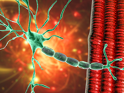
Neurodevelopment occurs at a rapid pace during the preschool years, research shows.
Measuring and tracking development in early childhood is important for understanding how to best support long-term positive health outcomes for children. However, the art of selecting measurement tools that are appropriate for a given study sample is complex; variations in language, cultural relevance and adaptations, availability of normative data, and more can impact the administration and interpretation of study findings.
In a recent study published in the Journal of Pediatric Psychology, the authors systematically review the standardized neurodevelopmental assessments used to study preschool-aged children’s cognitive development in Spanish-speaking Latin America.
“Thinking critically about the ways we measure these outcomes is an important first step in conducting ethical and accurate global research,” said Meagan Williams, M.S.P.H., C.C.R.C., senior clinical research coordinator for the division of Infectious Diseases and the Zickler Family Prenatal Pediatrics Institute at Children’s National Hospital and corresponding author of the study.
The hold-up in the field
The field of child development research is complicated by the presence and use of many different neurodevelopmental assessment tools, only some of which are standardized and use norms to allow researchers to compare their cohorts’ performance with population-level data.
“Many cognitive assessments, including some of the most commonly used assessments identified in our review of research conducted in Spanish-speaking Latin America, have only been officially normed in the United States,” Williams added. “Others have been translated into Spanish and normed in large countries such as Mexico or Spain, but Latin America is a large and culturally diverse region, and it is unclear whether these normative samples are generalizable and culturally relevant to other regions of Spanish-speaking Latin America.”
This systematic review outlined 41 different neurodevelopmental assessments used in 97 studies, including information about each study’s sample, use of each measurement tool and findings related to child cognitive development. The review goes into detail about the most widely used assessments, including background information of each assessment and trends in how authors of the studies in this review reported their methods and results.
Moving the field forward
When researchers use similar methods and valid outcome measures across studies, it becomes possible to combine data between cohorts and draw stronger conclusions about child outcomes. For example, The Wechsler Preschool and Primary Scale of Intelligence (WPPSI) was identified as the most used tool to measure intelligence of young children ages 2-6 in the systematic review. The WPPSI is also one of many assessments that Williams’s research team, led by Sarah Mulkey, M.D., is using to measure neurodevelopment among children exposed to Zika virus in utero.
“If researchers studying longitudinal outcomes after Zika virus exposure also use the WPPSI for their study samples – and if they openly report on their methods, adaptations, and sample characteristics – then it may be possible to harmonize data from multiple cohorts and draw stronger conclusions across studies,” Williams said.
But even the most popular and widespread assessments such as the WPPSI are not without their limitations. While norms exist for regions such as the United States and Mexico, additional work needs to be done to appropriately adapt and validate these tools for use in other populations.
Benefiting patients
Neurodevelopment occurs at a rapid pace during the preschool years. The earlier neurodevelopmental delays can be identified, the earlier experts can administer interventions to respond to these delays and help children get back on track with their peers.
This is particularly salient for research conducted in low- and middle-income countries, where socioeconomic, environmental and other exposures that are known to impact child development differ from those in higher-resource settings. Internationally, there appears to be significant variability in not only the assessment tools which are selected for use, but also in the methods reported across studies to conduct and interpret these assessments.
“By coming together as a field and prioritizing the use and validation of common and culturally appropriate assessments, we can better understand child outcome data on both an individual study level as well as on a broader population level, which will lead to a better understanding of the unique needs and strengths of the children we serve,” Williams said.
The team at Children’s National has been studying the neurodevelopment of children exposed to the Zika virus in Colombia since the beginning of the Zika epidemic.
“As our study on Zika virus has overlapped with the COVID-19 pandemic, we have had the opportunity to collect child outcome data at a very interesting time in history. We are excited that this review revealed that we chose a popular and well-studied assessment for our data collection in this region and we look forward to publishing our 5-year outcome data (which will include WPPSI data) in the coming year,” Williams and Mulkey concluded.
Dr. Mulkey is committed to studying the long-term neurodevelopmental impacts that viruses like Zika and SARS-CoV-2 have on infants born to mothers who were infected during pregnancy through research with the Congenital Infection Program at Children’s National and in collaboration with colleagues in Colombia.
Additional Children’s National authors include Madison Berl, Ph.D.


 The Children’s National Research Institute released its


 Advanced MRI visualization techniques to follow blood flow in the hearts of cardiac patients. Gene therapy for pediatric patients with Duchenne muscular dystrophy. 3D-printed casts for treating clubfoot. These were among the most popular articles we published on Innovation District in 2023. Read on for our full list.


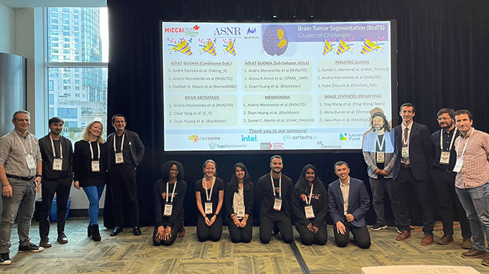
 Several experts from Children’s National Hospital will be sharing their knowledge at the upcoming