Pediatric ophthalmology celebrates 75th anniversary in Washington, D.C.
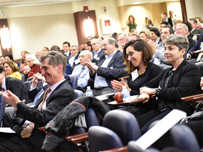
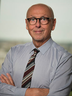
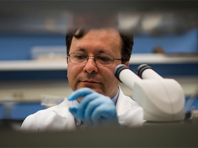
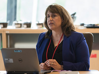
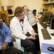
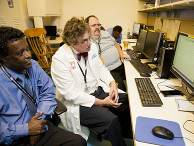
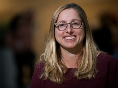
Angeline M. Parks Visiting Professor Sean P. Donahue, M.D., Ph.D., (front left) enjoys a light moment during the celebration of the 75th anniversary while Anthony Sandler, M.D., Children’s surgeon in chief, senior vice president of the Joseph E. Robert Jr. Center for Surgical Care and director of the Sheikh Zayed Institute, speaks to the group.
After 75 years dedicated to the eyes of children, the world’s pediatric ophthalmologists gathered in Washington, D.C., the specialty’s birthplace, to share the latest research and innovation in the field. The group gathered for a joint meeting of the International Strabismological Association (ISA) and the American Association for Pediatric Ophthalmology and Strabismus (AAPOS), which was held March 18-22, 2018.
“This year marks the 75th anniversary of our specialty, which was founded right here, at Children’s National, in Washington, D.C., when Dr. Frank Costenbader restricted his practice exclusively to children and began to train residents in the nuance of treating children’s eyes,” says Mohamad S. Jaafar, M.D., chief of the Division of Ophthalmology at Children’s National Health Center. “It is a tremendous honor to welcome my colleagues back to the birthplace of pediatric ophthalmology on this grand occasion.”
In advance of the larger meeting, Children’s Division of Ophthalmology welcomed some of the international attendees to Children’s National for a special gathering on Saturday, March 17, 2018.
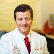
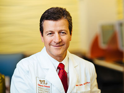
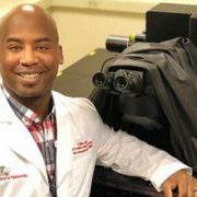
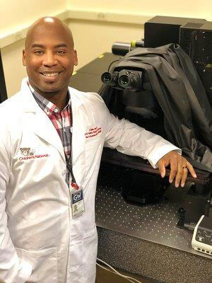
The event at Children’s featured a special lecture by this year’s Angeline M. Parks Visiting Professor, Sean P. Donahue, M.D., Ph.D. Dr. Donahue is the Sam and Darthea Coleman Chair in Pediatric Ophthalmology and Chief of Pediatric Ophthalmology at the Children’s Hospital at Vanderbilt. This Annual Visiting Professorship was established by the members of the Costenbader Society (The Children’s National Pediatric Ophthalmology Alumni Society) in memory of Angeline M. Parks, the wife of pediatric ophthalmologist Marshall M. Parks, M.D., to carry on her legacy of establishing a warm and supportive environment between physician and spouse, which benefits the physicians and their young patients.
Three former division chiefs of Ophthalmology at Children’s National, Drs. Costenbader, Parks and Friendly, have national lectureships established in their names to reflect their contributions to the field. Dr. Frank Costenbader, the society’s namesake, established the sub-specialty of pediatric ophthalmology. Dr. Parks founded the Children’s Eye Foundation and the AAPOS, and David S. Friendly, M.D., codified pediatric ophthalmology fellowship training across the United States.
Honor Awards for Children’s pediatric ophthalmologists at ISA-AAPOS
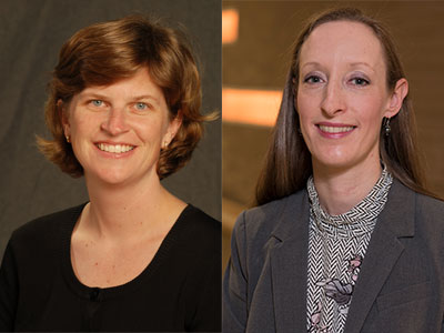
During the ISA-AAPOS meeting, two current Children’s National pediatric ophthalmologists were recognized with Honor Awards for their long-term dedication to pediatric ophthalmology, their patients, and their engagement in the AAPOS to advance the field.
William Madigan, M.D., vice chief of Ophthalmology at Children’s, a professor of surgery at the Uniformed Services University of the Health Sciences, and a clinical professor of Ophthalmology and Pediatrics at the George Washington University School of Medicine and Health Sciences. He was recognized by AAPOS for his long-time service, including:
- Chair of the organization’s audit committee and the Costenbader Lecture selection committee.
- Membership on the fellowship directors’ committee that developed nationwide requirements for pediatric ophthalmology fellowships and established the certification process to insure high quality and uniform education in the specialty.
- Invited lectures in Shanghai, China; Geneva, Switzerland; and Sao Paolo, Brazil, among others.
- Many posters and presentations about clinical and research topics of importance for members of the AAPOS and other distinguished professional societies.
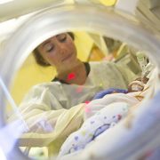
Marijean Miller, M.D., director of Neonatal Ophthalmology, division research director at Children’s National and clinical professor of Ophthalmology and Pediatrics at the George Washington University School of Medicine and Health Sciences, was recognized by AAPOS for her cumulative contributions to the society, including:
- Multiple memberships on vital committees, including AAPOS’s training and accreditation committee and audit committee.
- Presentation of original research via posters and oral presentations on topics including best practices in neonatal clinical care, innovative tools and applications and advocacy for patients and their families.
“We are so grateful to have a team that continues the tradition of excellence in pediatric ophthalmology here at Children’s National,” Dr. Jaafar says. “Drs. Madigan and Miller exemplify the dedication of our division to caring for the children we serve, and to advancing our field. Congratulations to both!”