Genetics & Rare Diseases

Virtual visits: A new house call for rare disease treatment

Dr. Natasha Shur shares “Genetics and Telemedicine: Extending Our Reach” at the Future of Pediatrics CME

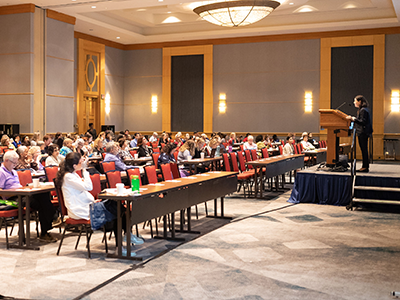
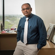
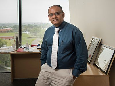
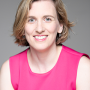
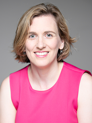
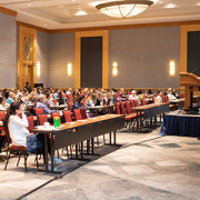
Natasha Shur, M.D., an attending clinical geneticist at Children’s National Health System, shares “Genetics and Telemedicine: Extending Our Reach” at the Future of Pediatrics CME symposium in Bethesda, Maryland, on June 20.

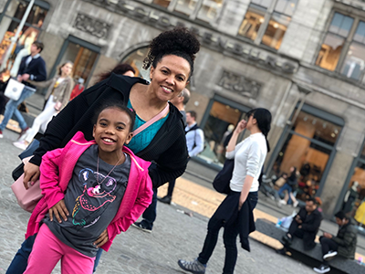
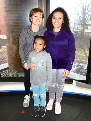
“For the first time it wasn’t autism, autism, autism,” Shannon Chin says after learning the reason her newborn daughter, Sariyah, who turned 3 in August, couldn’t feed like normal infants was due to a tiny deletion of chromosome 22. This atypical deletion, a variation of a genetic condition known as 22q11.2 deletion syndrome, left Sariyah unable to suck and obtain nourishment as an infant. She was born premature and relied on assisted feeding tubes, inserted through her nose, to help her grow.

At 22-weeks-old, Sariyah received the diagnosis, which affects 1 in 4,000 children born each year. Sariyah’s genetic tests encouraged Chin to follow up with a nagging question: What if her two sons, Rueben and Caleb, both of whom were diagnosed with autism spectrum disorder (ASD), had something else?

Debra Regier, M.D., a medical geneticist at Children’s National Health System, encouraged Chin to follow up with a genetic test to answer these questions and to confirm 22q11.2 deletion syndrome symptoms she observed in Rueben.

A microarray analysis recently revealed Rueben, 17, has atypical  22q11.2 deletion syndrome. Caleb, 5, took the test and has developmental delay and ASD, which is more likely to occur in children with 22q11.2 deletion syndrome. He tested negative for the same deletion as his siblings. Additional tests are underway.

As Chin juggles complex care for her children, she realizes the partial deletion of chromosome 22 presents differently in every child. Sariyah and Rueben share short stature; they fit into tiny clothes. That’s where the phenotypical clues stop. They don’t have a cleft palate or dysmorphic facial features, distinctive of typical cases of 22q11.2 deletion syndrome. Sariyah has physical symptoms. Her intestines merged together, which gastrointestinal surgery fixed. Rueben experiences behavioral and neurological symptoms, including picky eating, aggression and uncontrolled body movements, which led the Chin family to Dr. Regier. Sariyah, Rueben and Caleb all have neurodevelopmental delays that impact their speech and development.

Coordinating multiple visits with geneticists, specialists, surgeons, genetic counselors and pediatricians, while navigating insurance, is a lot for any parent, but especially for those, like Chin, who have special considerations. Her children are non-verbal, so she pays close attention to their physical cues. Simplifying this process is one reason why Natasha Shur, M.D., a medical geneticist at Children’s National, introduced virtual visits to her patients, including Rueben, who had challenges with in-person visits. She thought: How can we make medical care easier for patients and families?

In January, Dr. Shur expanded virtual visits into a pilot program for 50 to 60 patients, including Sariyah and Caleb, with the support of a grant from the Health Resources and Services Administration (HRSA), the division of telemedicine at Children’s National and the Rare Disease Institute (RDI), the medical home to thousands of pediatric patients living with rare or genetic conditions. This program lets patients with concern for or already diagnosed genetic conditions in Maryland, the District of Columbia and Virginia, where Dr. Shur is licensed to practice medicine, test out virtual visits. Patients can download the HIPAA-compliant app or click through a secure link on a digital device to connect with Dr. Shur or a pediatric subspecialist.

Dr. Shur shares the preliminary findings of a new virtual visits pilot program,

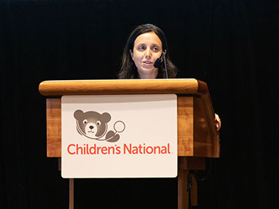
Dr. Shur shares the preliminary findings of a virtual visits pilot program, which 50-60 local patients have tested in conjunction with in-person visits as a flexible way to manage medical care for genetic conditions.

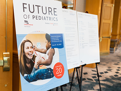
On June 20, Dr. Shur shared a presentation about the program, “Genetics and Telemedicine: Extending Our Reach,” with pediatricians attending the Children’s National Future of Pediatrics continuing medical education (CME) symposium in Bethesda, Maryland.

Instead of a formal pilot program launch and end date with data, Dr. Shur mentions she conducts quality improvement assessments with each patient. She asks what they like about virtual visits. Do they feel comfortable with the software and technology? What types of visits do they prefer to do at home? What works best at the hospital? Do they want to keep using this program?

For Chin and most participants, the answer is yes. These families appreciate saving time, mileage, and being in close access to pediatric subspecialists from the comfort of home.

Parents can conference call from separate locations and share screens with the doctors, which works well if one parent is at work and another is at home – or if they live apart. Children can maintain their normal routine, such as finishing breakfast, homework, playing or staying in bed if they don’t feel well, though it is important to see the child in the virtual visit.

Families can obtain virtual assessments about urgent conditions without taking time off from work or school. Currently, only 10 to 30% of virtual visit patients with concerns about genetic conditions need an in-person, follow-up appointment. Fortunately, many conditions are less urgent than thought at the time of referral. Dr. Shur and specialists also benefit from observing children in their natural environment.

At the symposium, Dr. Shur translates this into clinical terms: reduced no-show visits, the ability to schedule shorter, more flexible visits, the ability to quickly and accurately diagnose conditions and provide care, and the ability to keep children with compromised immune function out of public areas, including waiting rooms. She discussed building rapport with patients, almost all of whom like these flexible care models.

“The idea is that we’re trying to understand what is best done using virtual technology and what is better for those in-person connections. More detailed physical exams take place in person. There are some cases where eye-to-eye contact and sitting in the exam room together is important,” says Dr. Shur. “Virtual visits should never replace in-person care. It’s just a forward way of thinking about: How do we use our time best?”

Case study 1: Saving families time and miles

Dr. Shur notes that for some patients, distance is a deciding factor for scheduling care. One mother’s five-hour round-trip commute to the children’s hospital, without traffic, is now five minutes. As an air-traffic controller, her schedule changes. She values the flexibility of the new program. To connect with Dr. Shur, she logs into the app on her computer or smart phone and brings her 2-year-old son into the video. He has cardiofaciocutaneous syndrome (CFC), a condition that affects 200 to 300 people in the world. As a result of a MAP2K1 gene variant, one of four genes – BRAF, MAP2K1, MAP2K2 and KRAS – associated with CFC, he experiences feeding problems, reflux, constipation and developmental delays.

By scheduling more frequent, but shorter check-ins, Dr. Shur assesses how he responds to treatment and makes recommendations to the mother in real time, such as trying prune juice for digestive health. They talk about rearranging feeding measurements and intervals, including his 2 a.m. dose of a peptide formula, which the mom blends at home to support her son’s growth. This modification equates to more sleep for everyone.

If follow-up tests, such as an X-ray or a blood test are needed, Dr. Shur coordinates these exams with the family at the hospital or at a nearby medical center. Depending on the condition, Dr. Shur may refer the family to an ophthalmologist, cardiologist, neurologist or learning and development specialist.

As a parent, Dr. Shur appreciates the direct approach virtual visits deliver.

“As a mom, if I’m taking my child to the doctor for two hours, I want to know why I’m there,” Dr. Shur says. “What are all the options?”

Case study 2: Observing children at home

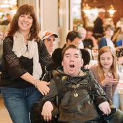
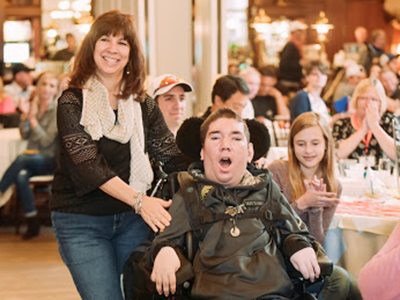
Chin, who was also featured in Dr. Shur’s CME presentation, appreciates virtual visits for their convenience and efficiency, but her favorite feature is letting doctors observe her children at home.

“Children act differently outside the home,” says Chin.

For example, instead of describing Rueben’s rapid, rhythmic arm movements, a flinging of the arms, Chin showed neurologists at a scheduled virtual home visit. For Marc DiFazio, M.D., a pediatric neurologist, it was evident that Reuben had a movement disorder commonly seen in children with ASD, which is responsive to medication. In five minutes, her son had a diagnosis. The involuntarily movement wasn’t a behavioral issue, as previously thought, but a movement disorder.

“The regular in-person visit has a beautiful role and it’s very important, but virtual visits bring a different focus,” says Dr. Shur. “We get to see what the child’s life is like, what the home setting is like and what their schedule is like. How can we make their day-to-day life easier?”

Phenylketonuria (PKU), a rare condition that prevents the body from breaking down phenylalanine (Phe), an amino acid in protein, is another condition that pairs well with virtual visits. PKU affects 1 in 10,000 to 15,000 newborns in the U.S. People with PKU often require medication, food-based formulas and a protein-restricted diet to help their body process or regulate Phe.

If a patient with PKU connects through a virtual visit, they (or their parents) can open the refrigerator, talk about low-protein foods, discuss potential barriers to following a low-Phe diet, show the team new supplements or over-the-counter medications they are taking, discuss reactions to new therapies and, for adults, discuss an injectable drug recently approved by the FDA that has side effects but may ultimately allow them to follow a regular diet. These observations may not warrant a traditional trip to the doctor but are important for geneticists and patients to discuss. The goal of these visits is to identify and work around potential health barriers, while preventing adverse health outcomes.

To support this model, a 60-minute in-person visit scheduled every six months to a year can be broken into 15-minute video appointments at more frequent intervals. The result, based on the same amount of clinical time, is a targeted and detailed assessment to support personalized treatment and to help the patient adapt to a low-Phe meal plan.

During the video call, Dr. Shur and the team may prescribe a different medication, order a diagnostic procedure or schedule a follow-up appointment, if necessary. Depending on the situation, the patient will still likely come in for in-person annual visits.

Program assessment: Evaluating visits for each patient

Despite the popularity of virtual visits, Dr. Shur mentions this program isn’t a good fit for everyone – depending on a patient’s preferences. There are also limitations to consider. If a parent is hesitant to try this platform or if the comprehensive physical examination is the first key step, they should schedule in-person visits. The goal is to give parents who are requesting or curious about virtual visits a chance to try the platform. Having a secure area, preferably a private space at home, is important. A Wi-Fi connection and a digital device are required, which may create barriers for some patients.

However, Dr. Shur finds the program can alleviate hurdles – such as transportation challenges. One patient lives two hours away and couldn’t make it in for routine medical visits due to car problems. Now she makes every virtual appointment. For the first time in her life, she can manage medical care for herself and for her children.

Most insurance companies Dr. Shur works with cover virtual visits. The key is to have the virtual connection, or video, so Dr. Shur can still physically see the patient. Otherwise, the visit doesn’t count. A grant from CareFirst covers the costs of visits for patients who are using Medicaid or who don’t have medical insurance.

Parallel trends are happening across the country and for other conditions. Officials at the Federal Communications Commission (FCC) are reviewing a three-year pilot to expand the use of connected care services, like virtual visits, for low-income Americans living in rural areas. The Rural Health Care Program, funded by the FCC, supports hospitals that implement telehealth programs.

The American Academy of Pediatrics (AAP) released a statement in 2015 about telemedicine technologies, noting that if these technologies are applied in a synergistic model under one health care system or are guided by a family doctor, they can transform pediatric health care.

The key is to avoid a fragmented virtual health system.

The AAP applauds virtual connections that support collaborations among pediatric physicians, subspecialists and surgeons, reduce travel burdens for families, alleviate physician shortages, improve the efficiency of health care and enhance the quality of care and quality of life for children with special health care needs.

Planning for the future, investing in physician-patient partnerships

A poster at the Future of Pediatrics conference

The American Academy of Pediatrics supports telemedicine technologies that enhance the quality of care and the quality of life for children with special health care needs.

“The feedback has been phenomenal,” Dr. Shur says about the future of virtual visits for genetics. “Virtual visits will never replace in-person visits. They will be used in conjunction with in-person visits to maximize care.”

Dr. Regier and Jamie Frasier, M.D., Ph.D., medical geneticists at Children’s National, are introducing virtual visits to their patients, and many providers plan to do so as the program expands.

Sarah Viall, PPCNP, a nurse practitioner and newborn screening specialist, works with Dr. Shur and the geneticists during some visits to explain non-urgent newborn screening results to parents through virtual connections. Some parents find it’s easier to dial in during lunch or while they are together at home.

To improve education for patients and families, the education and technology committees at the RDI – led by geneticists and genetic counselors in partnership with the Clinical and Translational Science Institute at Children’s National – launched a new smartphone app called BearGenes. Families can watch 15 videos about genetics on the pin-protected app or view them online. The interactive guide serves as a gene glossary for terms patients may hear in a clinical setting. Topics range from genetics 101, describing how DNA is encrypted in the body through four letters – A, T, C and G – to different types of genetic tests, such as whole exome sequencing, to look for differences in the spelling of genes, which the genetic counselors explain are genetic mutations.

“As we unite patients with virtual health platforms and new forms of technology, we want to see what works and what doesn’t. We want their feedback,” Dr. Shur reemphasizes. “Virtual visits are a dynamic process. These visits only work through patient partnership and feedback.”

As Chin navigates atypical 22q11.2 deletion syndrome and ASD, she continues to appreciate the virtual waiting room and the ease of access virtual visits provides.

Sharing screens during virtual visits enables Chin to examine and better understand her children’s abdomen and kidney sonograms, cardiology reports and hearing exams. It forces everyone in the visit to focus on one topic or image at a time, strengthening the connection.

Chin still has questions about her children’s DNA, but she’s getting close to having more answers. She’s eager to see Caleb’s genetic test results and to work with Hillary Porter, M.S., CGC, the family’s genetic counselor, to interpret the data.

“We’re all learning together,” Dr. Shur says about the new pilot program, which applies to genomics at large.

As research about 22q11.2 deletion syndrome advances, geneticists, pediatric subspecialists and pediatricians are unifying efforts to work as one diagnostic and treatment team. Virtual visits enable faster consultations and can shorten diagnostic odysseys, some of which may take up to five years for children with rare disorders.

Attendees at the Future of Pediatrics conference

Nearly 400 pediatricians attend the Children’s National Future of Pediatrics CME symposium to learn about the future of pediatrics and about ways to work together as a diagnostic and treatment team.

For Chin, by better understanding how a tiny fragment of a missing chromosome may influence her children’s growth and development, she is already making long-term plans and coordinating multidisciplinary medical treatment for each child.

She hopes that by sharing her story and knowledge about 22q11.2 deletion syndrome, she can help other parents navigate similar situations. Heradvice to parents is to follow up on lingering questions by bringing them up with your medical team.

Chin is optimistic and happy she did. She’s grateful for the virtual visits program, which simplifies complex care for her family. And she’s still waiting, but she hopes to learn more about her middle child’s DNA, unraveling another medical mystery.

Read more about the virtual visits pilot program at Becker’s Hospital Review and listen to an interview with Dr. Shur and Shannon Chin on WTOP.

Focusing on the “little brain” to rescue cognition

illustration of brain showing cerebellum

Research faculty at Children’s National in Washington, D.C., with colleagues recently published a review article in Nature Reviews Neuroscience that covers the latest research about how abnormal development of the cerebellum leads to a variety of neurodevelopmental disorders.

Cerebellum translates as “little brain” in Latin. This piece of anatomy – that appears almost separate from the rest of the brain, tucked under the two cerebral hemispheres – long has been known to play a pivotal role in voluntary motor functions, such as walking or reaching for objects, as well as involuntary ones, such as maintaining posture.

But more recently, says Aaron Sathyanesan, Ph.D., a postdoctoral research fellow at the Children’s Research Institute, the research arm of Children’s National  in Washington, D.C., researchers have discovered that the cerebellum is also critically important for a variety of non-motor functions, including cognition and emotion.

Sathyanesan, who studies this brain region in the laboratory of Vittorio Gallo, Ph.D., Chief Research Officer at Children’s National and scientific director of the Children’s Research Institute, recently published a review article with colleagues in Nature Reviews Neuroscience covering the latest research about how altered development of the cerebellum contributes to a variety of neurodevelopmental disorders.

These disorders, he explains, are marked by problems in the nervous system that arise while it’s maturing, leading to effects on emotion, learning ability, self-control, or memory, or any combination of these. They include diagnoses as diverse as intellectual disability, autism spectrum disorder (ASD), attention-deficit/hyperactivity disorder and Down syndrome.

“One reason why the cerebellum might be critically involved in each of these disorders,” Sathyanesan says, “is because its developmental trajectory takes so long.”

Unlike other brain structures, which have relatively short windows of development spanning weeks or months, the principal cells of the cerebellum – known as Purkinje cells – start to differentiate from stem cell precursors at the beginning of the seventh gestational week, with new cells continuing to appear until babies are nearly one year old.  In contrast, cells in the neocortex, a part of the brain involved in higher-order brain functions such as cognition, sensory perception and language is mostly finished forming while fetuses are still gestating in the womb.

This long window for maturation allows the cerebellum to make connections with other regions throughout the brain, such as extensive connections with the cerebral cortex, the outer layer of the cerebrum that plays a key role in perception, attention, awareness, thought, memory, language and consciousness. It also allows ample time for things to go wrong.

“Together,” Sathyanesan says, “these two characteristics are at the root of the cerebellum’s involvement in a host of neurodevelopmental disorders.”

For example, the review article notes, researchers have discovered both structural and functional abnormalities in the cerebellums of patients with ASD. Functional magnetic resonance imaging (MRI), an imaging technique that measures activity in different parts of the brain, suggests that significant differences exist between connectivity between the cerebellum and cortex in people with ASD compared with neurotypical individuals. Differences in cerebellar connectivity are also evident in resting-state functional connectivity MRI, an imaging technique that measures brain activity in subjects when they are not performing a specific task. Some of these differences appear to involve patterns of overconnectivity to different brain regions, explains Sathyanesan; other differences suggest that the cerebellums of patients with ASD don’t have enough connections to other brain regions.

These findings could clarify research from Children’s National and elsewhere that has shown that babies born prematurely often sustain cerebellar injuries due to multiple hits, including a lack of oxygen supplied by infants’ immature lungs, he adds. Besides having a sibling with ASD, premature birth is the most prevalent risk factor for an ASD diagnosis.

The review also notes that researchers have discovered structural changes in the cerebellums of patients with Down syndrome, who tend to have smaller cerebellar volumes than neurotypical individuals. Experimental models of this trisomy recapitulate this difference, along with abnormal connectivity to the cerebral cortex and other brain regions.

Although the cerebellum is a pivotal contributor toward these conditions, Sathyanesan says, learning more about this brain region helps make it an important target for treating these neurodevelopmental disorders. For example, he says, researchers are investigating whether problems with the cerebellum and abnormal connectivity could be lessened through a non-invasive form of brain stimulation called transcranial direct current stimulation or an invasive one known as deep brain stimulation. Similarly, a variety of existing pharmaceuticals or new ones in development could modify the cerebellum’s biochemistry and, consequently, its function.

“If we can rescue the cerebellum’s normal activity in these disorders, we may be able to alleviate the problems with cognition that pervade them all,” he says.

In addition to Sathyanesan and Senior Author Gallo, Children’s National study co-authors include Joseph Scafidi, D.O., neonatal neurologist; Joy Zhou and Roy V. Sillitoe, Baylor College of Medicine; and Detlef H. Heck, of University of Tennessee Health Science Center.

Financial support for research described in this post was provided by the National Institute of Neurological Disorders and Stroke under grant numbers 5R01NS099461, R01NS089664, R01NS100874, R01NS105138 and R37NS109478; the Hamill Foundation; the Baylor College of Medicine Intellectual and Developmental Disabilities Research Center under grant number U54HD083092; the University of Tennessee Health Science Center (UTHSC) Neuroscience Institute; the UTHSC Cornet Award; the National Institute of Mental Health under grant number R01MH112143; and the District of Columbia Intellectual and Developmental Disabilities Research Center under grant number U54 HD090257.

Children’s National ranked No. 6 overall and No. 1 for newborn care by U.S. News

Children’s National in Washington, D.C., is the nation’s No. 6 children’s hospital and, for the third year in a row, its neonatology program is No.1 among all children’s hospitals providing newborn intensive care, according to the U.S. News Best Children’s Hospitals annual rankings for 2019-20.

This is also the third year in a row that Children’s National has been in the top 10 of these national rankings. It is the ninth straight year it has ranked in all 10 specialty services, with five specialty service areas ranked among the top 10.

“I’m proud that our rankings continue to cement our standing as among the best children’s hospitals in the nation,” says Kurt Newman, M.D., President and CEO for Children’s National. “In addition to these service lines, today’s recognition honors countless specialists and support staff who provide unparalleled, multidisciplinary patient care. Quality care is a function of every team member performing their role well, so I credit every member of the Children’s National team for this continued high performance.”

The annual rankings recognize the nation’s top 50 pediatric facilities based on a scoring system developed by U.S. News. The top 10 scorers are awarded a distinction called the Honor Roll.

“The top 10 pediatric centers on this year’s Best Children’s Hospitals Honor Roll deliver outstanding care across a range of specialties and deserve to be nationally recognized,” says Ben Harder, chief of health analysis at U.S. News. “According to our analysis, these Honor Roll hospitals provide state-of-the-art medical expertise to children with rare or complex conditions. Their rankings reflect U.S. News’ assessment of their commitment to providing high-quality, compassionate care to young patients and their families day in and day out.”

The bulk of the score for each specialty is based on quality and outcomes data. The process also includes a survey of relevant specialists across the country, who are asked to list hospitals they believe provide the best care for patients with challenging conditions.

Below are links to the five specialty services that U.S. News ranked in the top 10 nationally:

The other five specialties ranked among the top 50 were cardiology and heart surgery, diabetes and endocrinology, gastroenterology and gastro-intestinal surgery, orthopedics, and urology.

Vittorio Gallo, Ph.D., inducted into Alpha Omega Alpha

Vittorio Gallo Alpha Omega Alpha Award

Vittorio Gallo, Ph.D., Chief Research Officer at Children’s National, was inducted into Alpha Omega Alpha (AΩA), a national medical honor society that since 1902 has recognized excellence, leadership and research in the medical profession.

“I think it’s great to receive this recognition. I was very excited and surprised,” Gallo says of being nominated to join the honor society.

“Traditionally AΩA membership is based on professionalism, academic and clinical excellence, research, and community service – all in the name of ‘being worthy to serve the suffering,’ which is what the Greek letters AΩA stand for,” says Panagiotis Kratimenos, M.D., Ph.D., an ΑΩΑ member and attending neonatologist at Children’s National who conducts neuroscience research under Gallo’s mentorship. Dr. Kratimenos nominated his mentor for induction.

“Being his mentee, I thought Gallo was an excellent choice for AΩΑ faculty member,” Dr. Kratimenos says. “He is an outstanding scientist, an excellent mentor and his research is focused on improving the quality of life of children with brain injury and developmental disabilities – so he serves the suffering. He also has mentored numerous physicians over the course of his career.”

Gallo’s formal induction occurred in late May 2019, just prior to the medical school graduation at the George Washington University School of Medicine & Health Sciences (GWSMHS) and was strongly supported by Jeffrey S. Akman, Vice President for Health Affairs and Dean of the university’s medical school.

“I’ve been part of Children’s National and in the medical field for almost 18 years. That’s what I’m passionate about: being able to enhance translational research in a clinical environment,” Gallo says. “In a way, this recognition from the medical field is a perfect match for what I do. As Chief Research Officer at Children’s National, I am charged with continuing to expand our research program in one of the top U.S. children’s hospitals. And, as Associate Dean for Child Health Research at GWSMHS, I enhance research collaboration between the two institutions.”

Detecting early signs of type 2 diabetes through microRNA

Robert J. Freishtat working in the lab

Obesity is a major risk factor for insulin resistance and type 2 diabetes. Now researchers understand the pathogenesis better among teens with mid-level obesity, thanks to clues released from circulating adipocyte-derived exosomes.

Researchers know that exosomes, tiny nanoparticles released from fat cells, travel through the bloodstream and body, regulating a variety of processes, from growth and development to metabolism. The exosomes are important in lean, healthy individuals in maintaining homeostasis, but when fat gets ‘sick’ – the most common reason for this is too much weight gain – it can change its phenotype, becoming inflammatory, and disrupts how our organs function, from how our skeletal muscle and liver metabolize sugar to how our blood vessels process cholesterol.

Robert J. Freishtat, M.D., M.P.H., the chief of emergency medicine at Children’s National Health System and a professor of precision medicine and genomics at the George Washington University School of Medicine and Health Sciences, and Sheela N. Magge M.D., M.S.C.E., who is now the director of pediatric endocrinology and an associate professor of medicine at the Johns Hopkins School of Medicine, were curious about what this process looked like in teens who fell in the mid-range of obesity.

Obesity is a major risk factor for insulin resistance and type 2 diabetes, but Dr. Freishtat and Dr. Magge wanted to know: Why do some teens with obesity develop type 2 diabetes over others? Why are some teens in this mid-range of obesity metabolically healthy while others have metabolic syndrome? Can fat in obese people become sick and drive disease?

To test this, Dr. Freishtat and Dr. Magge worked with 55 obese adolescents, ages 12 to 17, as part of a study at Children’s National. The participants – 32 obese normoglycemic youth and 23 obese hyperglycemic youth – were similar in age, sex, race, pubertal stage, body mass index and overall fat mass. The distinguishing factor: The hyperglycemic study participants, the teens with elevated blood sugar, differed in where they stored fat. They had extra visceral fat (or adipose tissue) storage, the type of fat that surrounds the liver, pancreas and intestines, a known risk factor for type 2 diabetes.

Dr. Magge and Dr. Freishtat predicted that circulating exosomes from the teens with elevated blood sugar are enriched for microRNAs targeting carbohydrate metabolism.

They used three tests to examine study participants’ metabolism, body composition and circulating exosomes. The first test, an oral glucose tolerance test, measures how efficiently the body metabolizes sugar; the second test is the whole body DXA, or dual-energy x-ray absorptiometry, which analyzes body composition, including lean tissue, fat mass and bone mineral density; and the third test, the serum adipocyte-derived exosomal microRNA assays, is an analysis of circulating fat signals in the bloodstream.

They found that teens with elevated blood sugar and increased visceral fat had different circulating adipocyte-derived exosomes. These study participants’ exosomes were enriched for 14 microRNAs, targeting 1,304 mRNAs and corresponding to 179 canonical pathways – many of which are directly associated with carbohydrate metabolism and visceral fat.

Dr. Magge will present this research, entitled “Changes in Adipocyte-Derived Exosomal MicroRNAs May Play a Role in the Progression from Obese Normoglycemia to Hyperglycemia/Diabetes,” as an oral abstract at the American Diabetes Association’s 79th Scientific Sessions on Saturday, June 8.

Dr. Freishtat envisions having this information will be especially helpful for a patient in a mid-range of obesity. Exosomes primarily consist of small non-coding RNAs. In the current study, the altered RNAs affect P13K/AKT and STAT3 signaling, vital pathways for metabolic and immune function.

“Instead of waiting until someone has the biochemical changes associated with type 2 diabetes, such as hyperglycemia, hyperlipidemia and insulin resistance, we’re hoping physicians will use this information to work with patients earlier,” says Dr. Freishtat. “Through earlier detection, clinicians can intervene when fat shows sign of illness, as opposed to when the overt disease has occurred. This could be intervening with diet and lifestyle for an obese individual or intervening with medication earlier. The goal is to work with children and teens when their system is more plastic and responds better to intervention.”

As this research evolves, Dr. Freishtat continues to look at the intergenerational effects of circulating adipocyte-derived exosomes. Through ongoing NIH-funded research in India, he finds these exosomes, similar in size to lipoproteins, can travel across the placenta, affecting development of the fetus in utero.

“What we’re finding in our initial work is that these exosomes, or ‘sick’ fat, cross the placenta and affect fetal development,” Dr. Freishtat says. “Some of the things that we’re seeing are a change in body composition of the fetus to a more adipose phenotype. Some of our work in cell cultures shows changes in stem cell function and differentiation, but what’s even more interesting to us is that if the fetus is a female sex that means her ovaries are developing while she’s in utero, which means a mother’s adipocyte-derived exosomes could theoretically be affecting her grandchild’s phenotype – influencing the health of three generations.”

While this research is underway, Dr. Freishtat is working with JPOD @ Boston, co-located with the Cambridge Innovation Center in Cambridge, Massachusetts, to develop a test to provide analyses of adipocyte-derived exosomal microRNAs.

“It’s important for families to know that these studies are designed to help researchers and doctors better understand the development of disease in its earliest stages, but there’s no need for patients to wait for the completion of our studies,” says Dr. Freishtat. “Reaching and maintaining a healthy body weight and exercising are important things teens and families can do today to reduce their risk for obesity and diabetes.”

Gustavo Nino, M.D., honored with national award from American Thoracic Society

Gustavo Nino

Gustavo Nino, M.D., a pulmonologist who directs the Sleep Medicine program at Children’s National, was honored by the American Thoracic Society with The Robert B. Mellins, M.D. Outstanding Achievement Award in recognition of his contributions to pediatric pulmonology and sleep medicine.

“I am humbled and pleased to be recognized with this distinction,” says Dr. Nino. “This national award is particularly special because it honors both academic achievements as well as research that I have published to advance the fields of pediatric pulmonology and sleep medicine.”

After completing a mentored career development award (K Award) from the National Institutes of Health (NIH), Dr. Nino established an independent research program at Children’s National funded by three different NIH R-level grants, an R01 research project grant; an R21 award for new, exploratory research; and an R4 small business/technology transfer award to stimulate research innovation.

The research team Dr. Nino leads has made important contributions to developing novel models to study the molecular mechanisms of airway epithelial immunity in newborns and infants. He also has pioneered the use of computer-based lung imaging tools and physiological biomarkers to predict early-life respiratory disease in newborns and infants.

Dr. Nino has published roughly 60 peer-review manuscripts including in the “Journal of Allergy and Clinical Immunology,” the “European Respiratory Journal,” and the “American Journal of Respiratory and Critical Care Medicine,” the three top journals in the field of respiratory medicine. He has been invited to chair sessions about sleep medicine during meetings held by the Pediatric Academic Societies, American College of Chest Physicians and the American Thoracic Society (ATS).

Dr. Nino also has served as NIH scientific grant reviewer of the Lung Cellular and Molecular Immunology Section; The Infectious, Reproductive, Asthma and Pulmonary Conditions Section; and The Impact of Initial Influenza Exposure on Immunity in Infants NIH/National Institute of Allergy and Infectious Diseases Special Emphasis Panel.

In addition to his research and academic contributions, over the past five years Dr. Nino has led important clinical and educational activities at Children’s National and currently directs the hospital’s Sleep Medicine program, which has grown to become one of the region’s largest programs conducting more than 1,700 sleep studies annually.

He has developed several clinical multidisciplinary programs including a pediatric narcolepsy clinic and the Advanced Sleep Apnea Program in collaboration with the Division of Ear, Nose and Throat at Children’s National. In addition, Dr. Nino started a fellowship program in Pediatric Sleep Medicine accredited by the Accreditation Council for Graduate Medical Education in collaboration with The George Washington University and has served as clinical and research mentor of several medical students, pediatric residents and fellows.

Spotlight on Suvankar Majumdar, M.D.

Suvankar Majumdar

As a provider with international experience, Suvankar Majumdar, M.D., joined Children’s National in August 2017 as chief of Children’s Division of Hematology within the Center for Cancer and Blood Disorders. Dr. Majumdar is excited to be at Children’s National because of the opportunities for growth, cutting-edge research and continuing education that our diverse population of patients can provide clinicians.

Born in Zambia, in southern Africa, and educated in the United Kingdom, Dr. Majumdar moved to Zimbabwe to study medicine, which he considers the turning point of his career. While in medical school, Dr. Majumdar oversaw and managed the treatment of patients with HIV and other chronic illnesses and determined that blood disorders, particularly sickle cell, was where he wanted to place his focus. Since then, he has served as the Director of the Comprehensive Pediatric Sickle Cell Program as well as Director of the Hemophilia Treatment Center at the University of Mississippi and is a recognized leader in hematology and sickle cell disease. It is this expertise, as well as his dedication to research studies, that have already made him an asset to Children’s National.

Within the Division of Hematology, Children’s providers focus on treating patients with blood disorders, bleeding and clotting disorders, red blood cell disorders (such as sickle cell) and more. Since coming to Children’s National, Dr. Majumdar has experienced a tremendous amount of dedication and enthusiasm from his colleagues. “I’m excited to build on what our faculty has accomplished so far. We’re already well poised to become a national leader in hematology,” he says. “I have no doubt that we will continue to accomplish our goals through collaboration and working toward a common life-saving cause.”

One of his immediate goals for the division is to focus on bringing improved patient care and accessibility in the surrounding Washington area. Additionally, Dr. Majumdar is currently conducting two research studies for sickle cell disease. As one of his studies enters the second phase, he’s focused on seeing the impact of an intravenous citrulline, a nitric oxide booster, on patients with sickle cell disease. Another study has begun to determine if specific genetic mutations that cause prolonged QT, or irregular heartbeats in patients, cause mortality, as sickle cell patients are predisposed to cardiac episodes.

It is Dr. Majumdar’s hope that the hematology team at Children’s National will also continue training the next generation of providers to advance research, education and clinical aspects of the field. To those looking to join the specialty, Dr. Majumdar suggests keeping an open mind when it comes to collaborating with colleagues. “My dad always said to my siblings and I that ‘to break one stick is easy, but to break three sticks is harder’ and really impressed upon us that we’re stronger together,” he says. “By working together, we’re more likely to produce the results that we’re looking for.”

Being located in the nation’s capital, providers at Children’s National are accustomed to seeing a diverse array of patients. For Dr. Majumdar, this presents a unique opportunity. “Meeting and interacting with different patients and families was really appealing when I decided to come to Children’s National. The variety of cases we see in the Division of Hematology can definitely present new challenges, but it’s also more rewarding,” he says.

Working with the pediatric population is also a passion of his. “Children are resilient and tend to bounce back quickly,” Dr. Majumdar says. “As a parent, I try to empathize with treatment concerns and always treat every child as if they were my own. I’m always going to make sure it’s the best level of care possible.”

Losing muscle to fat: misdirected fate of a multipotent stem cell drives LGMD2B

Fibro/adipogenic precursors (FAPs) control the onset and severity of disease in limb-girdle muscular dystrophy type 2 (LGMD2B)

Fibro/adipogenic precursors (FAPs) control the onset and severity of disease in limb-girdle muscular dystrophy type 2 (LGMD2B). a) Healthy and/or pre-symptomatic LGMD2B muscle contains resident FAPs. b) After myofiber injury, inflammatory cells invade and trigger FAP proliferation. c) In symptomatic LGMD2B muscle, there is a gradual accumulation of extracellular AnxA2, which prolongs the pro-inflammatory environment, causing excessive FAP proliferation. d) Blocking aberrant signaling due to AnxA2 buildup blocks FAP accumulation and thus preventing adipogenic loss of dysferlinopathic muscle. Credit: “Fibroadipogenic progenitors are responsible for muscle loss in limb girdle muscular dystrophy 2B.” Published online June 3, 2019, in Nature Communications. Marshall W. Hogarth, Aurelia Defour, Christopher Lazarski, Eduard Gallardo, Jordi Diaz Manera, Terence A. Partridge, Kanneboyina Nagaraju and Jyoti K. Jaiswal. https://rdcu.be/bFu9U.

Research led by faculty at Children’s National published online June 3, 2019, in Nature Communications shows that the sudden appearance of symptoms in limb-girdle muscular dystrophy type 2 (LGMD2B) is a result of impaired communication between different cell types that facilitate repair in healthy muscle. Of particular interest are the fibro/adipogenic precursors (FAPs), cells that typically play a helpful role in regenerating muscle after injury by removing debris and enhancing the fusion of muscle cells into new myofibers.

LGMD2B is caused by mutations in the DYSF gene that impair the function of dysferlin, a protein essential for repairing injured muscle fibers. Symptoms, like difficulty climbing or running, do not appear in patients until young adulthood. This late onset has long puzzled researchers, as the cellular consequences of dysferlin’s absence are present from birth and continue through development, but do not impact patients until later in life.

The study found that in the absence of dysferlin, muscle gradually increases the expression of the protein Annexin A2 which, like dysferlin, facilitates repair of injured muscle fiber. However, increasing Annexin A2 accumulates outside the muscle fiber and drives an increase in FAPs within the muscle as well as encourages these FAPs to differentiate into adipocytes, forming fatty deposits. Shutting down Annexin A2 or blocking the adipocyte fate of FAPs using an off-the-shelf medicine arrests the fatty replacement of dysferlinopathic muscle.

“We propose a feed-forward loop in which repeated myofiber injury triggers chronic inflammation which, over time, creates an environment that promotes FAPs to accumulate and differentiate into fat. This, in turn, contributes to more myofiber damage,” says Jyoti K. Jaiswal, MSc, Ph.D., a principal investigator in the Center for Genetic Medicine Research at Children’s National and the study’s senior author.

“Adipogenic accumulation becomes the nucleating event that results in an abrupt decline in muscle function in patients. This new view of LGMD2B disease opens previously unrealized avenues to intervene,” adds Marshall Hogarth, Ph.D., the study’s lead author.

Joyti Jaiswal

“We propose a feed-forward loop in which repeated myofiber injury triggers chronic inflammation which, over time, creates an environment that promotes FAPs to accumulate and differentiate into fat. This, in turn, contributes to more myofiber damage,” says Jyoti K. Jaiswal, MSc, Ph.D.

A research team led by Jaiswal collaborated with Eduard Gallardo and Jordi Diaz Manera, of Hospital de la Santa Creu in Barcelona, Spain, to examine muscle biopsies from people with LGMD2B who had mild to severe symptoms. They found that adipogenic deposits originate in the extracellular matrix space between muscle fibers, with the degree of accumulation tied to disease severity. They found a similar progressive increase in lipid accumulation between myofibers predicted disease severity in dysferlin-deficient experimental models. What’s more, this process can be accelerated by muscle injury, triggering increased adipogenic replacement in areas that otherwise would be occupied by muscle cells.

“Accumulation and adipogenic differentiation of FAPs is responsible for the decline in function for dysferlinopathic muscle. Reversing this could provide a therapy for LGMD2B, a devastating disease with no effective treatment,” predicts Jaiswal as the team continues research in this field.

Promising off-the-shelf drugs include batimastat, an anti-cancer drug that inhibits the extracellular matrix enzyme matrix metalloproteinase. This drug reduces FAP adipogenesis in vitro and lessens injury-triggered lipid formation in vivo. In experimental models, batimastat also increases muscle function.

In addition to Jaiswal, Hogarth, Gallardo and Diaz Manera, other study co-authors include Aurelia Defour, Christopher Lazarski, Terence A. Partridge and Kanneboyina Nagaraju, all of Children’s National.

Financial support for research described in this post was provided by the Muscular Dystrophy Association under awards MDA477331 and MDA277389, the National Institute of Arthritis and Musculoskeletal and Skin Diseases under award R01AR055686 and the National Institutes of Health under awards K26OD011171, R24HD050846 and P50AR060836.

Prenatal screening: the story of two siblings

Alex and Stephen Monaco

Stephen Monaco with his brother before a life-changing incident in 2001.

Jana and Tom Monaco have four children and two, Stephen and Caroline, were born with isovaleric acidemia (IVA) and secondary carnitine deficiency, a rare metabolic disorder. This genetic condition prevents the body from producing enzymes to break down the amino acid leucine, found in many proteins – from nuts and beans to chicken and fish. If undetected, the condition, which affects about one in 250,000 children, can be fatal. IVA can also lead to autism or severe brain damage. Fortunately, newborn screenings in every state now detect most IVA cases.

Eighteen years ago, a series of events happened with Stephen, age 3.5 at the time, which led to his diagnosis of having IVA and secondary carnitine deficiency. He celebrated his grandmother’s birthday with a family dinner on Memorial Day. The next day he woke up with symptoms of a stomach virus, which the family treated as such. The following morning he didn’t wake up at all. Jana went to his room to check on him and realized something was wrong. She called an ambulance and within 24 hours Stephen fell into a coma in her arms. He was immediately put on life support at a Virginia hospital.

Amy Lewanda, M.D., a geneticist, and Craig Futterman, M.D., an intensivist, both of whom now work at Children’s National Health System, delivered news about the condition: IVA is an inability for the IVD gene to create enzymes to break down protein. Within a 24- to 48-hour period, Stephen’s body flooded with isovaleric acid it couldn’t break down. Once the acid reached his brain he was paralyzed. Jana mentions you could find him in the emergency department of the hospital by following the odor: He reeked of ketones and isovaleric acid, which accumulated in his blood and body tissue. His blood glucose level was so low that he was practically in a diabetic coma.

Jana and Stephen Monaco

Jana and Stephen Monaco, at a charity golf tournament established in Stephen’s honor to raise awareness about and support for isovaleric acidemia (IVA).

If the Monaco family was able to get his blood checked locally at the hospital – which the clinicians did not yet have the ability to do because this condition is so rare – they may have been able to receive an early diagnosis, enabling them to intervene in infancy, as they did with their youngest daughter, Caroline.

After the diagnosis, in hindsight, Jana and Tom recognized Stephen’s symptoms as a toddler: picky eating, anemia, rejection of protein-rich foods, such as favoring jelly over peanut butter on a PB&J sandwich, opting for easy carbs, since they are easier for those with IVA to process, and breastfeeding longer, since breast milk is lower in protein. He had a peculiar odor trailing from his diaper, a common symptom of this condition. They also remembered he had a harder time recovering from a stomach virus, which left him weak and floppy, compared to one of his brothers, who had the same flu but bounced back faster. As parents, they did everything they could to promote healthy growth and development for their children – from properly installing  car seats to staying up-to-date on vaccines and enrolling everyone in activities, like Little League. They only wished they could have detected this condition earlier.

A second chance arrived six months after Stephen was diagnosed with IVA: Jana and Tom learned they were pregnant with Caroline. From studying Stephen’s condition, they knew Caroline had a 25 percent chance of having IVA and secondary carnitine deficiency. (Jana and Tom are recessive carriers for a mutated IVD gene, but remain asymptomatic.) They scheduled an amniocentesis, a prenatal test that provides information about a baby’s health from sample amniotic fluid, which can diagnose genetic defects and fetal infections. Caroline was just 16 weeks in utero, but abnormal metabolites from the amniotic fluid sample confirmed she had IVA and secondary carnitine deficiency.

Caroline Monaco

Caroline, a healthy teenager with IVA, is an example of the benefits of newborn screenings and early-life medical interventions.

Having advance knowledge about the condition enabled doctors and geneticists to create a plan for her delivery, which made a difference between her long-term prognosis and Stephen’s. After birth, she was transferred to the neonatal intensive care unit at Children’s National. She was fed a formula that prevented excess isovaleric acid build-up, part of an hour-by-hour protocol to ensure she stayed healthy. Caroline is now 16. She plays the viola in her school orchestra, rides horses and excels in school.

When Stephen was born, the state of Virginia, where the Monaco family lives, screened for eight prenatal conditions, such as PKU, a rare but more common condition. The state now screens for 31 conditions, thanks in part to Jana, Stephen and Caroline. The list grows as research evolves. Jana started advocating for these efforts in Richmond and on Capitol Hill when Caroline was 2. Her approach: Take Stephen and Caroline to her state capitol and to the U.S. Capitol to push for statewide newborn screenings – visually showing the same condition, but with two very different outcomes. How could anyone say no?

She worked with the Virginia Genetics Advisory Council and with the Health and Human Services Secretary Advisory Committee to pass the legislation, which helped detect other organic acidemias – inherited conditions that prevent babies from breaking down amino acids found in protein, creating potentially toxic situations, similar to Stephen’s. They advocated for adding other conditions to the panel, like severe combined immunodeficiency, commonly referred to as “bubble boy” syndrome. Stephan was the only newborn screening advocate in attendance with a disability. Now all 50 states have implemented these screenings.

Attendees of the charity golf event

The Monaco family raised $100,000 for the genetics division and ongoing IVA research at Children’s National Health System.

The family isn’t done yet. On Oct. 26, Stephen will celebrate his 22nd birthday and a fifth-annual golf tournament, created in his honor, to raise awareness about and support for IVA and similar conditions. The Monaco family started this tradition in 2015 on Stephen’s 18th birthday and have raised $100,000 for the genetics division at Children’s National. They hope Stephen’s legacy will leave others with a message they keep framed in their Virginia home: Learn from yesterday, live for today and hope for tomorrow.

They educate Caroline along the way, noting the annual golf tournament and their advocacy supports ongoing IVA research and care – ensuring that she and others with these rare metabolic conditions continue to live a long, healthy life, echoing their longstanding partnership with Children’s National to help children grow up stronger.

NCC-PDI announces medical device pitch winners

NCC-PDI Pitch Winners

Five pediatric medical device innovators each captured $50K in funding and access to a new pediatric device accelerator program in a competition hosted April 30, 2019 by National Capital Consortium for Pediatric Device Innovation that focused on orthopedic and spine devices. Clockwise from front left: Kolaleh Eskandanian, Children’s National Health System; Cristian Atria, nView Medical; John Barrett, Auctus Surgical Inc.; Paul Mraz, ApiFix; Dan Sands, AMB Surgical II; Anuradha Dayal, BabySteps, Children’s National Health System; Paul Grand, MedTech Innovator; (center) Bill Bentley, Robert E. Fischell Institute for Biomedical Devices, University of Maryland.

The National Capital Consortium for Pediatric Device Innovation (NCC-PDI) announced five winners of its “Make Your Medical Device Pitch for Kids!” competition held on April 30 at the University of Maryland. Each winner receives $50,000 in grant funding and gains access to the consortium’s first-of-its-kind “Pediatric Device Innovator Accelerator Program” led by MedTech Innovator.

NCC-PDI, one of five FDA Pediatric Device Consortia grant programs that support the development and commercialization of pediatric medical devices, is led by the Sheikh Zayed Institute for Pediatric Surgical Innovation at Children’s National Health System and the A. James Clark School of Engineering at the University of Maryland. The consortium recently added new accelerators BioHealth Innovation and MedTech Innovator and design firm partner, Smithwise.

A panel of 32 expert judges from business, healthcare, regulatory and legal sectors selected the winners based on the clinical significance and commercial feasibility of their medical devices for children. The competition focused solely on advancing care in the pediatric orthopedics and spine sector which the FDA identified as an emerging underserved specialty lacking innovation.

The competition winners are:

  • AMB Surgical, LLC, Dayton, Ohio – FLYTE, a device designed to reduce invasive and repetitive surgery in children and teens with orthopedic illnesses such as scoliosis and limb abnormalities
  • Auctus Surgical, Inc., San Francisco, Calif. – Auctus Surgical Dynamic Spinal Tethering System, a mechanism used to correct the scoliotic spine in pediatric patients through a tethering procedure
  • ApiFix Ltd, Boston, Mass. – ApiFix’s Minimally Invasive Deformity Correction (MID-C) System, a posterior dynamic deformity correction system for surgical treatment to provide permanent spinal curve correction while retaining flexibility
  • Children’s National Health System, Washington, D.C.– Babysteps platform to improve initial assessment of clubfoot deformity and predict the magnitude of correction
  • nView Medical, Salt Lake City, Utah – Surgical scanner using AI-based image creation to provide instant 3D imaging during surgery to improve imagery speed and accuracy

“All finalists are winners and we believe that, with NCC-PDI’s support, some of the awarded devices will be available to orthopedic and spine clinicians in the near future. That is vitally important since innovation has been stagnant in this area,” says Kolaleh Eskandanian, Ph.D., MBA, PMP, vice president and chief innovation officer at Children’s National and principal investigator of NCC-PDI. “This competition aims to increase the profile of companies by exposing them to a panel of industry leaders who may become future investors or strategic partners.”

Through the inaugural NCC-PDI “Pediatric Device Innovator Accelerator Program,” MedTech Innovator is providing winners with virtual in-depth, customized mentorship from some of the industry’s leading executives and investors. MedTech Innovator has a proven track record of identifying early-stage medical device companies with the key characteristics required for commercial success and accelerating their growth through its vast ecosystem of resources.

“As a pediatric orthopedic surgeon, I am encouraged by the innovations presented at this competition,” says Matthew Oetgen, M.D., division chief of Orthopaedic Surgery and Sports Medicine at Children’s National, who served on the judging panel. “We need more devices that compensate for the smaller size of children compared to adults and that can adapt as children’s bones continue to grow and develop. The finalists who competed fully embraced that challenge.”

This was NCC-PDI’s eighth competition in six years and a ninth competition is planned for fall 2019 that focuses on NICU. Including this recent round of winners, the consortium has supported 94 pediatric medical devices and helped five companies receive FDA or CE mark regulatory clearance.

To learn more about the winners and the fall 2019 pitch competition, visit the National Capital Consortium for Pediatric Device Innovation website.

Tracing the history of aggrecan gene mutations

M and her daughter

M takes a photo with her daughter in Washington, where they learned they have an ACAN gene mutation that causes short stature.

On Sunday, April 28, 2019 a team of researchers received the 2019 Human Growth Award at the Pediatric Endocrine Society’s Annual Meeting for their abstract, entitled “Clinical Characterization and Trial of Growth Hormone in Patients with Aggrecan Deficiency: 6 Month Data,” and presented this at the PES Presidential Poster Session.

Eirene Alexadrou, M.D., a fellow at Cincinnati Children’s Hospital Medical Center, accepted the award and honorarium, while ongoing research is underway. This study started in 2017, with the objective of characterizing the phenotypic spectrum and response to a standardized regimen of growth hormone in a small cohort of 10 patients and their families.

In 2017, Andrew Dauber, M.D., MMSc., the division chief of endocrinology at Children’s National Health System, led an international consortium of researchers in publishing a manuscript describing the phenotypic spectrum of 103 individuals – 70 adults and 33 children, including 57 females and 46 males – from 20 families with aggrecan gene (ACAN) mutations.

Dr. Dauber and his colleagues have established that short stature and accelerated bone age is common among people with ACAN mutations. In a review of retrospective data, including patients treated with a variety of growth-promoting therapies at varying doses, the research team found that over the first one, two and three years of treatment, the standard deviation scores (SDS) for height increased by .4, .7 and 1, respectively. The current abstract now describes seven children enrolled in a prospective standardized trial of growth hormone therapy. After six months of treatment, the children have increased their height SDS by an average of 0.46.

Additionally, the researchers are performing an in-depth look at the joint effects, including special MRIs of the knees. They found that two of the children had a problem with their knee cartilage called osteochondritis dissecans. They had not yet presented with clinical symptoms. The researchers hope that early intervention with physical therapy can help prevent significant joint disease in the future.

M and her mother and daughter in Cincinnati

M, her daughter, and M’s mother take a photo in Cincinnati, where they are participating in a clinical trial for aggrecan deficiency.

“Providing growth hormone therapy to children with ACAN gene mutations is relatively new in the field of pediatric endocrinology,” notes Dr. Dauber. “Previously, the assumption was that this was just short stature. We’ll continue to diagnose ACAN mutations in a clinical setting and work with families to reduce the risk of complications, such as joint problems or early-onset arthritis, which may co-occur with this gene mutation.”

As an example, Dr. Dauber met an 8-year-old patient several months ago who presented with symptoms of short stature. The patient is healthy, confident and still growing so her mother wasn’t worried about her but she made the appointment to see if there was an underlying cause to her daughter’s short stature. Her family history revealed clues to an ACAN mutation, which was later confirmed through genetic tests. Her mom, M, stands 4’8; her grandmother is 4’9. Her great grandmother was short and her great, great grandfather was 5’1. Short stature and joint problems run in the family. Once M mentioned she had osteochondritis dissecans and a hip replacement, she provided a textbook case study for carrying the ACAN mutation.

After the appointment, M shared the news with her mother about the possibility of having aggrecan deficiency. After taking genetic tests, M, her mother and M’s daughter learned they all have the ACAN mutation, and enrolled in the study that Dr. Dauber is guiding. Suddenly, it all made sense. After examining family photos, they traced the ACAN mutation back through four generations.

They could tell what relatives had an altered copy of the ACAN gene. M had it, while her two sisters did not. M’s mother was an only child, so she didn’t have aunts or uncles to compare her mother’s height to, but M’s grandmother was short, while her grandmother’s brother was average height. Although her mother’s family was from Germany, she learned that there is no specific ancestry associated with this mutation. It happens by chance and is passed down from a single parent to, on average, half of their children, a form of genetic inheritance called autosomal dominant transmission.

Ms great grandmother and grandfather

M’s great grandfather was noticeably shorter than her great grandmother, who was 5’4.

Through further research, M learned that the ACAN gene provides instructions for producing aggrecan protein, which is essential for bone growth, as well as for the stability of cartilage that lines bones and joints, explaining her recurring joint problems.

She also looked into the future, examining potential risk factors for her daughter: joint pain and bone conditions, which could contribute to arthritis, hip dysplasia and back problems.

The diagnosis now makes it easier for M and her daughter to favor bone-building activities that are easy on the joints, like swimming or water aerobics, instead of gymnastics and weight lifting. After having a hip replacement, M was careful to supplement with calcium and vitamin D. Now, she’ll take the same steps to ensure optimal bone health for her daughter. She’ll work with orthopedic specialists as her daughter grows into her pre-teen and adolescent years, carefully monitoring joint pain – altering activities that are tough on the joints, as necessary.

M let her daughter make a decision about growth hormone therapy, which she decided to try. The benefits of the treatment, increased height, carry inconveniences, such as taking daily shots, but they are sticking with it.

“We’re at the tip of the iceberg with research that explores this gene mutation,” says Dr. Dauber. “We’ll continue to study these families, and more, over time to assess growth patterns and  gene expression, which may reveal other mutations associated with short stature or joint problems, and guide future treatment options. It was a coincidence that this family had the ACAN mutation and scheduled an appointment, while we’re conducting this study. Otherwise, they may not have had an answer since this is fairly new research.”

M and her daughter are happy to be part of this study, which they will participate in for the next few years. M’s mother is also glad to participate. She made a different choice, decades ago, to reject hormone treatment when it was offered to her for undiagnosed short stature, but she’s sharing genetic clues, which may influence treatment options for her granddaughter and for her family’s next generation.

The original study, “Clinical Characterization of Patients with Autosomal Dominant Short Stature due to Aggrecan Mutations,” appeared in the Feb. 2017 issue of the Journal of Clinical Endocrinology and Metabolism, and published as an online advance on Nov. 21, 2016.

Thirty-six researchers collaborated on this original paper, which was funded by 16 international health institutes and foundations, including the Eunice Kennedy Shriver National Institute of Child Health and Human Development at the National Institutes of Health, the Swedish Research Council, the Swedish Governmental Agency for Innovation Systems, the Marianne and Marcus Wallenberg Foundation, the Stockholm County Council, the Swedish Society of Medicine, Byggmastare Olle Engkvist’s Foundation, the Sao Paulo Research Foundation, the Spanish Ministry of Education and Science, the Czech Health Research Council and the Ministry of Health, Czech Republic.

Using genomics to solve a 20-year case study

DNA Molecule

“The advent of different technologies and techniques over the years allowed pieces of her diagnosis to be made – and then brought all together,” says Andrew Dauber, M.D., MMSc.

After 20 years, a patient’s family received an answer to a decades-long genetic mystery. Their daughter had two rare disorders, Angelman syndrome and P450scc deficiency, which was detected after researchers found out she had uniparental disomy, two copies of chromosome 15 from one parent and none from another.

The research paper, entitled “Adrenal Insufficiency, Sex Reversal and Angelman Syndrome due to Uniparental Disomy Unmasking a Mutation in CYP11A1,” was published on March 22, 2018, and recognized as the best novel insight paper published by Hormone Research in Paediatrics in 2018, announced at the Pediatric Endocrine Society’s Annual Meeting in Baltimore on Saturday, April 27, 2019.

By using a variety of genetic tools, including whole-exome sequencing, microarray analyses and in-vitro modeling for gene splicing, the researchers were able to confirm this patient had uniparental disomy, a recessive genetic condition. They learned that after she received two impaired copies of chromosome 15 from her father, this woman developed a hormonal problem that led to adrenal insufficiency and sex reversal. This explained why she physically presented as a female, despite having testes and a Y-chromosome. It also explained other symptoms, including developmental delays and seizures.

“It’s a unique conglomeration of symptoms, manifested by the combination of these two very rare disorders,” says Andrew Dauber, M.D., MMSc., the division chief of endocrinology at Children’s National Health System and a guiding research author of this study. “The advent of different technologies and techniques over the years allowed pieces of her diagnosis to be made – and then brought together, commencing a 20-year diagnostic odyssey.”

For example, each of the conditions this patient has is known and rare: Angelman syndrome affects about one in 10 to 20,000 people in the U.S. Typical symptoms include those observed in this patient: delayed development, intellectual disability, speech impairment and seizures. Side-chain cleavage disorder, which leads to adrenal disorders and sex reversal, is also very rare. In 2005 the chances of survival with a P450scc defect were slim, but since then more than 28 infants have been diagnosed with this gene deficiency, which is required to convert cholesterol to pregnenolone, a hormone in the adrenal gland.

Dr. Dauber notes the chances of this occurring again are highly unlikely. The odds here are one in a gazillion. In this case, one disorder unmasked another, leaving researchers with new insights into the methodology for unraveling ultra-rare genetic disorders or for more common rare conditions.

“Knowing about the gene that caused the adrenal insufficiency and understanding this etiology won’t change medical care for this patient, but it will change the way researchers think about genetic detective work and about combining different technologies,” says Dr. Dauber. “We know that genetic disorders can be complex presentations of different disorders combined. This patient didn’t have one disorder, but three.”

When asked about the significance of the award, Dr. Dauber notes that, “It’s not that other people haven’t recognized this concept before, but this case is a striking example of it. Different technologies will unveil different types of genetic changes, which is why you have to use the right technology or the right technologies in the right combination to piece together the whole picture.”

Ahlee Kim, M.D., the lead study author and a clinical research fellow at Cincinnati Children’s Hospital Medical Center, will receive the award and the honorarium.

Additional study authors include Masanobu Fujimoto, Ph.D., Vivian Hwa, Ph.D., and Philippe Backeljauw, M.D., from Cincinnati Children’s Hospital.

The research was supported by grant K23HD07335, awarded to Dr. Dauber, from the Eunice Kennedy Shriver National Institute of Child Health and Human Development of the National Institutes of Health (NIH). Additional funding included grant 1UL1TR001425 from the NIH’s National Center for Advancing Translational Sciences.

Decoding cellular signals linked to hypospadias

DNA Molecule

“By advancing our understanding of the genetic causes and the anatomic differences among patients, the real goal of this research is to generate knowledge that will allow us to take better care of children with hypospadias,” Daniel Casella, M.D. says.

Daniel Casella, M.D., a urologist at Children’s National, was honored with an AUA Mid-Atlantic Section William D. Steers, M.D. Award, which provides two years of dedicated research funding that he will use to better understand the genetic causes for hypospadias.

With over 7,000 new cases a year in the U.S., hypospadias is a common birth defect that occurs when the urethra, the tube that transports urine out of the body, does not form completely in males.

Dr. Casella has identified a unique subset of cells in the developing urethra that have stopped dividing but remain metabolically active and are thought to represent a novel signaling center. He likens them to doing the work of a construction foreman. “If you’re constructing a building, you need to make sure that everyone follows the blueprints.  We believe that these developmentally senescent cells are sending important signals that define how the urethra is formed,” he says.

His project also will help to standardize the characterization of hypospadias. Hypospadias is classically associated with a downward bend to the penis, a urethra that does not extend to the head of the penis and incomplete formation of the foreskin. Still, there is significant variability among patients’ anatomy and to date, no standardized method for documenting hypospadias anatomy.

“Some surgeons take measurements in the operating room, but without a standardized classification system, there is no definitive way to compare measurements among providers or standardize diagnoses from measurements that every surgeon makes,” he adds. “What one surgeon may call ‘distal’ may be called ‘midshaft’ by another.” (With distal hypospadias, the urethra opening is near the penis head; with midshaft hypospadias, the urethra opening occurs along the penis shaft.)

“By advancing our understanding of the genetic causes and the anatomic differences among patients, the real goal of this research is to generate knowledge that will allow us to take better care of children with hypospadias,” he says.

Parents worry about lingering social stigma, since some boys with hypospadias are unable to urinate while standing, and in older children the condition can be associated with difficulties having sex. Surgical correction of hypospadias traditionally is performed when children are between 6 months to 1 year old.

When reviewing treatment options with family, “discussing the surgery and postoperative care is straight forward. The hard part of our discussion is not having good answers to questions about long-term outcomes,” he says.

Dr. Casella’s study hopes to build the framework to enable that basic research to be done.

“Say we wanted to do a study to see how patients are doing 15-20 years after their surgery.  If we go to their charts now, often we can’t accurately describe their anatomy prior to surgery.  By establishing uniform measurement baselines, we can accurately track long-term outcomes since we’ll know what condition that child started with and where they ended up,” he says.

Dr. Casella’s research project will be conducted at Children’s National under the mentorship of Eric Vilain, M.D., Ph.D., an international expert in sex and genitalia development; Dolores J. Lamb, Ph.D., HCLD, an established leader in urology based at Weill Cornell Medicine; and Marius George Linguraru, DPhil, MA, MSc, an expert in image processing and artificial intelligence.

40 years, 8 editions: Writing “Children With Disabilities”

Mark Batshaw

Forty years ago, Mark L. Batshaw, M.D., almost singlehandedly wrote a 23-chapter first edition that ran about 300 pages. Now Dr. Batshaw’s tome, “Children With Disabilities,” is in its eighth edition, and this new volume is almost 1,000 pages, with 42 chapters, two co-editors and over 35 authors from Children’s National.

Back in 1978, Mark L. Batshaw, M.D., was a junior faculty member at John’s Hopkins University School of Medicine. In the evenings he taught a course in the university’s School of Education  titled “The Medical and Physical Aspects of the Handicapped Child,” for Master’s level special education students. Because no textbook at that time focused on that specific topic, Batshaw developed his own slide set.

“At the end of the first year of teaching the course my students said ‘You really ought to consider writing a text book based on your slides to help us move forward,’ ” Dr. Batshaw recalls. The father of three carved out time by writing on weekends and at night, cutting back on sleep.

His first goal was to create a textbook that would serve as a curriculum for a series of courses that would be taught at universities to specialists who work with children with disabilities, including social workers, physical and occupational therapists, speech and language pathologists, special education teachers, nurses, doctors and dentists.

“I wanted to cover the whole range of disabilities and divided the book initially into a series of sections, including embryology, to help students understand what can go wrong in fetal development to lead to a developmental disability; and chapters on each developmental disability, including autism, attention-deficit/hyperactivity disorder (ADHD), cerebral palsy, learning disabilities and traumatic brain injury,” he says. “The third section was devoted to available treatments, including occupational and physical therapy, speech language therapy, nutrition and medications. The final section focused on outcomes.”

His second aim was for the book to serve as a reference text for professionals in the field. The 33-year-old contacted a brand-new new publisher, Paul H. Brookes Publishing Co., that focused on special education. “They took a chance on me, and I took a chance on them,” he says.

Forty years ago, he almost singlehandedly produced a 23-chapter first edition that ran about 300 pages. Now Dr. Batshaw’s tome is in its eighth edition, and this new volume is almost 1,000 pages. And, rather than being its sole author, Dr. Batshaw enlisted two co-editors and at least five dozen authors who contributed specialty expertise in genetic counseling, social work, physical and occupational therapy, medicine and nursing. His daughter, Elissa, a special education teacher and school psychologist, authored a chapter about special education services, and his son, Drew, an executive at a start-up company, contributed autobiographical letters about the effect ADHD has had on his life.

The book, “Children With Disabilities,” also includes:

  • A glossary of medical terms so that as the reader reviews patient reports they can easily look up an unfamiliar term
  • An appendix on commonly used drugs to treat children with disabilities in order to look up the medicine by name and see the range of doses
  • An appendix devoted to different syndromes children might have
  • A reference section with organizations and foundations that help children with disabilities
  • A web site with sections designed for students and other content designed for teachers with thought questions to guide practical use of information in each chapter and more than 450 customizable PowerPoint slides for download
  • Call-out boxes for interdisciplinary team members, such as genetic counselors, explaining the roles they serve and their educational background, and
  • Excerpts of recent research articles.

“The students say they don’t sell the book. Usually when students have a textbook, they try to sell it second hand after the course ends,” explains Dr. Batshaw, now Executive Vice President, Physician-in-Chief and Chief Academic Officer at Children’s National. “Instead, students keep it and use it as a practical reference as they become professionals in their field. It has had the impact I had hoped for both as a textbook and a reference book: They say they refer to it when they have patients with a particular disorder they’re not used to treating to read up on it.”

Now a bestseller, there are more than 200,000 copies in print, including Portuguese and Ukrainian translations. “It didn’t start that way. It grew organically,” he says.

In addition to Dr. Batshaw, Children’s contributors to “Children With Disabilities” include Nicholas Ah Mew, M.D., pediatric geneticist; Nickie N. Andescavage, M.D., neonatologist; Mackenzie E. Brown, D.O., fellow in Pediatric Rehabilitation Medicine; Justin M. Burton, M.D., chief, Division of Pediatric Rehabilitation Medicine; Gabrielle Sky Cardwell, BA, clinical research assistant; Catherine Larsen Coley, PT, DPT, PCS, physical therapist; Laurie S. Conklin, M.D., pediatric gastroenterologist; Denice Cora-Bramble, M.D., MBA, executive vice president and chief medical officer; Heather de Beaufort, M.D., pediatric ophthalmologist; Dewi Frances T. Depositario-Cabacar, M.D., pediatric neurologist; Lina Diaz-Calderon, M.D., fellow in Pediatric Gastroenterology; Olanrewaju O. Falusi, M.D., associate medical director of municipal and regional affairs, Child Health Advocacy Institute; Melissa Fleming, M.D., pediatric rehabilitation specialist; William Davis Gaillard, M.D., chief Division of Epilepsy, Neurophysiology and Critical Care; Satvika Garg, Ph.D., occupational therapist; Virginia C. Gebus, R.N., MSN, APN, CNSC, nutritionist; Monika K. Goyal, M.D., MSCE, assistant chief, Division of Emergency Medicine; Andrea Gropman, M.D., chief, Division of Neurodevelopmental Pediatrics and Neurogenetics, geneticist and Neurodevelopmental pediatrician; Mary A. Hadley, BS, senior executive assistant; Susan Keller, MLS., MS-HIT, research librarian; Lauren Kenworthy, Ph.D., director, Center for Autism Spectrum Disorders; Monisha S. Kisling, MS, CGC, genetic counselor; Eyby Leon, M.D., pediatric geneticist; Erin MacLeod, Ph.D., RD, LD, director, Metabolic Nutrition; Margaret B. Menzel, MS, CGC, genetic counselor; Shogo John Miyagi, Ph.D., PharmD, BCPPS, Pediatric Clinical Pharmacology fellow; Mitali Y. Patel, DDS, program director, Pediatric Dentistry; Deborah Potvin, Ph.D., neuropsychologist; Cara E. Pugliese, Ph.D., clinical psychologist; Khodayar Rais-Bahrami, M.D., neonatologist and director, Neonatal-Perinatal Medicine Fellowship Program; Allison B. Ratto, Ph.D., clinical psychologist; Adelaide S. Robb, M.D., chief, Division of Psychiatry and Behavioral Sciences; Joseph Scafidi, D.O., neonatal neurologist; Erik Scheifele, D.M.D., chief, Division of Oral Health; Rhonda L. Schonberg, MS, CGC, genetic counselor; Billie Lou Short, M.D., chief, Division of Neonatology; Kara L. Simpson, MS, CGC, genetic counselor; Anupama Rao Tate, D.M.D., MPH, pediatric dentist; Lisa Tuchman, M.D., MPH, chief, Division of Adolescent and Young Adult Medicine; Johannes N. van den Anker, M.D., Ph.D., FCP, chief, Division of Clinical Pharmacology, Vice Chair of Experimental Therapeutics; Miriam Weiss, CPNP-PC, nurse practitioner; and Tesfaye Getaneh Zelleke, M.D., pediatric neurologist.

Research and Education Week honors innovative science

Billie Lou Short and Kurt Newman at Research and Education Week

Billie Lou Short, M.D., received the Ninth Annual Mentorship Award in Clinical Science.

People joke that Billie Lou Short, M.D., chief of Children’s Division of Neonatology, invented extracorporeal membrane oxygenation, known as ECMO for short. While Dr. Short did not invent ECMO, under her leadership Children’s National was the first pediatric hospital to use it. And over decades Children’s staff have perfected its use to save the lives of tiny, vulnerable newborns by temporarily taking over for their struggling hearts and lungs. For two consecutive years, Children’s neonatal intensive care unit has been named the nation’s No. 1 for newborns by U.S. News & World Report. “Despite all of these accomplishments, Dr. Short’s best legacy is what she has done as a mentor to countless trainees, nurses and faculty she’s touched during their careers. She touches every type of clinical staff member who has come through our neonatal intensive care unit,” says An Massaro, M.D., director of residency research.

For these achievements, Dr. Short received the Ninth Annual Mentorship Award in Clinical Science.

Anna Penn, M.D., Ph.D., has provided new insights into the central role that the placental hormone allopregnanolone plays in orderly fetal brain development, and her research team has created novel experimental models that mimic some of the brain injuries often seen in very preterm babies – an essential step that informs future neuroprotective strategies. Dr. Penn, a clinical neonatologist and developmental neuroscientist, “has been a primary adviser for 40 mentees throughout their careers and embodies Children’s core values of Compassion, Commitment and Connection,” says Claire-Marie Vacher, Ph.D.

For these achievements, Dr. Penn was selected to receive the Ninth Annual Mentorship Award in Basic and Translational Science.

The mentorship awards for Drs. Short and Penn were among dozens of honors given in conjunction with “Frontiers in Innovation,” the Ninth Annual Research and Education Week (REW) at Children’s National. In addition to seven keynote lectures, more than 350 posters were submitted from researchers – from high-school students to full-time faculty – about basic and translational science, clinical research, community-based research, education, training and quality improvement; five poster presenters were showcased via Facebook Live events hosted by Children’s Hospital Foundation.

Two faculty members won twice: Vicki Freedenberg, Ph.D., APRN, for research about mindfulness-based stress reduction and Adeline (Wei Li) Koay, MBBS, MSc, for research related to HIV. So many women at every stage of their research careers took to the stage to accept honors that Naomi L.C. Luban, M.D., Vice Chair of Academic Affairs, quipped that “this day is power to women.”

Here are the 2019 REW award winners:

2019 Elda Y. Arce Teaching Scholars Award
Barbara Jantausch, M.D.
Lowell Frank, M.D.

Suzanne Feetham, Ph.D., FAA, Nursing Research Support Award
Vicki Freedenberg, Ph.D., APRN, for “Psychosocial and biological effects of mindfulness-based stress reduction intervention in adolescents with CHD/CIEDs: a randomized control trial”
Renee’ Roberts Turner for “Peak and nadir experiences of mid-level nurse leaders”

2019-2020 Global Health Initiative Exploration in Global Health Awards
Nathalie Quion, M.D., for “Latino youth and families need assessment,” conducted in Washington
Sonia Voleti for “Handheld ultrasound machine task shifting,” conducted in Micronesia
Tania Ahluwalia, M.D., for “Simulation curriculum for emergency medicine,” conducted in India
Yvonne Yui for “Designated resuscitation teams in NICUs,” conducted in Ghana
Xiaoyan Song, Ph.D., MBBS, MSc, “Prevention of hospital-onset infections in PICUs,” conducted in China

Ninth Annual Research and Education Week Poster Session Awards

Basic and Translational Science
Faculty:
Adeline (Wei Li) Koay, MBBS, MSc, for “Differences in the gut microbiome of HIV-infected versus HIV-exposed, uninfected infants”
Faculty: Hayk Barseghyan, Ph.D., for “Composite de novo Armenian human genome assembly and haplotyping via optical mapping and ultra-long read sequencing”
Staff: Damon K. McCullough, BS, for “Brain slicer: 3D-printed tissue processing tool for pediatric neuroscience research”
Staff: Antonio R. Porras, Ph.D., for “Integrated deep-learning method for genetic syndrome screening using facial photographs”
Post docs/fellows/residents: Lung Lau, M.D., for “A novel, sprayable and bio-absorbable sealant for wound dressings”
Post docs/fellows/residents:
Kelsey F. Sugrue, Ph.D., for “HECTD1 is required for growth of the myocardium secondary to placental insufficiency”
Graduate students:
Erin R. Bonner, BA, for “Comprehensive mutation profiling of pediatric diffuse midline gliomas using liquid biopsy”
High school/undergraduate students: Ali Sarhan for “Parental somato-gonadal mosaic genetic variants are a source of recurrent risk for de novo disorders and parental health concerns: a systematic review of the literature and meta-analysis”

Clinical Research
Faculty:
Amy Hont, M.D., for “Ex vivo expanded multi-tumor antigen specific T-cells for the treatment of solid tumors”
Faculty: Lauren McLaughlin, M.D., for “EBV/LMP-specific T-cells maintain remissions of T- and B-cell EBV lymphomas after allogeneic bone marrow transplantation”

Staff: Iman A. Abdikarim, BA, for “Timing of allergenic food introduction among African American and Caucasian children with food allergy in the FORWARD study”
Staff: Gelina M. Sani, BS, for “Quantifying hematopoietic stem cells towards in utero gene therapy for treatment of sickle cell disease in fetal cord blood”
Post docs/fellows/residents: Amy H. Jones, M.D., for “To trach or not trach: exploration of parental conflict, regret and impacts on quality of life in tracheostomy decision-making”
Graduate students: Alyssa Dewyer, BS, for “Telemedicine support of cardiac care in Northern Uganda: leveraging hand-held echocardiography and task-shifting”
Graduate students: Natalie Pudalov, BA, “Cortical thickness asymmetries in MRI-abnormal pediatric epilepsy patients: a potential metric for surgery outcome”
High school/undergraduate students:
Kia Yoshinaga for “Time to rhythm detection during pediatric cardiac arrest in a pediatric emergency department”

Community-Based Research
Faculty:
Adeline (Wei Li) Koay, MBBS, MSc, for “Recent trends in the prevention of mother-to-child transmission (PMTCT) of HIV in the Washington, D.C., metropolitan area”
Staff: Gia M. Badolato, MPH, for “STI screening in an urban ED based on chief complaint”
Post docs/fellows/residents:
Christina P. Ho, M.D., for “Pediatric urinary tract infection resistance patterns in the Washington, D.C., metropolitan area”
Graduate students:
Noushine Sadeghi, BS, “Racial/ethnic disparities in receipt of sexual health services among adolescent females”

Education, Training and Program Development
Faculty:
Cara Lichtenstein, M.D., MPH, for “Using a community bus trip to increase knowledge of health disparities”
Staff:
Iana Y. Clarence, MPH, for “TEACHing residents to address child poverty: an innovative multimodal curriculum”
Post docs/fellows/residents:
Johanna Kaufman, M.D., for “Inpatient consultation in pediatrics: a learning tool to improve communication”
High school/undergraduate students:
Brett E. Pearson for “Analysis of unanticipated problems in CNMC human subjects research studies and implications for process improvement”

Quality and Performance Improvement
Faculty:
Vicki Freedenberg, Ph.D., APRN, for “Implementing a mindfulness-based stress reduction curriculum in a congenital heart disease program”
Staff:
Caleb Griffith, MPH, for “Assessing the sustainability of point-of-care HIV screening of adolescents in pediatric emergency departments”
Post docs/fellows/residents:
Rebecca S. Zee, M.D., Ph.D., for “Implementation of the Accelerated Care of Torsion (ACT) pathway: a quality improvement initiative for testicular torsion”
Graduate students:
Alysia Wiener, BS, for “Latency period in image-guided needle bone biopsy in children: a single center experience”

View images from the REW2019 award ceremony.

Getting to know SPR’s future President, Beth Tarini, M.D., MS

Beth Tarini

Quick. Name four pillar pediatric organizations on the vanguard of advancing pediatric research.

Most researchers and clinicians can rattle off the names of the Academic Pediatric Association, the American Academy of Pediatrics and the American Pediatric Society. But that fourth one, the Society for Pediatric Research (SPR), is a little trickier. While many know SPR, a lot of research-clinicians simply do not.

Over the next few years, Beth A. Tarini, M.D., MS, will make it her personal mission to ensure that more pediatric researchers get to know SPR and are so excited about the organization that they become active members. In May 2019 Dr. Tarini becomes Vice President of the society that aims to stitch together an international network of interdisciplinary researchers to improve kids’ health. Four-year SPR leadership terms begin with Vice President before transitioning to President-Elect, President and Past-President, each for one year.

Dr. Tarini says she looks forward to working with other SPR leaders to find ways to build more productive, collaborative professional networks among faculty, especially emerging junior faculty. “Facilitating ways to network for research and professional reasons across pediatric research is vital – albeit easier said than done. I have been told I’m a connector, so I hope to leverage that skill in this new role,” says Dr. Tarini, associate director for Children’s Center for Translational Research.

“I’m delighted that Dr. Tarini was elected to this leadership position, and I am impressed by her vision of improving SPR’s outreach efforts,” says Mark Batshaw, M.D., Executive Vice President, Chief Academic Officer and Physician-in-Chief at Children’s National. “Her goal of engaging potential members in networking through a variety of ways – face-to-face as well as leveraging digital platforms like Twitter, Facebook and LinkedIn – and her focus on engaging junior faculty will help strengthen SPR membership in the near term and long term.”

Dr. Tarini adds: “Success to me would be leaving after four years with more faculty – especially junior faculty – approaching membership in SPR with the knowledge and enthusiasm that they bring to membership in other pediatric societies.”

SPR requires that its members not simply conduct research, but move the needle in their chosen discipline. In her research, Dr. Tarini has focused on ensuring that population-based newborn screening programs function efficiently and effectively with fewer hiccups at any place along the process.

Thanks to a heel stick to draw blood, an oxygen measurement, and a hearing test, U.S. babies are screened for select inherited health conditions, expediting treatment for infants and reducing the chances they’ll experience long-term health consequences.

“The complexity of this program that is able to test nearly all 4 million babies in the U.S. each year is nothing short of astounding. You have to know the child is born – anywhere in the state – and then between 24 and 48 hours of birth you have to do testing onsite, obtain a specific type of blood sample, send the blood sample to an off-site lab quickly, test the sample, find the child if the test is out of range, get the child evaluated and tested for the condition, then send them for treatment. Given the time pressures as well as the coordination of numerous people and organizations, the fact that this happens routinely is amazing. And like any complex process, there is always room for improvement,” she says.

Dr. Tarini’s research efforts have focused on those process improvements.

As just one example, the Advisory Committee on Heritable Disorders in Newborns and Children, a federal advisory committee on which she serves, was discussing how to eliminate delays in specimen processing to provide speedier results to families. One possible solution floated was to open labs all seven days, rather than just five days a week. Dr. Tarini advocated for partnering with health care engineers who could help model ways to make the specimen transport process more efficient, just like airlines and mail delivery services. A more efficient and effective solution was to match the specimen pick-up and delivery times more closely with the lab’s operational times – which maximizes lab resources and shortens wait times for parents.

Conceptual modeling comes so easily for her that she often leaps out of her seat mid-sentence, underscoring a point by jotting thoughts on a white board, doing it so often that her pens have run dry.

“It’s like a bus schedule: You want to find a bus that not only takes you to your destination but gets you there on time,” she says.

Dr. Tarini’s current observational study looks for opportunities to improve how parents in Minnesota and Iowa are given out-of-range newborn screening test results – especially false positives – and how that experience might shake their confidence in their child’s health as well as heighten their own stress level.

“After a false positive test result, are there parents who walk away from newborn screening with lingering stress about their child’s health? Can we predict who those parents might be and help them?” she asks.

Among the challenges is the newborn screening occurs so quickly after delivery that some emotionally and physically exhausted parents may not remember it was done. Then they get a call from the state with ominous results. Another challenge is standardizing communication approaches across dozens of birthing centers and hospitals.

“We know parents are concerned after receiving a false positive result, and some worry their infant remains vulnerable,” she says. “Can we change how we communicate – not just what we say, but how we say it – to alleviate those concerns?”

Genetics 101: Rare diseases aren’t rare

The Rare Disease Institute staff on Rare Disease Day

Children’s National Health System is home to the Rare Disease Institute, the National Organization for Rare Disease’s first Center of Excellence, the largest clinical genetics program in the United States.

With the advent of DNA databanks, informatics, new technology, pediatric consortiums and global partnerships, clinical researchers have never been in a better position to diagnose and treat rare diseases. A rare disease is categorically defined as a condition that affects less than 200,000 people. However, 25 to 30 million Americans, about one in 10, have a rare disease.

Accelerations in genetic research and diagnostic criteria remain one of the most significant accomplishments in medicine, but these breakthroughs invite new challenges: How will researchers provide ongoing care and treatment for patients navigating a rare disease? How can doctors and researchers multiply themselves to ensure everyone has the latest information and resources they need? How can researchers use existing trials to augment other fields? How can we diagnose, catalogue and treat hundreds of new rare diseases each year, while accelerating the research and care of 7,000 existing rare conditions?

If these questions intrigue you, excite you and make you want to collaborate with scientific peers, welcome to the field of genetics. A common theme researchers and families talk about is that rare diseases affect a small proportion of the population, but have a huge impact.

On April 10, 1,200 international researchers, lawmakers, scientists and drug developers from 50 countries will meet in Oxon Hill, Md., 10 miles south of Washington, for a three-day summit, the World Orphan Drug Congress USA, to discuss how to unify efforts to enhance and maximize care for rare disease patients.

Here are eight themes to keep in mind:

    1. Rare diseases are chronic diseases. The human genome project has enabled the molecular mapping of 8,000 diseases with genetic underpinnings. Of these diseases, 600 diseases have therapies. A child born with a urea cycle disorder had a 5% chance of surviving the disease 40 years ago. Now the survival rate is 95%. Helping children survive is essential, but we need to think about the best treatments and standards for long-term care.
    2. Rare diseases are expensive. In Western Australia, according to the 2010 Western Australia Population Cohort, rare diseases account for less than 5% of hospital visits but for 10% of hospital costs. Similar data from Cleveland finds one-third of pediatric hospital visits have a genetic link but account for half of hospital costs.
    3. Rare diseases share common links. We’ve diagnosed 7,000 rare diseases but there are more to unravel. For example, breast cancer has over 30 molecular subtypes – some of which turn into rare diseases. By better understanding these molecular pathways, we may be able to inform common fields of medicine.
Marshall Summar's Rare Disease 101 presentation

Dr. Marshall Summar, a medical geneticist, speaks about the future of rare disease research and treatment at a Rare Disease 101 lecture hosted by the Rare Disease Congressional Caucus on Capitol Hill on Feb. 27. To sustain discoveries, Dr. Summar mentions a digital-first, flexible mindset is essential. Standard language and scalable, universal reference structures are required.

  1. Global partnerships create research repositories. Gold-standard research models – double blind, controlled studies with numerous participants – aren’t possible if five people in the world share the same disease. To increase the number of study participants, global partnerships and longitudinal registries are essential.
  2. Standard language helps. To avoid replicating existing research and to help teams quickly reference findings, we need to adopt standardized language to quantify measurements. Researchers from Berlin and Brazil may help inform the etiology of and future treatments for PKU, but they need to manage, store, access and share their collective findings, while remaining flexible.
  3. The science is here. The FDA is approving more drugs for rare diseases than ever before including gene therapy and micro organs, or Rare Diseases-on-chip models. The challenge with treating so many rare diseases isn’t developing new research, but creating therapies and studies to accommodate this patient volume. About 250 rare disease discoveries happen each year. At the current rate, it will take 2,000 years to treat them all.
  4. Progress is here. The Orphan Drug Act fast-tracked approval for rare disease treatments and therapies, and nearly half of all drugs coming in for FDA approval are for rare diseases. However, only 5% of rare diseases have FDA-approved drugs.
  5. We need to replicate geneticists. To provide optimal care, doctors need to standardize education models and use new forms of technology, such as artificial intelligence and deep learning, to share resources faster via patient education portals, resources for families, CME courses and virtual connections with pediatricians or families.

If you live in Washington, D.C., follow the genetics team and consider working with us as we move into a new home, the Children’s National Research and Innovation Campus, in 2020.

$2M NIH grant for treating disease linked to APOL1

Zhe Han

Children’s researcher Zhe Han, Ph.D., has received a $2 million award from the National Institutes of Health (NIH) to study new approaches to treat kidney disease linked to inheriting Apolipoprotein L1 (APOL1) risk alleles. These risk alleles are particularly common among persons of recent African descent, and African Americans are disproportionately affected by the increased risk in kidney disease associated with these risk alleles.

Han, an associate professor in Children’s Center for Genetic Medicine Research, has established a leading research program that uses the fruit fly Drosophila as a model system to study how genetic mutations lead to disease.

Drosophila is a very basic model, but studies in the fly have led to major breakthroughs in understanding fundamental biological processes that underlie health and disease in humans,” Han says. “Since coming to Children’s National five years ago, I have focused a significant part of my research studying particular fly cells called nephrocytes that carry out many of the important roles of human kidney glomeruli, units within the kidney where blood is cleaned. Working together with clinician colleagues here, we have demonstrated that these Drosophila cells can be used to very efficiently study different types of renal disease caused by genetic mutations.”

The APOL1 risk alleles are genetic variants, termed G1 and G2, found almost exclusively in people of African ancestry and can lead to a four-fold higher risk of end-stage kidney disease, the last of five stages of chronic kidney disease. Exactly how inheriting these risk alleles increases the risk of kidney disease remains an unanswered question and the focus of considerable research activity. Han’s laboratory has developed a Drosophila model of APOL1-linked renal disease by producing the G1 and G2 forms of APOL1 specifically in nephrocytes. This led to defects in fly renal cells that strikingly overlap with disease-associated changes in experimental model and human kidney cells expressing APOL1 risk alleles.

The new NIH award will fund large-scale screening and functional testing to identify new treatment targets and new drugs to treat kidney disease linked to APOL1. Using a genetic screening approach, Han’s lab will identify nephrocyte “modifier” genes that interact with APOL1 proteins and counter the toxic effects of risk-associated G1 and G2 variants.

The team also will identify nephrocyte genes that are turned on or off in the presence of APOL1 risk alleles, and confirm that such “downstream” APOL1-regulated genes are similarly affected in experimental model and human kidney cells. The potential of the newly identified “modifier” and “downstream” genes to serve as targets of novel therapeutic interventions will be experimentally tested in fly nephrocytes in vivo and in cultured mammalian kidney cells.

Finally, the Drosophila model will be used as a drug screening platform for in vivo evaluation of positive “hits” from a cell-based APOL1 drug screening study in order to identify compounds that are most effective with the fewest side effects.

“These types of studies can be most efficiently performed in Drosophila,” Han adds.  “They take advantage of the speed and low cost of the fly model system and the amazing array of well-established, sophisticated genetic tools available for the fly. Using this model to elucidate human disease mechanisms and to identify new effective therapies has truly become my research passion.”

NUP160 genetic mutation linked to steroid-resistant nephrotic syndrome

DNA strands on teal background

Mutations in the NUP160 gene, which encodes one protein component of the nuclear pore complex nucleoporin 160 kD, are implicated in steroid-resistant nephrotic syndrome, an international team reports March 25, 2019, in the Journal of the American Society of Nephrology. Mutations in this gene have not been associated with steroid-resistant nephrotic syndrome previously.

“Our findings indicate that NUP160 should be included in the gene panel used to diagnose steroid-resistant nephrotic syndrome to identify additional patients with homozygous or compound-heterozygous NUP160 mutations,” says Zhe Han, Ph.D., an associate professor in the Center for Genetic Medicine Research at Children’s National and the study’s senior author.

The kidneys filter blood and ferry waste out of the body via urine. Nephrotic syndrome is a kidney disease caused by disruption of the glomerular filtration barrier, permitting a significant amount of protein to leak into the urine. While some types of nephrotic syndrome can be treated with steroids, the form of the disease that is triggered by genetic mutations does not respond to steroids.

The patient covered in the JASN article had experienced persistently high levels of protein in the urine (proteinuria) from the time she was 7. By age 10, she was admitted to a Shanghai hospital and underwent her first renal biopsy, which showed some kidney damage. Three years later, she had a second renal biopsy showing more pronounced kidney disease. Treatment with the steroid prednisone; cyclophosphamide, a chemotherapy drug; and tripterygium wilfordii glycoside, a traditional therapy, all failed. By age 15, the girl’s condition had worsened and she had end stage renal disease, the last of five stages of chronic kidney disease.

An older brother and older sister had steroid-resistant nephrotic syndrome as well and both died from end stage kidney disease before reaching 17. When she was 16, the girl was able to receive a kidney transplant that saved her life.

Han learned about the family while presenting research findings in China. An attendee of his session said that he suspected an unknown mutation might be responsible for steroid-resistant nephrotic syndrome in this family, and he invited Han to work in collaboration to solve the genetic mystery.

By conducting whole exome sequencing of surviving family members, the research team found that the mother and father each carry one mutated copy of NUP160 and one good copy. Their children inherited one mutated copy from either parent, the variant E803K from the father and the variant R1173X, which causes truncated proteins, from the mother. The woman (now 29) did not have any mutations in genes known to be associated with steroid-resistant nephrotic syndrome.

Some 50 different genes that serve vital roles – including encoding components of the slit diaphragm, actin cytoskeleton proteins and nucleoporins, building blocks of the nuclear pore complex – can trigger steroid-resistant nephrotic syndrome when mutated.

With dozens of possible suspects, they narrowed the list to six variant genes by analyzing minor allele frequency, mutation type, clinical characteristics and other factors.

The NUP160 gene is highly conserved from flies to humans. To prove that NUP160 was the true culprit, Dr. Han’s group silenced the Nup160 gene in nephrocytes, the filtration kidney cells in flies. Nephrocytes share molecular, cellular, structural and functional similarities with human podocytes. Without Nup160, nephrocytes had reduced nuclear volume, nuclear pore complex components were dispersed and nuclear lamin localization was irregular. Adult flies with silenced Nup160 lacked nephrocytes entirely and lived dramatically shorter lifespans.

Significantly, the dramatic structural and functional defects caused by silencing of fly Nup160 gene in nephrocytes could be completely rescued by expressing the wild-type human NUP160 gene, but not by expressing the human NUP160 gene carrying the E803K or R1173X mutation identified from the girl’s  family.

“This study identified new genetic mutations that could lead to steroid-resistant nephrotic syndrome,” Han notes. “In addition, it demonstrates a highly efficient Drosophila-based disease variant functional study system. We call it the ‘Gene Replacement’ system since it replaces a fly gene with a human gene. By comparing the function of the wild-type human gene versus mutant alleles from patients, we could determine exactly how a specific mutation affects the function of a human gene in the context of relevant tissues or cell types. Because of the low cost and high efficiency of the Drosophila system, we can quickly provide much-needed functional data for novel disease-causing genetic variants using this approach.”

In addition to Han, Children’s co-authors include Co-Lead Author Feng Zhao, Co-Lead Author Jun-yi Zhu, Adam Richman, Yulong Fu and Wen Huang, all of the Center for Genetic Medicine Research; Nan Chen and Xiaoxia Pan, Shanghai Jiaotong University School of Medicine; and Cuili Yi, Xiaohua Ding, Si Wang, Ping Wang, Xiaojing Nie, Jun Huang, Yonghui Yang and Zihua Yu, all of Fuzhou Dongfang Hospital.

Financial support for research described in this post was provided by the Nature Science Foundation of Fujian Province of China, under grant 2015J01407; National Nature Science Foundation of China, under grant 81270766; Key Project of Social Development of Fujian Province of China, under grant 2013Y0072; and the National Institutes of Health, under grants DK098410 and HL134940.

A new algorithm: Using genomics and EHR to detect severe growth disorders

Test tube that says IGF-1 test

Andrew Dauber, M.D., MMSc., a pediatric endocrinologist and the chief of endocrinology at Children’s National, guided research presented at ENDO 2019, the Endocrine Society’s annual meeting, enabling clinicians and researchers to understand the genetic underpinnings of certain pediatric growth disorders, while using electronic health record (EHR) algorithms to screen for presenting symptoms in the exam room. In some cases, this prompts further genetic testing and shortens the diagnostic odyssey for pediatric growth disorders – such as Turner syndrome.

Here is a summary of the research findings, delivered as two oral abstracts and a poster session.

ABSTRACT 1: Presented on Saturday, March 23, at 12:30 p.m. CST

Healthy childhood growth cohort provides insight into PAPPA2 and IGF-1 relationship, revealing a new level of complexity to the biology of growth with implications for the study and treatment of severe growth disorders

Program: Growth, puberty, and insulin action and resistance

Session OR07-5: A Cross-Sectional Study of IGF-I Bioavailability through Childhood: Associations with PAPP-A2 and Anthropometric Data

Background: Insulin-like growth factor 1 (IGF-1) is a hormone essential for human growth and is often bound to IGFBP-3, an IGF binding protein. Pregnancy Associated Plasma Protein-A2 (PAPP-A2) cleaves intact IGFBP-3, freeing IGF-1 to support normal growth functions. This is the first study, led by Dr. Andrew Dauber with collaborators from Cincinnati Children’s Hospital Medical Center, to track PAPP-A2 and intact IGFBP-3 concentrations throughout childhood. The research team studied 838 healthy children, ages 3-18, in the Cincinnati Genomic Control Cohort, to better understand patterns of growth and development by examining the relationship between PAPPA2 and IGF-1 bioavailability.

Study results: Free IGF-1 increased with age. PAPP-A2, a positive modulator of IGF-1 bioavailability, decreased with age, which surprised the researchers, and is not positively associated with absolute levels of free IGF-1. However, higher levels of PAPP-A2 cleave IGFBP-3 resulting in lower levels of intact IGFBP-3, and consequently, increasing the percentage of free to total IGF-1. This demonstrates that PAPP-A2 is a key regulator of IGF-1 bioavailability on a population-wide scale.

Impact: This research may help endocrinologists create unique, targeted treatment for children with PAPPA2 mutations and could help stratify patients with potential risk factors, such as IGF-1 resistance due to increased binding of IGF-1, associated with severe growth and height disorders. See adjoining study below.

Watch: Video interview with Dr. Dauber

ABSTRACT 2: Presented on Saturday, March 23, at 12:45 p.m. CST

Electronic health records can alert physicians to patients who could benefit from genetic testing to identify severe growth disorders

Program: Growth, puberty, and insulin action and resistance

Session OR07-6: Integrating Targeted Bioinformatic Searches of the Electronic Health Records and Genomic Testing Identifies a Molecular Diagnosis in Three Patients with Undiagnosed Short Stature

Background: Despite referrals to pediatric endocrinologists and extensive hormonal analysis, children with short stature due to a genetic cause, may not receive a diagnosis. Electronic health records may help identify patients – based on associated phenotypes and clinical parameters – who could benefit from genetic testing.

Study results: Researchers from three children’s hospitals – Boston Children’s Hospital, Children’s Hospital of Philadelphia and Cincinnati Children’s Hospital Medical Center – gathered data, starting small, with a known variable, or phenotype, associated with severe growth disorders: insulin-like growth factor 1 (IGF-1) resistance. A targeted bioinformatics search of electronic health records led the team to identify 39 eligible patients out of 234 candidates who met the criteria for a possible genetic-linked growth disorder. Participants were included if their height fell below two standard deviations for age and sex and if their IGF-1 levels rose above the 90th percentile. Patients who had a chronic illness, an underlying genetic condition or precocious puberty were excluded. Whole-exome sequencing (WES) was performed on DNA extracted from willing participants, including 10 patients and their immediate family members. The research team identified new genetic causes in three out of 10 patients with severe growth disorders, who were previously missed as having a genetic-linked growth disorder.

Note: Two patients had two novel IGF1R gene variants; a third had a novel CHD2 variant (p. Val540Phe). The two patients with IGF1R variants had a maternally inherited single amino acid deletion (p.Thr28del) and a novel missense variant (p. Val1013Phe).

Impact: Similar EHR algorithms can be replicated to identify pediatric patients at risk for or thought to have other genetic disorders, while expanding genetic research and improving patient care.

Watch: Video interview with Dr. Dauber

POSTER: Presented on Monday, March 25, at 1 p.m. CST

Electronic health record alerts could help detect Turner syndrome, shorten diagnostic odyssey for girls born with a missing or partially-deleted X chromosome

Program: Session P54. Pediatric puberty, ovarian function, transgender medicine and obesity

Poster Board #MON-249: Algorithm-Driven Electronic Health Record Notification Enhances the Detection of Turner Syndrome

Background: Turner syndrome (TS) results from a complete or partial loss of the second X chromosome and affects about one in every 2,500 female births. TS is common in females with unexplained short stature, but the diagnosis is often not made until late childhood (8-9 years), leading to delays in treatment and screening for comorbidities, such as heart conditions, chronic ear infections, vision problems and challenges with non-verbal learning. Using electronic health record (EHR) alarms can help clinicians screen for and diagnose TS patients earlier in life.

Study results: Researchers from Cincinnati Children’s Hospital Medical Center searched EHRs for female patients with idiopathic short stature who met the team’s selection criteria: Their height fell below two standard deviations from the mean for age as well as one standard deviation below the mid-parental height, had a BMI greater than 5 percent and did not have a chronic illness. The search produced 189 patients who met the diagnostic criteria, 72 of whom had not received prior genetic testing. Out of genetic samples available, 37 were compatible for a microarray analysis – which helped the team identify two cases of TS and a third chromosomal abnormality, all of which were missed by routine clinical evaluation.

Impact: DNA samples may not be available for all patients, but clinicians and researchers can identify and integrate tools into EHR’s – creating their own algorithms. An example includes setting up alerts for specific growth parameters, which helps identify and screen patients for TS.

The abstracts Dr. Dauber and his team discuss at ENDO 2019 support ongoing research, including a partnership among four leading children’s hospitals – Children’s National Health System, Boston Children’s Hospital, Children’s Hospital of Philadelphia and Cincinnati Children’s Medical Center – funded by an R01 grant to study how electronic health records can detect and identify novel markers of severe growth disorders.

The researchers hope their findings will also identify and help screen for comorbidities associated with atypical growth patterns, supporting multidisciplinary treatment throughout a child’s life. The study started in August 2018 and includes three sets of unique diagnostic criteria and will analyze WES from dozens of patients over five years.

Read more about Dr. Dauber’s research presented at ENDO 2019 in Endocrine Today and watch his video commentary with Medscape.