The multi-disciplinary team who implanted the first AVEIR leadless pacemakers at Children’s National.
Two new devices being used in adults with heart rhythm disorders — atrial and dual chamber leadless pacemakers and extravascular defibrillators — were successfully implanted in pediatric patients for the first time at Children’s National. These devices represent the latest technology in pacing and defibrillating the heart to maintain its rhythm. Though they are smaller in size, have fewer complications and longer battery life than most of the devices currently available for young patients, they have not been available for use in these younger patients until recently.
“For the first time, we’re bringing these devices that are smaller, smarter, less painful and more flexible to children and teenagers who can really benefit from them,” says Elizabeth Sherwin, MD, a pediatric cardiologist and electrophysiologist at Children’s National who led the teams completing these minimally invasive procedures.
The patient benefit
Offering implantation of these devices gives more children and adults with congenital heart disease access to the latest technologies in implantable heart rhythm devices, which may offer unique benefits for these groups.
Abbott AVEIR dual chamber leadless pacemaker is the newest FDA approved leadless pacemaker. It uses electricity to maintain heart rhythm and can be used to pace both the top and bottom chambers of the heart, which is particularly important for pediatric and adult congenital patients. These devices also:
Are designed to be removed and replaced after battery runs down, which is ideal for children and young adults who will have multiple replacements over a lifetime.
Long battery life, so fewer replacements may be necessary.
Can be placed minimally invasively
Dr. Sherwin says that the minimally invasive procedure and the lack of leads on these devices are particularly key for younger people because these factors remove or reduce the risk of complications commonly experienced with pacemakers in children. There is a reduced risk of bleeding, infection, lead movement or fracture, and long-term problems with the veins. Even better, because they are placed directly in the heart, there are no scars on the chest or visible signs of a pacemaker present.
Medtronic Aurora EV-ICD is an extravascular implantable cardioverter-defibrillator (ICD), which is implanted under the skin (subcutaneous) with a generator on the left chest wall and a lead that goes under the breastbone (sternum). The design includes:
A smaller generator.
No need to go through chest muscle, leading to less painful implantation and more comfort long term.
Emergency heart pacing through the substernal lead – both to try to terminate a fast arrhythmia to avoid need for a shock, and to treat in case the heartbeat is too slow.
Longer battery life (projected 11 years).
Avoids the need to have leads in the blood vessels, with the many potential complications that go along with transvenous leads.
For both of these newer devices, the patient’s size, weight and medical history will help determine if they are a good candidate.
The big picture
The Electrophysiology team is the first and only pediatric and congenital cardiology team trained to implant AVEIR leadless pacemakers and the Aurora EV-ICD for eligible individuals in the mid-Atlantic region. Dr. Sherwin, Charles Berul, MD, and Tom (Nak) Choi, MD, are trained to provide these procedures for people in Washington, DC, Virginia and Maryland. For both devices, Children’s National is among only a handful of children’s hospitals in the U.S. with the training and expertise to offer access to these technologies.
“This is a game-changer for kids with rhythm disorders and adults with congenital heart disease,” Dr. Sherwin says. “We are really excited to be among the first to offer these options for patients who need them.”
The Children’s National Heart Center team led activities designed to encourage skin-to-skin contact between parents and infants in the Cardiac Intensive Care Unit and Heart and Kidney Unit.
Clinicians at Children’s National Hospital and Children’s Hospital Orange County are leading a nationwide event to encourage families to practice more skin-to-skin, or kangaroo, care with newborn infants who have congenital heart disease, including throughout hospitalization.
Thirty-one hospitals across the United States will participate in this congenital heart disease focused “Skin-to-skin-a-thon,” that will include family and clinical care provider activities and education throughout pediatric cardiac intensive care units and step-down units.
The event will celebrate the tremendous benefits that research shows both families and infants gain from physical contact early in life.
Early skin-to-skin care has been shown to:
Reduce stress in both baby and the parent
Help with baby’s physiologic stability including regulating vital signs like temperature, heart rate, and blood pressure
Provide infant pain relief
Improve infant digestion and weight gain
Support good sleep/wake cycles in babies.
Increase oxytocin for mothers, which can help improve milk production/support breastfeeding
Most studies showing these benefits have included pre-term babies or those born after a healthy term. The idea of encouraging family skin-to-skin care in the hospital setting has been widely adopted in neonatal intensive care units but is not done routinely in cardiac intensive care units. One study estimated that only 6% of parents whose babies were hospitalized for congenital heart disease reported any skin-to-skin care during their stay, with most stays averaging 22 days.
“Research shows so many benefits for all infants and their parents — and our congenital heart newborns stand to gain even more from this type of contact, but often receive it far less,” says Sarah Schlatterer, MD, PhD, medical director of Neurocardiac Critical Care at Children’s National. “This awareness effort is designed to help families understand how to do this safely and also empower our bedside care providers to encourage skin-to-skin care as much as they can every day.”
The event overall is inspired and supported by the Cardiac Newborn Neurodevelopmental Network SIG of the Cardiac Neurodevelopmental Outcomes Collaborative, who planted the seed of the idea and assisted with dissemination of information and coordinating between participating hospitals.
https://innovationdistrict.childrensnational.org/wp-content/uploads/2025/05/skin-to-skin-group-photo-feature.jpg300400Innovation Districthttps://innovationdistrict.childrensnational.org/wp-content/uploads/2023/12/innovationdistrict_logo-1-1030x165.pngInnovation District2025-05-15 16:34:092025-05-15 16:35:21Children’s National co-leads efforts to increase skin-to-skin care for babies with congenital heart disease
An 11-year-old boy is the first in the world to have an artificial heart valve replaced with a live tissue valve from a donated heart through a partial heart transplant. The procedure took place at Children’s National Hospital in Washington, D.C. The successful surgery, performed by the cardiac surgery team, is also the region’s first partial heart transplant.
“I am honored this family trusted our hospital and our team’s expertise to perform this life-changing first-of-its-kind procedure for Preston,” says Cardiac Surgery Chief Yves d’Udekem, MD, PhD. “I look forward to hearing about all the new activities and adventures he and his family can do once he is completely recovered from surgery.”
“Everyone is ecstatic with his progress so far,” says Lauren Porter, who is the patient’s mother. “We hope having this surgery will give him a lot more freedom to do the things he loves in his life, and we hope that by sharing our story we are helping to make procedures like this more available to kids who need them in the future.”
Artificial heart valves are the standard of care for a failing valve in a child born with congenital heart disease. But Dr. d’Udekem says they are exceptionally difficult in children. First, a traditional artificial tissue valve lasts only about a decade, so children like Preston who have their first valve inserted before age 2 will inevitably face at least two to three additional open-heart surgeries before age 40. Additionally, just like adults, an artificial mechanical heart valve requires the patient to take blood thinners and major precautions against injury for their entire lives. Research has also shown that the placement of an artificial heart valve causes the heart to change shape over time, impacting heart function later in life and leading to a shortened life span.
Replacing this valve with a living transplanted valve will give Preston freedom from a lifetime of blood-thinning medication. Research also shows these live tissue implants should grow along with him, greatly decreasing the likelihood of future open-heart surgeries.
“Everyone is ecstatic with his progress so far,” says Lauren Porter, who is the patient’s mother. “We hope having this surgery will give him a lot more freedom to do the things he loves in his life, and we hope that by sharing our story we are helping to make procedures like this more available to kids who need them in the future.”
Children’s National is the first hospital to remove a child’s previously implanted artificial valve and replace it with a live working valve from a donated heart through a relatively new procedure called a partial heart transplant. The Children’s National partial heart transplant replaced the heart’s mitral valve, which is the valve between the left upper chamber (left atrium) and the left lower chamber (left ventricle) of the heart. In general, partial heart transplants are rare. Prior to this surgery, four U.S. hospitals have used partial heart transplants to replace a failing, living heart valve with a valve from a donor heart, but no organization to date has ever replaced a prosthetic valve with a real one.
“Making this procedure an option for certain children who need a heart valve replacement is critical to having patients live their best lives and to providing hope to their family as they grow into adolescence and adulthood,” says Wayne J. Franklin, MD, senior vice president of the Children’s National Heart Center and a congenital cardiology specialist. “I am proud of our team that conducts such important research to innovate better clinical solutions for all of our patients with congenital heart disease.”
Live tissue partial heart transplants also offer an additional benefit. Donated hearts that do not qualify for use in a total heart transplant may have healthy components, like valves, that can be used for patients who don’t require total replacement. Candidates and potential donors are listed in a registry and matched according to biological factors including blood type, similar to the process for determining full heart transplant candidates.
Children’s National Hospital hosted its fifteenth annual Research, Education and Innovation Week from March 31–April 4, 2025, bringing together clinicians, scientists, educators and innovators from across the institution to celebrate discovery and collaboration. This year’s theme, “Empowering the Future in Pediatric Research and Innovation with Equity, Technology and a Global Reach,” served as a call to action for advancing science that improves child health both locally and around the world.
Each day of the week-long event featured thought-provoking lectures — now available to watch — dynamic panel discussions, interactive workshops and vibrant poster sessions, all highlighting the diverse and interdisciplinary work taking place across Children’s National.
Centering the patient and the planet
REI Week began on Monday with a powerful keynote lecture from Lynn R. Goldman, MD, MS, MPH, Michael and Lori Milken dean of the Milken Institute School of Public Health at the George Washington University. In her talk, “Children: Uniquely vulnerable to climate-related threats,” Dr. Goldman underscored the urgent need to protect children from the environmental hazards of a changing climate and to integrate climate science into pediatric care and advocacy.
At mid-morning, Mary-Anne “Annie” Hartley, MD, PhD, MPH, director of the LiGHT Laboratory at École Polytechnique Fédérale de Lausanne, introduced the “MOOVE” platform — Massive Open Online Validation and Evaluation of clinical LLMs. Her talk demonstrated how artificial intelligence, when rigorously validated, has the potential to transform clinical decision-making and global health equity.
Monday’s final keynote, “Zinc and childhood diarrhea,” was presented by Christopher Duggan, MD, MPH, director of the Division of Nutrition at Harvard Medical School. Dr. Duggan highlighted the global health impact of zinc supplementation in reducing childhood mortality — a reminder that simple, evidence-based interventions can save millions of lives.
In that first day, the first poster session of the week showcased projects in adolescent medicine, global health, infectious diseases, oncology and more. The session reflected the full breadth of research taking place across Children’s National.
Ambroise Wonkam, MD, PhD, professor of genetic medicine at Johns Hopkins University, then delivered Tuesday’s Global Health Keynote Lecture, “Harnessing our common African genomes to improve health and equity globally.” His work affirmed that inclusive genomics is key to building a healthier world.
Later, the Global Health Initiative event and GCAF Faculty Seminar encouraged attendees to pursue collaborative opportunities at home and abroad, reflecting the growing global footprint of Children’s National research programs.
Transforming education and care delivery
On Wednesday, Larrie Greenberg, MD, professor emeritus of pediatrics, kicked off the day with a Grand Rounds keynote on educational transformation: “Shouldn’t teachers be more collaborative with their learners?” He followed with a CAPE workshop exploring the effectiveness of case-based learning.
In the Jill Joseph Grand Rounds Lecture, Deena J. Chisolm, PhD, director of the Center for Child Health Equity at Nationwide Children’s Hospital, challenged attendees to move beyond dialogue into action in her talk, “Health equity: A scream to a whisper?,” reminding researchers and clinicians that advocacy and equity must be foundational to care.
The day continued with a poster session spotlighting medical education, neonatology, urology and neuroscience, among other fields.
Posters and pathways to progress
Throughout the week, poster sessions highlighted cutting-edge work across dozens of pediatric disciplines. These sessions gave attendees the opportunity to engage directly with investigators and reflect on the shared mission of discovery across multiple disciplines, including:
The REI Week 2025 Awards Ceremony celebrated outstanding contributions in research, mentorship, education and innovation. The winners in each category were:
POSTER SESSION AWARDS
Basic & Translational Research
Faculty: Benjamin Liu, PhD
“Genetic Conservation and Diversity of SARS-CoV-2 Envelope Gene Across Variants of Concern”
Faculty: Steve Hui, PhD
“Brain Metabolites in Neonates of Mothers with COVID-19 Infection During Pregnancy”
Faculty: Raj Shekhar, PhD
“StrepApp: Deep Learning-Based Identification of Group A Streptococcal (GAS) Pharyngitis”
Post docs/Fellows/Residents: Dae-young Kim, PhD
“mhGPT: A Lightweight Domain-Specific Language Model for Mental Health Analysis”
Post docs/Fellows/Residents: Leandros Boukas, MD, PhD
“De Novo Variant Identification From Duo Long-Read Sequencing: Improving Equitable Variant Interpretation for Diverse Family Structures”
Staff: Naseem Maghzian
“Adoptive T Lymphocyte Administration for Chronic Norovirus Treatment in Immunocompromised Hosts (ATLANTIC)”
Graduate Students: Abigail Haffey
“Synergistic Integration of TCR and CAR T Cell Platforms for Enhanced Adoptive Immunotherapy in Brain Tumors”
High School/Undergraduate Students: Medha Pappula
“An ADHD Diagnostic Interface Based on EEG Spectrograms and Deep Learning Techniques”
Clinical Research
Faculty: Folasade Ogunlesi, MD
“Poor Air Quality in Sub-Saharan Africa is Associated with Increase Health Care Utilization for Pain in Sickle Cell Disease Patients”
Faculty: Ayman Saleh, MD
“Growth Parameters and Treatment Approaches in Pediatric ADHD: Examining Differences Across Race”
Post docs/Fellows/Residents: Nicholas Dimenstein, MD, MPH
“Pre-Exposure Prophylaxis (PrEP) Eligibility in the Pediatric Emergency Department”
Staff: Tayla Smith, MPH
“The Public Health Impact of State-Level Abortion and Firearm Laws on Health Outcomes”
Graduate Students: Natalie Ewing
“Patterns of Bacteriuria and Antimicrobial Resistance in Patients Presenting for Primary Cloacal Repair: Is Assisted Bladder Emptying Associated with Bacteriuria?”
Graduate Students: Manuela Iglesias, MS
“Exploring the Relationship Between Child Opportunity Index and Bayley-III Scores in Young Children”
High School/Undergraduate Students: Nicholas Lohman
“Preliminary Findings: The Efficacy, Feasibility and Acceptability of Group Videoconference Cognitive Behavioral Therapy with Exposure and Response Prevention for Treating Obsessive-Compulsive Disorder Among Children and Young People”
Community-Based Research
Faculty: Sharon Shih, PhD “Assessing Pediatric Behavioral Health Access in DC using Secret Shopper Methodology”
Post docs/Fellows/Residents: Georgios Sanidas, MD “Arrested Neuronal Maturation and Development in the Cerebellum of Preterm Infants”
Staff: Sanam Parwani
“Intersectionality of Gender and Sexuality Diversity in Autistic and Non-Autistic Individuals”
Graduate Student: Margaret Dearey “Assessing the Burden of Period Poverty for Youth and Adolescents in Washington, DC: A Pilot Study”
Quality and Performance Improvement
Faculty: Nichole L. McCollum, MD
“A Quality Improvement Study to Increase Nurse Initiated Care from Triage and Improve Timeliness to Care”
Post docs/Fellows/Residents: Hannah Rodriguez, MD
“Reducing Unnecessary Antibiotic Use in a Level IV NICU”
Staff: Amber K. Shojaie, OTD, OTR/L
“Implementing Dynamic Axilla Splints in a Large Burn Patient”
Meleah Boyle, PhD, MPH
“Understanding and Addressing Environmental Sustainability to Protect the Health of the Children’s National and Global Communities”
Eiman Abdulrahman, MD
“Research Capacity Building to Improve Pediatric Emergency and Critical Care in Ethiopia”
Pilot Awards
Alexander Andrews, MD
“EEG as a Diagnostic and Prognostic Marker in Severe Pediatric Malaria, Blantyre Malawi”
Daniel Donoho, MD & Timothy Singer, MD
“Feasibility Study of a Novel Artificial Intelligence-Based Educational Platform to Improve Neurosurgical Operative Skills in Tanzania”
Hasan Syed, MD
“Bridging the Gap an Educational Needs Assessment for Pediatric Neurosurgery Training in Pakistan”
Sofia Perazzo, MD & Lamia Soghier, MD, MEd, MBA
“QI Mentorship to Improve Pediatric Screening and Follow-up in Rural Argentina”
Benjamin Liu, PhD
“AI-Empowered Real-Time Sequencing Assay for Rapid Detection of Schistosomiasis in Senegal”
Rae Mittal, MD
“Assessment and Enhancement of Proficiency in Emergency Child Neurology Topics for Post-Graduate Emergency Medicine Trainees in India”
Innovation Day ignites bold thinking
Thursday, REI Week shifted to the Children’s National Research & Innovation Campus for Innovation Day, a celebration of how bold ideas and collaborative culture can accelerate progress in pediatric medicine.
REI Week 2025 reaffirmed the values that define Children’s National: a commitment to excellence, collaboration and equity in pediatric research and care. As discoveries continue to emerge from our hospital and our research campuses, the connections built and ideas sparked during this week will help shape the future of pediatric health — locally and globally.
By elevating voices from the bedside to the bench, with the support of the executive sponsors Nathan Kuppermann, MD, MBChB, Catherine Bollard, MBChB, MD, Kerstin Hildebrandt, MSHS, Linda Talley, MS, RN, NE-BC and David Wessel, MD, REI Week demonstrated that we must embrace the community in all aspects of our work. Because we know that there are answers we can only get from the patients that we serve—and we need to be their voice.
Research, Education & Innovation Week will be back next year on April 13-17, 2026.
Posters at the REI Week 2025 Monday, March 31 poster session.
Panelists discuss innovation during REI Week 2025.
Global Health Initiative community engagement event during REI Week 2025.
Chris Rees presents his REI Week 2025 lecture.
Nathan Kuppermann listens to a presenter during the REI Week 2025 Tuesday, April 1, poster session.
Michelle Riley-Brown, Nathan Kuppermann, Catherine Bollard and Naomi Luban on stage during the REI Week 2025 awards ceremony.
Brandy Salmon presents on innovation programs at Virginia Tech during the REI Week 2025 Innovation Day.
Catherine Bollard listens to a presenter during the REI Week 2025 Monday, March 21 poster session.
Ambroise Wonkman poses for a picture with Children’s National staff.
Tanzeem Choudhury presenting during REI Week 2025.
https://innovationdistrict.childrensnational.org/wp-content/uploads/2025/04/REI-Week-2025-Monday-Poster-Session-CNRI.jpg385685Innovation Districthttps://innovationdistrict.childrensnational.org/wp-content/uploads/2023/12/innovationdistrict_logo-1-1030x165.pngInnovation District2025-04-22 10:31:052025-06-10 12:20:52REI Week 2025 empowers the future in pediatric research and innovation
Dr. Franklin’s talk offered his observations of how the administrative backbone behind clinical care supports a thriving center for infants and children with congenital heart disease and their families.
Wayne Franklin, MD, FACC, senior vice president of Children’s National Heart Center, joined a panel discussion at Cardiology 2025: The 28th Annual Update on Pediatric and Congenital Heart Disease. The panel, Healthcare Administration in Pediatric and Congenital Cardiovascular Disease: Sharing Challenges and Creating Solutions, sought to identify the traits that successful U.S. healthcare programs, and especially pediatric cardiovascular programs, have in common.
Dr. Franklin’s talk, “Structuring Administration for Pediatric & Congenital Cardiovascular Care,” offered his observations of how the administrative backbone behind clinical care supports a thriving center for infants and children with congenital heart disease and their families.
The big picture
Dr. Franklin noted that the best programs are finding successful combinations of the right ingredients to make the “secret sauce” — focus on high quality care being delivered in ways that are financially sustainable. More than ever, this is hard to accomplish.
Today’s pediatric programs are often organized in an academic model where clinicians are employed by a university or medical school, typically within a Department of Pediatrics, with pediatric subspecialties all falling together under that department. While the academic model has been successful up to this point, there may be valuable lessons to learn and opportunities for further success by looking at outcomes from other models in the broader adult healthcare sector.
Key takeaways
Dr. Franklin offered several examples of key models for pediatric heart centers to consider. They include:
A multi-disciplinary, “service-line centered” structure: All doctors, nurses, advanced practice providers and support staff aligned together under a “center” or “institute” model, similar to the Cleveland Clinic’s Institutes of Excellence.
A blended, “privademic” structure for clinicians, where they are direct employees of a hospital, but not of a larger university or healthcare system.
Clinician leadership and engagement in business administration structure and function, to make sure that patients remain front and center in business decisions.
Dedicated quality and safety teams that are driven by data and outcomes, foster frequent and early communication and ensure care providers actively engage with these efforts.
A model that supports innovations in care and investments in research to continue advancing best practices for patients and families.
A system for education and training to make sure the next generation can effectively carry on the established culture of excellence.
What matters most
No matter the structure, the most important and common theme among successful health systems, hospitals and even specific heart programs, is steadfast, organization-wide dedication to decision making driven by what is best for patients. This approach should drive a focus on early detection and/or prevention, and lead to positive outcomes, which ultimately brings financial sustainability.
A pacemaker modified in a novel way to work better for the smallest children, including newborns, is safe and effective to stabilize heart rhythms for at least two years, according to a study published in Circulation: Arrhythmia and Electrophysiology, a journal of the American Heart Association.
The study includes the findings from 29 infants who received the novel pediatric pacemaker, which is no bigger than a AAA battery, at multiple institutions in the United States. A majority of them (79%) were born premature, weighing less than five pounds (2.3 kg).
The devices remain stable, with effective pacing, normal electrical parameters and battery longevity aligned with projections for up to two years. This design and application provides a viable alternative to standard-size generators and addresses a vital unmet need for these small patients. In fact, though the study includes data from the first 29 cases, the number of children who have received these devices across the United States today has doubled to nearly 60.
The specially modified pediatric-sized implantable pacemaker includes a Medtronic Micra sub-assembly that connects to an epicardial lead. While this makes the leadless pacemaker into one that uses leads, the resulting device is significantly smaller than any commercially available pacemaker previously on the market in the U.S.
The novel pediatric implantable pulse generator is about a quarter of the size of a traditional pacemaker.
Why it matters
“The need for an urgent permanent pacemaker in newborns is quite rare, but when needed, it is often an emergency,” said lead author Charles Berul, MD, a cardiologist and electrophysiologist at Children’s National Hospital in a press release from the American Heart Association. “Babies who were very small often cannot get a permanent pacemaker and must undergo multiple temporary pacing wires or other techniques in the hopes of getting them big enough to undergo a standard pacemaker placement.”
Dr. Berul also notes that a smaller pacemaker may also help frail elderly patients and be a better choice for some children and adults.
What’s next: Better delivery
Innovating smaller devices is a good start. However, when a newborn or young child needs any pacemaker or defibrillator, they face open chest surgery. Their arteries and veins are just too small for even the smallest size transvenous pacemaker catheter.
In December of 2024, a team that included experts from Children’s National Hospital traveled to Uganda to continue work on a pilot program applying artificial intelligence (AI) to the diagnosis of rheumatic heart disease (RHD). Ugandan health care providers have been trained and equipped to acquire echocardiograms for their patients but lack expertise in consistently being able to diagnose RHD by detecting leaky heart valves. The team created a tool that uses AI to predict RHD by identifying leaky heart valves on handheld ultrasound devices, then prompts a referral for a full echocardiogram.
The goal is to find ways to help people in Uganda diagnose RHD early, before a patient is in need of surgery, and initiate antibiotics so their heart can return to normal. The team of researchers, including fellow Kelsey Brown, MD, helped to implement additional steps toward this goal in December. According to Dr. Brown, the results were excellent. After four days of seeing patients, over 450 people were screened. The AI tool has an 86% accuracy rating. After returning from Uganda, the research team plans to work on the AI tool and further improve its accuracy rating. Eventually, the vision is that this tool can roll out on a larger scale for more places around the world to access it.
Craig Sable, MD, Marius Linguraru, DPhil, MA, MSc, and Pooneh Roshanitabrizi, PhD, from our Sheikh Zayed Institute, who developed the AI algorithms, worked in partnership with the Rheumatic Heart Disease Research Collaborative (RRCU) in Uganda. This trip was also made possible thanks to a grant funded through the Children’s National Global Health Initiative. Special thank you to our AI partner, US2.AI, who made the deployment of the AI models onto a tablet that provided real-time results, possible.
https://innovationdistrict.childrensnational.org/wp-content/uploads/2025/02/RHD-AI-Uganda-CNRI.jpg385685Innovation Districthttps://innovationdistrict.childrensnational.org/wp-content/uploads/2023/12/innovationdistrict_logo-1-1030x165.pngInnovation District2025-02-26 10:43:202025-05-06 13:49:45Children’s National brings AI into the RHD early diagnosis equation
Drs. d’Udekem and Franklin at the 28th Annual Update on Pediatric and Congenital Cardiovascular Disease.
The 28th Annual Update on Pediatric and Congenital Cardiovascular Disease took place from February 19-23, 2025, at the Disney Yacht & Beach Club Resort in Lake Buena Vista, Florida. The conference, themed “Hope, Heal, Learn,” emphasized critical advancements and practices in pediatric cardiovascular care. Children’s National Heart Center experts presented their latest research findings, insights and innovations during the conference.
Speaker: Does the Nature and Size of the Fontan Pathway Make a Difference?
Speaker: 3rdAnnual Thomas L. Spray Surgery Lecture
Awards and recognitions
3rd Annual Thomas L. Spray Surgery Lecture in Pediatric and Congenital Cardiovascular Surgery Selected Speaker: And We Thought That the Fontan Was the Last Operation, Yves d’Udekem, MD, PhD
Top 8 Nursing Abstracts: Early Outcomes of Lost to Follow Up Outreach in Pediatric Congenital Heart Disease Survivors, Arielle Scarpati, NP
Poster and abstract presentations
An Unusual Case of Partial Anomalous Pulmonary Venous Connection with Dual Connection to the Superior Vena Cava and Left Atrium, David Finkelstein, MD, MS
Early Outcomes of Lost to Follow Up Outreach in Pediatric Congenital Heart Disease Survivors, Arielle Scarpati, NP
Pseudoaneurysm of Mitral-Aortic Intervalvular Fibrosa with Left Ventricular Dilation of Arrhythmias, David Finkelstein, MD
https://innovationdistrict.childrensnational.org/wp-content/uploads/2025/02/CHOP-conference-feature.jpg300400Innovation Districthttps://innovationdistrict.childrensnational.org/wp-content/uploads/2023/12/innovationdistrict_logo-1-1030x165.pngInnovation District2025-02-25 13:01:172025-02-25 13:03:20Heart Center experts contribute to Annual Update on Pediatric and Congenital Cardiovascular Disease
Children’s National is ranked one of the top 10 pediatric hospitals in the nation by U.S. News & World Report. Our faculty and staff are proud of the impact made on the lives of children and families in our community. Your participation in the U.S. News & World Report annual reputational survey validates the quality of care we provide and reflects the mutual respect and trust we share as healthcare professionals.
How to determine your voting eligibility
Voting for the U.S. News & World Report Best Children’s Hospitals rankings can be done only through Doximity.
To participate, physicians must:
Be board-certified and meet the eligibility criteria for the voting categories.
For child and adolescent psychologists, your account must be up to date with your specialty and subspecialty correctly marked.
Be a credential-verified member of Doximity (you must have an active and claimed Doximity profile).
Have all certifications and board documents currently up-to-date in your Doximity profile.
You have to claim your profile on Doximity.com to participate in the online survey. If you have not yet claimed your Doximity profile, go to Doximity.com, and click “Find My Profile.”
Once your profile has been claimed, you must confirm your email address and board certifications.
Verified Doximity members will receive an email inviting them to participate in the U.S. News survey.
For more information on how to claim your profile, visit Doximity.com
How to update and verify existing Doximity account information
Your Doximity profile must have up-to-date licenses, certifications and board documents.
Once you are logged in, your profile will automatically be in “Edit Mode.” You are able to add new items or edit existing information.
Update your Doximity profile and ensure your information is current.
Once registered, users wishing to participate in the online survey should:
Watch for an email from Doximity about the annual member survey.
Even if you don’t see the email, if you are a registered Doximity user, you can still vote by logging in to Doximity.com with your username and password during the voting period.
Once logged in, look for a U.S. News graphic or button on the homepage and click on it.
The survey asks users to name the hospitals that provide the best care in your respective specialty, without consideration to location or cost. Pediatric specialists will list 10 hospitals. The order in which you list the hospitals does not matter.
Please note: Children’s National Hospital is listed as “Children’s National Hospital Washington, DC” on the survey.
Visit Doximity’s FAQs if you have issues or questions about registration or claiming your profile.
How to cast your vote
In February 2025 when voting opens, all survey-eligible physicians will receive a notification on the Doximity app for Android or iOS. If you do not use the Doximity app, you will receive an email when voting opens.
Log in to your Doximity account at doximity.com or via the mobile app.
Click the Notifications icon or tap the “Submit your Nominations” button on the homepage. You can also search for “U.S. News Best Hospitals”
Select 10 hospitals in your respective specialty that you believe provide the best care in the United States.
Submit your vote
Having technical issues?
If you have difficulty registering with Doximity or completing the survey, please visit Doximity support for assistance.
Vote
The 2025 U.S. News & World Report Best Children’s Hospitals reputation voting will open in mid-February. Look for your Doximity notification to vote.
Children who had heart surgery and come from less advantaged neighborhoods in the Washington, D.C., region are much more likely to die in the long term than those from neighborhoods with more wealth and opportunity.
Children who had heart surgery and come from less advantaged neighborhoods in the Washington, D.C., region are much more likely to die in the long term than those from neighborhoods with more wealth and opportunity. The finding was part of a presentation, Socioeconomic Disadvantage Is Associated with Higher Long-Term Mortality After Cardiac Surgery, presented by Jennifer Klein, MD, MPH, cardiologist at Children’s National Hospital, during the Society of Thoracic Surgeons (STS) Annual Meeting in Los Angeles.
What is it?
Clinical and demographic data were collected for 2,546 people who underwent corrective surgery for congenital heart disease over a 15-year period (2007-2022) in a single-center retrospective analysis. Using the Child Opportunity Index (COI), an index that uses environmental/health, educational, and socioeconomic domains to assess patients’ neighborhoods, the two lowest COI quintile neighborhoods were designated as “disadvantaged” while the two highest COI quintile neighborhoods were designated as “advantaged.” Multivariable mixed model analyses were conducted to understand the factors associated with overall mortality.
The research showed that patients from disadvantaged neighborhoods suffered in a great proportion both overall mortality [n=168 (13.6%) vs 66 (8.4%), p< 0.001] and major complications [n=168 (13.6%) vs 81 (10.3%), p< 0.001]. The positive association between neighborhood disadvantage and long-term mortality was maintained in stepwise logistic regression multivariable analysis after controlling for differences between surgical complexity and other clinical risk factors.
What it means
“Previous research has shown disparities like this,” says Dr. Klein. “What’s significant here is that the disparity for children from disadvantaged neighborhoods grows over time. That means corrective interventions won’t be implemented in the operating room or within the first 30 days after surgery. They need to happen in a cardiologist’s office, in the schools, and in the community long after a child goes home from the hospital.”
The findings from this particular study are also noteworthy as they are the first to include significantly greater numbers of patients who are more likely to experience this risk (48.5% of the study group). Previous research has sought to look at this research question but analysis may have been skewed by an over-representation of socioeconomically advantaged patients.
What’s next
Differences in mortality risk continue to grow greater as more time passes, the authors note, indicating that the disparity is not a product of surgical technique or disparities in care delivery at the time of surgery.
Instead, their outcomes are being influenced by factors outside the operating room. This opens a new avenue of research focused on identifying the “why.”
“What happens after they go home?” Klein asks. “Can they access medications? Do they come back for follow-up appointments? Can they afford food?”
https://innovationdistrict.childrensnational.org/wp-content/uploads/2025/01/cardiac-surgery-feature.jpg300400Innovation Districthttps://innovationdistrict.childrensnational.org/wp-content/uploads/2023/12/innovationdistrict_logo-1-1030x165.pngInnovation District2025-01-30 11:43:322025-01-30 11:45:32Socioeconomic disadvantage associated with higher long-term mortality after heart surgery
Denver D. Brown, MD, nephrologist at Children’s National Hospital, presented at Kidney Week 2024 on the connection between metabolic acidosis and cardiovascular disease risk in children with chronic kidney disease (CKD).
Denver D. Brown, MD, nephrologist at Children’s National Hospital, presented at Kidney Week 2024 on the connection between metabolic acidosis and cardiovascular disease risk in children with chronic kidney disease (CKD). This research aimed to investigate whether untreated metabolic acidosis could potentially contribute to cardiovascular outcomes in children with CKD, a group at high risk for cardiovascular disease and death from cardiovascular complications. Dr. Brown explains her motivation, findings and future directions for this critical research.
Q: Why did you choose to research this topic?
A: My interest in this area stems from my research interest in pediatric CKD outcomes, with a specific focus on the consequences of metabolic acidosis as well as my larger passion which is to improve the quality and longevity of life for children with CKD. There are several adult focused studies investigating theorized links between untreated metabolic acidosis and poor outcomes such as CKD progression, worse bone health/growth and higher cardiovascular risk. However, in the pediatric CKD population, the consequences of chronic metabolic acidosis remain largely unexplored but is of importance since data shows that nearly one third of children with metabolic acidosis are not receiving treatment to correct their acidosis.
Cardiovascular disease is the number one cause of death in children with CKD. Even though overt cardiovascular disease often does not manifest until adulthood, we do see risk factors emerging during childhood such as high blood pressure and abnormal cholesterol levels. So, if metabolic acidosis contributes to cardiovascular disease risk, it’s crucial to identify and treat it — especially since metabolic acidosis is very treatable
This research was in collaboration with the Chronic Kidney Disease in Children (CKiD) study, which is the largest cohort study of pediatric CKD across North America. It provided robust data on laboratory values, blood pressure, cardiovascular measurements and echocardiograms which allowed me to look deeper into the potential cardiovascular implications of metabolic acidosis.
Q: Where do you see this research going?
A: My ultimate goal is to conduct clinical trials focused on the pediatric CKD population. Children are not well represented in CKD trials as pediatric CKD is relatively rare, making recruitment for studies challenging. However, we can’t keep applying adult data to children because the causes and manifestations of CKD in kids are different.
It is my hope that this research serves as evidence that can be used for a pediatric clinical trial that investigates the benefits of alkali therapy, the treatment for metabolic acidosis. I envision studying how alkali therapy impacts not only cardiovascular outcomes but also growth and other important health domains. The data gathered from my study could ultimately be used in a multi-site trial, aiming to test alkali therapy on a broader scale across various pediatric CKD centers.
Q: Is any work being done between nephrologists and cardiologists to address this issue?
A: This specific study was more exploratory. In adults, the data on the link between metabolic acidosis and cardiovascular disease is mixed. Some studies suggest a connection, while others find no such effect when treating metabolic acidosis. My current work is focused on gathering data to determine if there’s a potential link in the pediatric population and whether it should be examined further in a future trial.
I haven’t collaborated directly with cardiologists on this research. However, this could lead to collaboration with cardiologists down the line.
Q: How is Children’s National Hospital leading the way in this research?
A: Although my research data wasn’t exclusively from Children’s National, the hospital played a pivotal role in the CKiD study, both as an enrollment site and through the involvement of Marva Moxey-Mims, M.D., chief of Nephrology at Children’s National, who played a key role in the CKiD study design and initiation.
Children’s National actively participates in, and encourages, novel and innovative research studies. Being at an institution that prioritizes and contributes to research that advances the health of children has been instrumental in my research career.
https://innovationdistrict.childrensnational.org/wp-content/uploads/2021/10/Denver-Brown.png300400Innovation Districthttps://innovationdistrict.childrensnational.org/wp-content/uploads/2023/12/innovationdistrict_logo-1-1030x165.pngInnovation District2025-01-29 09:45:292025-01-29 09:47:25The link between metabolic acidosis and cardiovascular disease in children with CKD
The Children’s National Heart Center is leading the way to find new and better treatments for cardiac problems as early as possible, during pregnancy and just after birth.
These critical stages are vital for a child’s lifelong heart health. “The innovations we are pursuing have the potential to transform the landscape of cardiac treatment,” says Wayne Franklin, M.D., F.A.C.C., Heart Center senior vice president.
“By focusing on the earliest stages of life, we can significantly alter the trajectory of children’s heart health, creating a lifetime of possibilities.”
Mending a Broken Heart
Abby with her sister and parents.
Abby, who just turned 1, is a smiley baby who loves to play peek-a-boo with her sister, Ruby. “We marvel that she is perfectly healthy,” says her father, Dan. He and wife Kelsey love to linger over ordinary moments. Her given name, Abigail, means “a father’s joy.”
When Kelsey was 18 weeks pregnant, she and Dan learned their baby had signs of heart injury, which led to a dangerous rhythm problem called “complete heart block.” The previous year, their infant son died from the same condition, which was discovered too late. The family prepared for another loss. But early detection of the problem and advanced care that started in the womb made all the difference for Abby. Children’s National prenatal cardiology experts began monitoring Abby’s development from the earliest possible moment and were able to intervene before devastating injury occurred.
Before Abby’s diagnosis, in light of the previous pregnancy, Kelsey enrolled in a clinical trial. The research sought a better way to identify and treat the heart condition Abby was at risk for. Anita Krishnan, M.D., a pediatric cardiologist and clinician scientist, met the family during their initial visit and arranged a monitoring plan that included frequent visits to make sure Abby’s heart was working normally. Soon after Kelsey’s first visit, doctors noted a problem.
Mary Donofrio, M.D., F.A.A.P., F.A.C.C., F.A.S.E., a leading pediatric and fetal cardiologist and The Van Metre Companies Professor of Fetal Cardiology, led the team that initiated lifesaving in utero therapy, followed Abby’s progress in the womb and planned for her arrival. The goal was to extend the pregnancy for as long as possible so she would survive birth and the heart surgery that would follow.
Abby’s prognosis improved as weeks passed. As a newborn, she would be a candidate for an infant pacemaker the size of a penny. It would help regulate her heartbeat and enable her to live a “normal” life.
Pericardial port for pediatric pacemaker delivery, developed by Dr. Charles Berul and team.
Kelsey and Dan met with Charles Berul, M.D., emeritus chief of Cardiology and The Van Metre Companies Professor of Cardiology, and his team. There was uncertainty about whether Abby’s heart was too damaged for the pacemaker to work, but Dr. Berul, who has spent decades refining designs for this type of device, expressed confidence. Abby would be the world’s 27th infant, and the fifth at Children’s National, to have one implanted.
“To have him say, ‘We’ve developed this device, we know what we’re doing, all of the other babies who have had this are doing well and we’ll be right here in the room with you,’ was pretty incredible,” Dan says.
Kelsey’s monitoring in the clinical trial and the innovative therapy that started before birth likely helped Abby survive until she was born at 32 weeks at MedStar Washington Hospital Center. Dr. Donofrio and the Children’s National care team were in the delivery room and rushed Abby to our Cardiac Intensive Care Unit. Dr. Donofrio arranged for mom and daughter to pass in the hall on the way. “I heard her cry and felt relief for the first time,” Kelsey says.
Abby’s pacemaker enabled her heart to beat properly on its own. She soon moved to our Neonatal Intensive Care Unit. In two months, she went home with her family. Kelsey and Dan monitor her pacemaker with a handheld device that sends reports to her team at the hospital.
“We’re lucky to have doctors nearby who are at the forefront of this lifesaving research. Children’s National took care of us with a great deal of humanity. Now we can focus on being a family,” says Dan.
In 2024, Children’s National Hospital continued to make remarkable strides across diverse areas of pediatric medicine, from groundbreaking technological innovations to critical health advocacy. The following compilation showcases ten significant stories that demonstrate the breadth and depth of the hospital’s impact, as featured in major national news outlets including NBC Nightly News, CNN, The Washington Post, The New York Times, NPR, The Today Show, Healio, and POLITICO. Delve into our 2024 news highlights for more.
Charles Berul, M.D., and a patient family talk about the pill-sized pacemaker that saved the life of Abby, an infant born with deadly heart defects. (NBC Nightly News)
Sivabalaji Kaliamurthy, M.D., addiction psychiatrist and director of the Addictions Program, spoke to CNN about the impact of drug addiction on teen health and the lack of resources available to treat opioid use disorder. (CNN)
Susma Vaidya, M.D., M.P.H., associate medical director of the IDEAL Clinic, shared her concerns about childhood obesity treatment recommendations issued today by a leading panel of independent U.S. health experts. (The Washington Post)
Shideh Majidi, M.D., M.S.C.S., and Emily Frymark, clinical dietitian, spoke about how the food pharmacy, created in partnership with the Capital Area Food Bank, benefits patients with diabetes and other chronic conditions. (The Washington Post)
Kendric Cromer, a 12-year-old boy being treated at Children’s National Hospital, became the first person in the world with sickle cell disease to begin a commercially approved gene therapy that may cure the condition. “This is a big effort,” says David Jacobsohn, M.D., ScM, M.B.A. (The New York Times)
Mikael Petrosyan, M.D., associate chief of General and Thoracic Surgery, discusses the stress medical staff face when treating young victims of gun violence. (NPR)
Landon, an 11-year-old patient, rang the bell at Children’s National Hospital with family, friends, doctors and nurses cheering after finishing his final round of chemotherapy. (The Today Show)
Monika Goyal, M.D., M.S.C.E., pediatric emergency medicine specialist and co-director of the Center for Translational Research, emphasized the need for awareness in addressing period poverty in teenagers and young adults. (Healio)
Kolaleh Eskandanian, Ph.D., M.B.A., P.M.P., vice president and chief innovation officer, participates in a panel discussion covering AI data collection, associated risks, reliance and other topics related to artificial intelligence. (POLITICO)
Children’s National patient Kendric Cromer, 12, became one of the first children ever to be treated with a newly approved gene therapy that will free him from the sickle cell disease that has stolen his childhood. (The New York Times)
https://innovationdistrict.childrensnational.org/wp-content/uploads/2024/12/2024-News-Logo-Collage-feature.jpg300400Innovation Districthttps://innovationdistrict.childrensnational.org/wp-content/uploads/2023/12/innovationdistrict_logo-1-1030x165.pngInnovation District2024-12-26 11:34:102024-12-26 11:49:26Children’s National in the News: 2024
Experts from Children’s National Heart Center presented and shared their latest research findings at this year’s American Heart Association (AHA) Scientific Sessions, held in Chicago, Illinois, in mid-November.
The annual AHA Scientific Sessions are attended by scientists, clinicians, researchers and other health care professionals from around the globe who have an interest in cardiovascular disease. Children’s National Hospital experts highlighted work focused on caring for the full spectrum of people who live with congenital heart disease — from tiny neonates through adulthood.
Presentations
Transvenous cardiac re-synchronization: When is it effective in CHD? Charles Berul, M.D.
Leveling the Playing Field: Creating Equity within Pediatric Cardiology Leadership and Salary, Wayne Franklin, M.D.
Debate: Patients with small coronary artery aneurysms SHOULD be discharged – US experience, Ashraf Harahsheh, M.D.
Science of Engagement: Inclusion of Adults with Congenital Heart Disease Living with Neurodevelopmental Disability in PCOR, Anitha John, M.D., Ph.D.
100 Years of AHA Leading Global Health, Craig Sable, M.D.
Best Oral Abstract: Safety of Discontinuing Secondary Antibiotic Prophylaxis After Echocardiographic Normalization in Early Rheumatic Heart Disease, GOAL-Post Study, Craig Sable, M.D., co-author
Su2032│CMR can discriminate need for biopsy and rejection therapy in children post heart transplant, Ravi Vamsee Vegulla, M.D.
Posters and poster presentations
Minimally-Invasive Intrapericardial Injections under Direct Visualization via Thoracic Cavity Access in Infant and Pediatric-sized Pre-clinical Model, Charles Berul, M.D., Ryan O’Hara, Ph.D.
Early total cfDNA, but not donor fraction, predicts late events after heart transplantation, Shriprasad Deshpande, M.D.
Impact of Angiotensin Receptor Neprilysin Inhibitor on Chronic Heart Failure with Reduced Ejection, Shriprasad Deshpande, M.D.
Fraction in Adult Congenital Heart Disease Patients: A Systematic Review and Meta-analysis, Shriprasad Deshpande, M.D.
Trough Level Prediction of Major Adverse Transplant Events: A Report from the TEAMMATE Trial, Shriprasad Deshpande, M.D.
Thrombocytosis is Prevalent and Associated with Greater Inflammation and Coronary Artery Involvement in Both Kawasaki Disease and Multisystem Inflammatory Syndrome in Children Associated with COVID-19, Ashraf Harahsheh, M.D.
Social Determinants of Health: Impact on Mortality and Care Status for Adults with CHD, Jamie Jackson, Ph.D.; Anitha John, M.D., Ph.D., co-author
Loss to Follow-Up Among Adults with Congenital Heart Defects: A Report from Congenital Heart Disease Project to Understand Lifelong Survivor Experience (CHD PULSE), Anitha John, M.D., Ph.D., co-author
Determining the Physiologic Effect of the Cavopulmonary Connection on Caval Flows Using 4D Flow MRI , Vasupradha Suresh Kumar, M.D.
Shape Variations in Right Ventricular 3D Geometry are associated with adverse outcomes in Hypoplastic Left Heart Syndrome Patients: A Fontan Outcomes Registry using CMR Examination (FORCE) Study, Yue-Hin Loke, M.D.
Matrix Metalloproteinases and Tissue Inhibitors of Metalloproteinases as Biomarkers in Duchenne Muscular Dystrophy Cardiomyopathy, Christopher Spurney, M.D., co-author
Duchenne Muscular Dystrophy Boys Have Diastolic Dysfunction Based on Cardiac Magnetic Resonance, Christopher Spurney, M.D., co-author
https://innovationdistrict.childrensnational.org/wp-content/uploads/2024/12/AHA-Meeting-Logo-feature.jpg300400Innovation Districthttps://innovationdistrict.childrensnational.org/wp-content/uploads/2023/12/innovationdistrict_logo-1-1030x165.pngInnovation District2024-12-26 11:06:552025-02-28 12:33:54Children’s National Hospital at American Heart Association Scientific Sessions 2024
2024 marked another groundbreaking year for Children’s National Hospital, showcasing remarkable advances across the spectrum of pediatric medicine, research and healthcare innovation. From pioneering surgical procedures to breakthrough artificial intelligence applications, the institution continued to push the boundaries of what’s possible in children’s healthcare. Read on for our list of the most popular articles we published on Innovation District in 2024.
A study led by researchers at Children’s National Hospital showed that babies born during the COVID-19 pandemic have differences in the size of certain structures in the brain, compared to infants born before the pandemic. The findings suggest that exposure to the coronavirus and being pregnant during the pandemic could play a role in shaping infant brain development. (3 min. read)
Children’s National Hospital was ranked as a top hospital in the nation by the U.S. News & World Report 2024-25 Best Children’s Hospitals annual rankings. This marks the eighth straight year Children’s National has made the Honor Roll list. The Honor Roll is a distinction awarded to only 10 children’s hospitals nationwide. (2 min. read)
In January 2023, a team of multidisciplinary doctors performed the first case in the world of using bilateral high intensity focused ultrasound (HIFU) pallidotomy on Jesus, a 22-year-old patient with dyskinetic cerebral palsy. The procedure is part of a clinical trial led by Chima Oluigbo, M.D., pediatric neurosurgeon at Children’s National Hospital. (3 min. read)
A novel ultrasound device developed by Bloom Standard received the Food and Drug Administration’s valued breakthrough device designation with the help of Children’s National Hospital. The device that enables autonomous, hands-free ultrasound scans to be performed anywhere, by any user. (2 min. read)
Understanding the effects of Lyme disease on the developing fetal brain is essential to ensure timely prenatal and postnatal treatments to protect the fetus and newborn. In response to this need, Children’s National Hospital is leading a pilot study to establish the groundwork needed for a larger study to determine the effect of in utero exposure to Lyme disease on pregnancy and early childhood neurodevelopmental outcomes. (3 min. read)
Five years ago, Cayden was born 6 weeks early weighing less than four pounds and at risk of dying from her critical congenital heart disease. Today, she’s a happy five-year-old. Early diagnosis of her hypoplastic right ventricle, double inlet left ventricle and critical coarctation of the aorta allowed for the team at Children’s National Hospital to create a careful plan for safe delivery and to offer an innovative hybrid HLHS surgical approach at the hospital within 24 hours after she was born. (1 min. read)
Children’s National Hospital appointed Wayne J. Franklin, M.D., F.A.C.C., as the new senior vice president (SVP) of the Children’s National Heart Center. In this role, Dr. Franklin oversees the full spectrum of heart care services including cardiac imaging and diagnostics, interventional cardiology, electrophysiology, cardiac anesthesia, cardiac surgery and cardiac intensive care. (2 min. read)
By pioneering artificial intelligence (AI) innovation programs at Children’s National Hospital, Marius George Linguraru, D.Phil., M.A., M.Sc., and the AI experts he leads are ensuring patients and families benefit from a coming wave of technological advances. The team is teaching AI to interpret complex data that could otherwise overwhelm clinicians. (4 min. read)
Painting a sobering picture, a research team led by Children’s National Hospital culled years of data demonstrating that maternal mental illness is an under-recognized contributor to the death of new mothers. They called for urgent action to address this public health crisis. (3 min. read)
Children’s National Hospital appointed Nathan Kuppermann, M.D., M.P.H., as its new executive vice president, chief academic officer and chair of Pediatrics. In this role, Dr. Kuppermann oversees research, education and innovation for the Children’s National Research Institute as well as academic and administrative leadership in the Department of Pediatrics at George Washington University School of Medicine & Health Services. (2 min. read)
Researchers from Children’s National Hospital presented findings from the first clinical trial of the medication vosoritide for children with hypochondroplasia – a rare genetic growth disorder. During the phase 2 trial, researchers found vosoritide increased the growth rate in children with hypochondroplasia, allowing them to grow on average an extra 1.8 cm per year. (2 min. read)
Since its establishment in July 2023, the Center for Prenatal, Neonatal & Maternal Health Research at Children’s National Hospital has gained recognition through high-impact scientific publications, featuring noteworthy studies exploring the early phases of human development. (3 min. read)
https://innovationdistrict.childrensnational.org/wp-content/uploads/2024/12/2024-with-lightbulb-CNRI.jpg385685Innovation Districthttps://innovationdistrict.childrensnational.org/wp-content/uploads/2023/12/innovationdistrict_logo-1-1030x165.pngInnovation District2024-12-19 13:29:592024-12-19 13:33:52The best of 2024 from Innovation District
Mary Donofrio, MD, medical director of Prenatal Cardiology at Children’s National, and other dedicated pediatric cardiologists working in this evolving specialty have spent most of the last two decades defining the field.
Congenital heart disease (CHD) can be detected in utero with precision and accuracy. With advanced technology, identification of a problem happens earlier than ever, including identifying details that predict whether a baby may be dangerously sick at birth. This gives fetal and pediatric cardiologists time to make plans for delivery and specialized care immediately after birth. These critical first moments can be the key to survival for infants with the most complicated defects.
Mary Donofrio, MD, medical director of Prenatal Cardiology at Children’s National Hospital, and other dedicated pediatric cardiologists working in this evolving specialty have spent most of the last two decades defining the field and demonstrating the importance of making sure every child with a congenital heart defect is diagnosed as early as possible to give them the best chance for a healthy life.
Children’s National leads the way
Children’s National performs more than 4,000 fetal ultrasounds each year to detect and manage the unborn child with congenital heart disease, making it one of the most experienced centers in the United States at finding these conditions and planning for their care.
For more than 20 years, every fetus diagnosed with congenital heart disease at Children’s National following an obstetrician referral has their anticipated level of delivery room care assigned by a fetal cardiologist. Protocols were created at Children’s National and validated to establish specialized delivery room management for each patient. The management plan includes specifics about the time and place of delivery and which delivery room staff members are required for stabilization and care after birth based on the severity of the condition.
The outcomes from this approach were published in a landmark 2013 study showing the impact on improving outcomes for infants with the most serious forms of congenital heart disease. Since then, these protocols have become part of more extensive fetal cardiology care guidelines that are in use both at Children’s National and around the world.
“The guidelines we wrote include recommendations about who should get a fetal echo, how to do a fetal echo, how to manage babies in utero including when a fetal intervention might be necessary, and finally how to decide the level of cardiology care that should be present in the delivery room,” according to Donofrio, who served as lead author.
In Washington, D.C., approximately 60 to 75% of congenital heart defects are diagnosed before a baby is born, giving doctors and other care providers critical days, weeks and months to plan how best to protect the fragile infant during their transition into the world from the safe haven of their mother’s body.
What’s next
Fetal imaging guidelines tell obstetricians which expecting mothers should be referred for a fetal ultrasound given a higher level of risk for CHD over the population risk. However, most women do not have any risk factors that will trigger additional testing beyond obstetrical screening. Also, many families even if referred are far from a center that is qualified to perform a fetal echocardiogram to detect these conditions.
Research at Children’s National, led by Anita Krishan, MD, and Dr. Donofrio in collaboration with the Fetal Heart Society, an international research collaborative, showed that in the U.S., factors such as socio-economic status, ethnicity and geography are important barriers to detection of severe congenital heart diseases such as hypoplastic left heart syndrome and transposition of the great arteries.
In a follow-up study by Jennifer Klein, MD, and Dr. Krishnan, distance was not the only barrier to detecting CHD, however. Geo-mapping technology using zip codes allowed the team at Children’s National to pinpoint “hot spots” where detection is decreased, even in places where care should be available. The Heart Center team is hoping to work with providers in these neighborhoods to improve access to care and help educate local clinic providers about how to image and when to refer for further testing.
Donofrio and colleagues are also working to develop ways to improve the diagnosis of fetal heart disease in places that are far from the Heart Center. This includes exploring more portable diagnostic tools and applying telehealth strategies to connect fetal heart experts with local care providers to make an action plan, before a baby arrives potentially in distress. In addition, a phone-based application is under development to help sonographers to identify abnormal images in real time during routine scans in remote locations. Improved detection rates have also opened the doors to powerful new studies investigating how maternal health and stress impacts brain development in fetuses with congenital heart disease. Ongoing research looks at ways to better support expecting mothers, with the goal of helping moms cope with stress during pregnancy so her baby has the best chance possible to be born healthy and strong.
Donofrio says she won’t stop until in utero detection of congenital heart disease is 100%. “Where you live, your neighborhood, your life experience or how far you live from the Heart Center, should not decrease our ability to do everything possible to care for every baby and achieve the best outcome possible,” she says.
https://innovationdistrict.childrensnational.org/wp-content/uploads/2024/12/Donofrio-feature.jpg300400Innovation Districthttps://innovationdistrict.childrensnational.org/wp-content/uploads/2023/12/innovationdistrict_logo-1-1030x165.pngInnovation District2024-12-16 13:31:362025-01-16 13:13:27Fetal detection, risk stratified care algorithms give infants with CHD their best chance to thrive
The American Academy of Pediatrics (AAP) has updated their clinical recommendations for a crucial heart screening protocol for newborn infants. The simple, noninvasive screening using pulse oximetry has been part of the U.S. Recommended Uniform Screening Panel since 2011. Today, it is a required part of newborn screening in all 50 states.
Protocol updates reduce time from screening to intervention
The AAP recommendations refine the already successful protocol to ensure the screening is applied consistently, accurately and efficiently. Changes include:
A more simplified CCHD screening algorithm that eliminates a second verification re-screen so treatments can start sooner when intervention is urgent.
Oxygen saturation measurements of 95% or greater in both the right hand and either foot for an infant to pass. Previously 95% or greater oxygen saturation measurements in the right hand or either foot would pass.
A lifesaving unintentional benefit
The report also highlights an important, unintended benefit of pulse-oximetry screening that became evident after long-term implementation: The screening protocol also detects critical noncardiac conditions such as sepsis and pneumonia that benefit from early identification and treatment in vulnerable newborns. In fact, the authors note that for every case of critical congenital heart disease detected, four or five cases of infections or respiratory causes of low oxygen saturation are identified.
Recommendations for the next decade
The clinical report authors also note several recommendations for continued implementation of the screening algorithm.
Screening with pulse oximetry is best when paired with fetal ultrasounds and newborn examination. It should not be used alone to determine whether an infant has critical congenital heart disease.
Data collection, data sharing and improved access to care, including electronic data exchanges and stronger collaborations between birth hospitals and public health programs, are critical for these screening protocols to serve as tools to improve outcomes for children born with congenital heart disease.
“The earlier we can detect these conditions, the earlier we can treat these babies during their first days of life,” said Gerard R. Martin, M.D., M.A.C.C., F.A.H.A., F.A.A.P., senior author and pediatric cardiologist at Children’s National Hospital. “The timely coordination of care saves lives and has proven to be cost-efficient. The routine and uniform use of screening at every medical center is essential to ensure equity. We want all newborns born with critical congenital heart disease to benefit from screening no matter where they were born.”
Children’s National leads the way
Dr. Martin and colleagues across the country continue work to research and refine the algorithm with a focus on standardizing the application for more uniform results and raising awareness of the need for continued collaboration and cross-institutional data sharing to improve outcomes nationwide.
Since 2009, Dr. Martin and nurses at Children’s National have been part of the national cohort of clinicians who advocate for this screening for newborns in every birthing hospital. Findings from long-term implementation studies conducted by Children’s National and Holy Cross Health in Maryland have helped further refine the algorithm and establish the scientific evidence for its benefits and effectiveness.
Jones presented seven key takeaways from the early development and implementation of the NeuroCardiac Critical Care Program.
“Neurodevelopmental care is not a decision on a day-to-day basis. It is a series of micro-decisions embedded in our practice every single day,” says Melissa Jones, MSN, APRN, CPNP-AC, director of the NeuroCardiac Critical Care Program at Children’s National Hospital.
Several years ago, Jones and colleagues in the Cardiac Intensive Care Unit (CICU) at Children’s National launched the program, which involved team education, implementation of evidence-based best practices, research and quality improvement efforts with the goal of optimizing brain neurodevelopment for patients in cardiac intensive care.
More than 1,000 people around the world registered for a recent virtual educational webinar hosted by the Congenital Heart Academy focused on the topics of neuroprotection for children with congenital heart disease. During the webinar the team outlined the process and lessons learned from developing this important, novel program.
What it is
The NeuroCardiac Critical Care Program at Children’s National is an integrated, multidisciplinary group of clinicians focused on eliminating secondary brain injury, optimizing brain development and promoting healthy family bonding in the CICU.
The team prioritizes several key areas, including:
Weekly neurodevelopmental rounds
Environmental changes
Pain and sedation management
Parent and caregiver engagement
Neuromonitoring guidelines
Jones presented the multidisciplinary team-oriented approach that led to the launch of the program, which continues to evolve and grow in the CICU today. She also offered a series of lessons learned, such as:
Even the patients who are the most fragile can receive developmentally supportive care.
Elevating and disseminating the expertise of the team is key.
Continuous staff educations and buy-in is essential.
Working with existing resources (people and material) is important.
Early and small wins can build traction for the team.
Taking care of parents is developmental care.
Culture change takes time, patience and persistence.
Children’s National leads the way
Children’s National is a national leader in the study of neurodevelopment across the lifespan of children born with congenital heart disease. This includes cutting edge work to understand the fetal brain, earlier diagnosis and intervention for heart disease and how congenital heart anomalies affect growth and development in utero, studies of neuroprotection strategies for use in the operating room, neurologically supportive approaches in cardiac critical care, and neuropsychological support systems as these children grow up and into adulthood.
Cardiac critical care and telehealth experts at Children’s National have been longstanding contributors to the knowledge sharing efforts of the Congenital Heart Academy from its beginning, including leading a precursor international, multi-disciplinary knowledge sharing telehealth series for critical care strategies started during the COVID-19 pandemic.
https://innovationdistrict.childrensnational.org/wp-content/uploads/2024/11/Neurodevelopmental-lessons-learned-feature.jpg300400Innovation Districthttps://innovationdistrict.childrensnational.org/wp-content/uploads/2023/12/innovationdistrict_logo-1-1030x165.pngInnovation District2024-11-05 12:47:532024-11-05 12:48:53Sharing development of the NeuroCardiac Critical Care Program through Congenital Heart Academy
An evolving, continuous surveillance telecritical care model in the pediatric Cardiac Intensive Care Unit (CICU) at Children’s National Hospital has demonstrated early findings pointing to its ability to act as an additional virtual layer of safety for patient care that supports bedside providers by identifying concerning health trends based on a patient’s data.
Children who are hospitalized with congenital heart disease are more likely to experience cardiac arrest than children without cardiovascular diseases. Though these children are more likely to survive cardiac arrest today than a decade ago thanks to improvements in treatment options, survival after a cardiac arrest while in the hospital is still low. Additional solutions to minimize this serious complication are sorely needed.
An evolving, continuous surveillance telecritical care model in the pediatric Cardiac Intensive Care Unit (CICU) at Children’s National Hospital has demonstrated early findings pointing to its ability to act as an additional virtual layer of safety for patient care that supports bedside providers by identifying concerning health trends based on a patient’s data. The model aims to minimize cardiac arrest, ensure clear and effective communication, support escalation of care when appropriate and – simultaneously – be minimally disruptive to the bedside teams’ workflow.
What this means
The Board of Visitors Telehealth Command Center, housed within the CICU at Children’s National, recently reported its initial experience after its first four years of operation, successfully conducting 18,171 virtual surveillance activities on children admitted to its CICU – analyzing data from remote monitoring, video camera feed from patient rooms, data from electronic medical records and an artificial intelligence (AI) prediction tool dashboard. This work led to 248 critical communications with bedside teams, who subsequently provided interventions that may have prevented or decreased the severity or length of time of a patient’s cardiac arrest.
This study showed that the models tested in this large dataset have successfully blended AI and remote clinician expertise to capture concerning trends in the health of critically ill pediatric patients and then share vital information with bedside care providers. The study also shows the importance of adapting any telecritical care system to ensure it works in concert with highly trained professionals. These professionals rightly remain the first line of defense against any concerning trend in a patient’s status.
Children’s National leads the way
This is the first report in the pediatric critical care setting using a continuous care model to support a pediatric CICU to prevent cardiac arrest in children with critical heart disease. Most previous reports of telecritical care in children describe a model based on physician-to-physician communication used to connect rural and isolated populations or international cases, with clinical expertise provided from a remote distance.
What’s next
Ongoing research is being conducted to explore direct connections between tele-critical care communications and patient outcomes, such as reducing cardiac arrest in children after congenital heart surgery.
Continued refinement of virtual surveillance workflows and AI tools will allow for earlier detection, communication and intervention – in the hopes of identifying concerning trends earlier and intervening sooner.
Development of automated triggers for virtual surveillance and communications, helped by more AI tools, to remove the need for the personal assessment of a physician or nurse at the tele-critical care unit to trigger communications.
https://innovationdistrict.childrensnational.org/wp-content/uploads/2024/10/telehealth-command-center-feature.jpg300400Innovation Districthttps://innovationdistrict.childrensnational.org/wp-content/uploads/2023/12/innovationdistrict_logo-1-1030x165.pngInnovation District2024-10-28 10:52:072024-10-28 10:53:13New evidence: Virtual and AI support predict and prevent cardiac arrest
Six medical technology innovators focused on pediatric cardiology were selected to receive grants of $50,000 each in the “Make Your Medical Device Pitch for Kids!TM” competition in Toronto. The funds will help awardees bring their devices to the market and improve care for children with heart conditions.
The awardees, selected from a highly competitive field of ten finalists, are:
Sibel Health, Chicago – Hospital-to-home monitoring for pediatric heart conditions
The competition is presented by the Alliance for Pediatric Device Innovation (APDI), a nonprofit consortium led by Children’s National Hospital and funded through the Food and Drug Administration (FDA), and Additional Ventures, a nonprofit focused on accelerating research progress and improving clinical care for individuals born with single ventricle heart defects. Along with grant funding, awardees gain access to support services and technical expertise provided by APDI and Additional Ventures in areas that include engineering, regulatory, reimbursement, clinical trials study design and data science services.
According to the Centers for Disease Control and Prevention, about 40,000 children are born annually with a congenital heart defect. Children with heart conditions need medical devices tailored to their specific physiological needs. There is a significant unmet need for pediatric devices designed to monitor and treat young patients effectively in cardiology, interventional cardiology, cardiac surgery and electrophysiology. This competitive grant program is designed to identify and support the development and commercialization of devices addressing these needs.
“Congratulations to our awardees, whose innovative technologies show great promise in advancing care for pediatric heart patients,” said Kolaleh Eskandanian, Ph.D., M.B.A., vice president and chief innovation officer at Children’s National and APDI program director and principal investigator. “We are thrilled to welcome this new cohort into our pediatric device accelerator, where they will have the opportunity to collaborate with clinician-scientists at Children’s National and connect to Additional Ventures’ network. Along with these collaborations, the awardees will benefit from a full range of APDI wraparound services designed to support the development of devices specifically for pediatric patients, helping them navigate the complex path to market.”
The competition was held in conjunction with the 12th Annual Symposium on Pediatric Device Innovation, presented by Children’s National and co-located with The MedTech Conference powered by AdvaMed. Focused on transforming pediatric care with exclusive innovations for children, this year’s symposium featured panel discussions and keynote presentations with leading experts in pediatrics and medical technology to exchange information and ideas on critical issues in pediatric device development and pediatric healthcare innovation gaps.
“Additional Ventures is thrilled to support this new class of innovators whose products will make a profound impact in the management and care of pediatric heart patients,” said Additional Ventures CEO Kristie Keller, Ph.D. “We welcome them to our growing community of inventors, researchers and clinicians, and we look forward to working together with our awarded teams and ADPI to bring these products to market. We hope that this competition both inspires and activates the community and brings much-needed new entrants and new ideas to pediatric-first device development.”
APDI is one of five nonprofit consortia in the FDA’s Pediatric Device Consortia grant program. It receives funding to provide a platform of services, expertise and grants that support pediatric innovators in bringing medical devices to the market that specifically address the unmet needs of children. Led by Children’s National, APDI partners include Johns Hopkins University, CIMIT at Mass General Brigham, Tufts Medical Center, MedStar Health Research Institute, MedTech Color and OrthoPediatrics Corp.
https://innovationdistrict.childrensnational.org/wp-content/uploads/2024/10/CNH-Cardio-Pitch-Winners-2024-CNRI.jpg385685Innovation Districthttps://innovationdistrict.childrensnational.org/wp-content/uploads/2023/12/innovationdistrict_logo-1-1030x165.pngInnovation District2024-10-17 10:29:132025-03-10 14:03:04Winners announced in pediatric medical device competition focused on cardiology
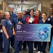
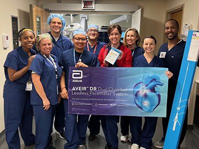
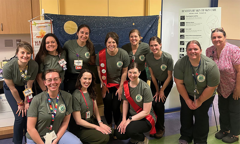
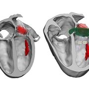
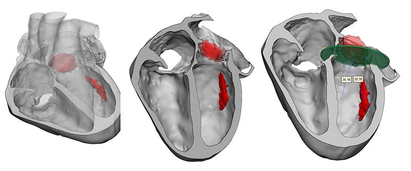
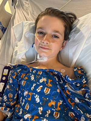
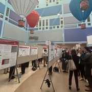


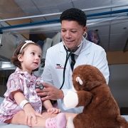
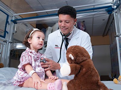
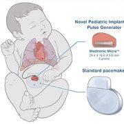
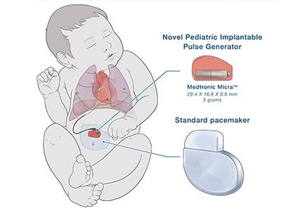
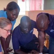
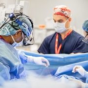
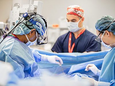

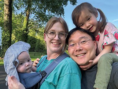
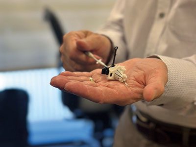

 2024 marked another groundbreaking year for Children’s National Hospital, showcasing remarkable advances across the spectrum of pediatric medicine, research and healthcare innovation. From pioneering surgical procedures to breakthrough artificial intelligence applications, the institution continued to push the boundaries of what’s possible in children’s healthcare. Read on for our list of the most popular articles we published on Innovation District in 2024.
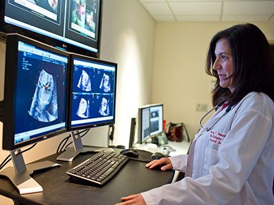

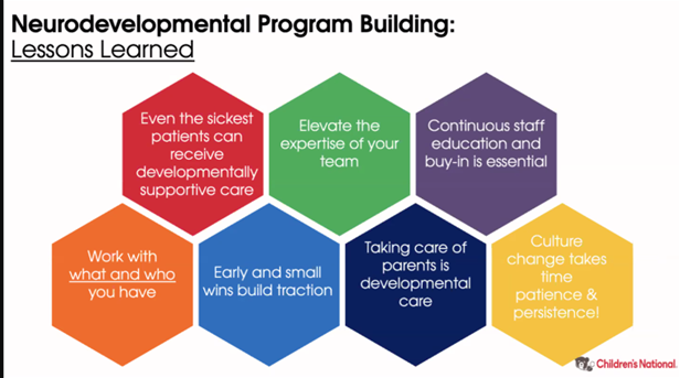


 Six medical technology innovators focused on pediatric cardiology were selected to receive grants of $50,000 each in the “Make Your Medical Device Pitch for Kids!TM” competition in Toronto. The funds will help awardees bring their devices to the market and improve care for children with heart conditions.