Building resilient kids through healthy adults

ECIN Briefing

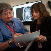
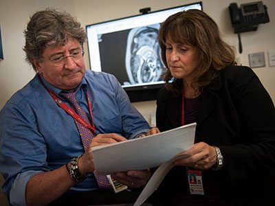
Mr. Lane, Dr. Hodgkinson, Dr. Biel, and Dr. Beers provided a briefing at the Washington, D.C., City Council in July about the Early Childhood Innovation Network, which takes evidence-based national models for early childhood mental health interventions and adds components designed to address Washington, D.C.’s unique needs.

Exposures to adverse childhood experiences are the single biggest predictor of outcomes for physical health, mental health, social functioning and academic achievement in children and into adulthood. There is evidence that negative experiences – such as poverty, housing insecurity, having a parent with untreated mental illness or actively engaged in substance abuse – have biological impacts on a child’s brain size and function.

Conversely, during the critical first few years of life, safe, stable and nurturing relationships from adult caregivers build healthy brains, even in the midst of adversity. Additionally, the ability of a child’s brain to absorb experience and to change means that early intervention to reduce exposure to or impact of these negative events can be particularly effective for young children. In a briefing for the Washington, D.C., City Council, leaders from the Early Childhood Innovation Network (ECIN) shared these facts and outlined how ECIN’s local collaborative of health, education and social service providers promotes resilient families and children through interventions designed to work best for each family.

“We are taking evidence-based practices from other places, and then personalizing them to our communities in D.C.,” says Lee Beers, M.D., co-director of the ECIN and the medical director for Municipal and Regional Affairs within the Child Health Advocacy Institute at Children’s National Health System. “We spent a lot of time seeking input and advice from primary care doctors, social services providers and community leaders, to make sure that we bring programs to clinics like the Children’s Health Center at Anacostia that are useful, sustainable and measurable for the children and families who live there.”

The network’s other co-director, Matthew G. Biel, M.D., chief of the Division of Child and Adolescent Psychiatry at MedStar Georgetown University Hospital continues, “We know that the best way to help these kids is by addressing challenges across generations – we can’t reach children without first helping the adults. In addition to evaluating for risk factors, we also need to screen for protective factors – how families can best buffer these young children from the toxic effects of adverse childhood experiences. Then, in a non-confrontational setting such as a routine primary care visit, we can provide them with additional tools to enhance those protective factors.”

A working example: HealthySteps D.C.

Drs. Beers and Biel cited the implementation of the HealthySteps program, an evidence-based intervention with a national network of over 100 pediatric and family practice sites across 15 states, locally in D.C. as one example of ECIN’s approach. The program, now underway at the Children’s Health Center at Anacostia and recently launched at the Children’s Health Center at THEARC, embeds specially trained HealthySteps specialists into the primary care team to provide parents and professionals with skills and tools that nurture healthy development in young children.

Nationwide, HealthySteps has been shown to have a significant impact on children, families and practices at relatively low cost, providing services within the primary care setting such as:

  • Early identification and access to effective interventions for development delays
  • Coaching on age-appropriate parent-child interactions and child social-emotional development
  • Support for parental depression, domestic violence, substance abuse, food, housing and other social determinants
  • Creating better integration between pediatric primary care and early childhood systems

ECIN’s D.C.-based version takes this successful national model and adds additional D.C. needs-based specific activities:

  • Each family is assigned a Family Champion who identifies and addresses specific resource needs, including mental health services, parent training, or support groups and basic needs such as insurance, housing or employment
  • HealthySteps specialists offer brief interventions within the primary care setting to address pressing needs such as maternal depression, grief and loss and child behavior management
  • HealthySteps specialists deliver specialized training to providers on child behavioral and developmental health

“Even in the short time since we implemented HealthySteps, we’re seeing significant impact around care coordination and case management for the families at our Children’s Health Center at Anacostia,” says Stacy Hodgkinson, Ph.D., a licensed clinical psychologist at Children’s National who serves as a HealthySteps specialist at the Children’s Health Center at Anacostia.

HealthySteps D.C. is the first of several initiatives under development by the Early Childhood Innovation Network. The group is also working together with additional community partners such as Educare, Martha’s Table, LIFT, and MedStar Washington Hospital Center to explore, implement and evaluate the effectiveness of programs in areas such as building social-emotional skills in young children, financial literacy and mental health support for mothers-to-be.

Community connections and coordination

“So many children with needs do not get connected to services, and the Early Childhood Innovation Network addresses this challenge. Even better, there has been a genuineness from ECIN to engaging community and earning buy-in for programs from the very beginning. They’ve made community leaders and parents an integral part of the network’s program design and implementation,” adds Ambrose Lane, Jr., chair and founder of the Health Alliance Network and chairman at the D.C. Department of Health Chronic Disease Citywide Collaborative.

Daily tasks harder for girls with ASD

Little girl eating

Researchers found that girls with autism struggle with day-to-day functioning and independence skills more than boys.

Researchers at the Center for Autism Spectrum Disorders at Children’s National found something surprising in their recent study of executive function and adaptive skills. Girls, who often score well on direct assessments of communication skills, struggle more than boys with crucial tasks such as making a plan, getting organized and following through, as well as basic daily tasks like getting up and getting dressed, or making small talk.

“When parents were asked to rate a child’s day-to-day functioning, it turns out that girls were struggling more with these independence skills. This was surprising because in general, girls with ASD have better social and communication skills during direct assessments. The natural assumption would be that those communication and social skills would assist them to function more effectively in the world, but we found that this isn’t always the case,” says Allison Ratto, Ph.D., a psychologist in the Center for Autism Spectrum Disorders and one of the study’s authors. “Our goal was to look at real world skills, not just the diagnostic behaviors we use clinically to diagnose ASD, to understand how people are actually doing in their day to day lives.”

Conducted by a team within the Center for Autism Spectrum Disorders, the National Institute of Mental Health and The George Washington University, the study is the largest to date examining executive function and adaptive skills in women and girls with autism spectrum disorders (ASD).

The study collected parent-reported data from several rating scales of executive function and adaptive behavior, including the Behavior Rating Inventory of Executive Function, Parent Form (BRIEF) and the Vineland Adaptive Behavior Scales-II (VABS-II). The group included 79 females and 158 males meeting clinical criteria for autism spectrum disorders, ranging in ages from 7 to 18 years old. The groups were matched for intelligence, age and level of autism and ADHD symptoms.

Little is known about autism in females

The findings are part of a growing body of research focused on how ASD may affect females differently than males. The ratio of girls to boys with autism is approximately one to three. As a result of the larger numbers of males, existing data is predominantly focused on traits and challenges in that population. This is especially true in clinical trials, where enrollment is overwhelmingly male.

“Our understanding of autism is overwhelmingly based on males, similar to the situation faced by the medical community once confronted with heart disease research being predominantly male,” notes Lauren Kenworthy, Ph.D., director of the Center for Autism Spectrum Disorders and the study’s senior author. “We know how to identify signs, symptoms and treatments for autism in males, but we know very little about unique aspects of it in females.”

The historical lack of specific discovery around how autism presents in females may contribute to misdiagnosis or delay, and prevent implementation of necessary interventions. Such delays can have a major impact on outcomes, as recent research has demonstrated the critical importance of early diagnosis and intervention in ASD.

“Our focus in caring for children with autism is equipping all of them with strategies and skills to allow them to function and succeed in day-to-day living,” Dr. Kenworthy continues. “This study highlights that some common assumptions about the severity of challenges faced by girls with ASD may be wrong, and we may need to spend more time building the adaptive and executive function skills of these females if we want to help them thrive.”

“Enhancing our understanding of how biological differences change the presentation of autism in the long term is crucial to giving every person with ASD the tools they need to succeed in life,” she concludes.

Giving children a voice in clinical trials

Pamela Hinds

“When experimental cancer drugs are studied, researchers collect details about how these promising therapies affect children’s organs, but rarely do they ask the children themselves about symptoms they feel or the side effects they experience,” says Pamela S. Hinds, Ph.D., R.N.

Children as young as 8 years old with incurable cancer can reliably characterize the impact an experimental therapy has on their symptoms and quality of life – even at the earliest stages of drug development – making self-reported patient outcomes a potential new clinical trial endpoint, according to a longitudinal validity study led by Children’s National Health System researchers.

Cancer is the No. 1 disease-related cause of death in U.S. children aged 1 to 19, and roughly 25 percent of the 12,400 children newly diagnosed with cancer will die of their disease, the study authors write.

“When experimental cancer drugs are studied, researchers collect details about how these promising therapies affect children’s organs, but rarely do they ask the children themselves about symptoms they feel or the side effects they experience,” says Pamela S. Hinds, Ph.D., R.N., director of Nursing Research and Quality Outcomes at Children’s National and lead author of study published June 5, 2017 in the journal Cancer. “Without this crucial information, the full impact of the experimental treatment on the pediatric patient is likely underreported and clinicians are hobbled in their ability to effectively manage side effects.”

To demonstrate the feasibility of children self-reporting outcomes, Hinds and colleagues recruited children and adolescents aged 8 to 18 with incurable or refractory cancers who were enrolled in Phase 1 safety trials or Phase 2 efficacy trials at four cancer settings: Children’s National, Seattle Children’s Hospital, Children’s Hospital of Philadelphia and Boston Children’s Hospital. Using a validated instrument to measure symptoms, function and other aspects of quality of life reported by patients, as well as four open-ended interview questions, researchers were able to better understand what aspects of symptoms and quality of life were most important to patients at this point in their lives and cancer treatment.

Of the 20 study participants, most were male (60 percent), adolescents (65 percent) and white (70 percent). Thirteen (65 percent) had solid tumors. Patients could describe “a good day” as having fewer side effects from the experimental therapy and fewer interruptions to their lives. “Bad days” were marked by interruptions to their usual activities and missing out on spending time with family and friends due to being at the hospital. A few study participants suggested that researchers add questions related to being away from home, family and friends and the ripple effect of treatment on other family members.

“Only by measuring and understanding self-reported symptoms and function in children and adolescents with incurable cancer can we adequately address threats to their quality of life and improve symptom control and supportive care,” Hinds and co-authors conclude. “By giving children a voice in the process, clinicians will be able to better anticipate and manage symptoms and thereby improve life for patients and their families.”

A brain’s protector may also be its enemy

Kazue Hashimoto Torii

By looking back to the earliest moments of embryonic brain development, Kazue Hashimoto-Torii, Ph.D. and her collaborators sought to explain the molecular and cellular bases for complex congenital brain disorders that can result from exposure to harmful agents.

When the brain is exposed to an environmental stressor all is not immediately lost. Brain cells have mechanisms that protect them against the ravages of alcohol and other toxic substances. One of these is a protein the cells make, known as Heat Shock Factor 1 (Hsf1), which helps to shield them from damage. The fetal brain also can make Hsf1, which protects its particularly vulnerable cells from environmental stressors that pregnant mothers are exposed to during gestation.

However, a new study suggests that this system is not perfect. Research led by Children’s National Health System scientists suggests that when too much Hsf1 is produced, it actually can impair the brain during development. While this finding was made in a preclinical model, it raises questions about neural risks for human infants if their mothers drink alcohol in the first or second trimester of pregnancy.

When fetuses are chronically exposed to harmful agents such as alcohol, ethanol or methyl mercury in utero, the experience can negatively affect fetal brain development in unpredictable ways. Some fetal brains show little or no damage, while others suffer severe damage. By looking at the earliest moments of embryonic brain development, an international research team that includes five Children’s National authors sought to explain the molecular and cellular bases for complex congenital brain disorders that can result from exposure to such harmful agents.

“From a public health perspective, there is ongoing debate about whether there is any level of drinking by pregnant women that is ‘safe,’ ” says Kazue Hashimoto-Torii, Ph.D., principal investigator in the Center for Neuroscience Research at Children’s National and senior author of the paper published May 2 in Nature Communications. “We gave ethanol to pregnant preclinical models and found their offspring’s neural cells experienced widely differing responses to this environmental stress. It remains unclear which precise threshold of stress exposure represents the tipping point, transforming what should be a neuroprotective response into a damaging response. Even at lower levels of alcohol exposure, however, the risk for fetal neural cells is not zero,” Hashimoto-Torii adds.

The cerebral cortex – the thin outer layer of the cerebrum and cerebellum that enables the brain to process information – is particularly vulnerable to disturbances in the womb, the study authors write. To fend off insult, neural cells employ a number of self-preservation strategies, including launching the protective Hsf1-Heat shock protein (Hsp) signaling pathway that is used by a wide range of organisms, from single-cell microbes to humans. Developing fetuses activate Hsf1-Hsp signaling upon exposure to environmental stressors, some to no avail.

To help unravel the neurological mystery, the researchers used a method that allows a single molecule to fluoresce during stress exposure. They tapped specific environmental stressors, such as ethanol, hydrogen peroxide and methyl mercury – each of which are known to produce oxidative stress at defined concentrations. And, using an experimental model, they examined the Hsf1 activation pattern in the developing cerebral cortex by creating a marker, an encoding gene tagged with a type of fluorescent protein that makes it glow bright red.

“Our results suggest that heterogeneous events of abnormal brain development may occur probabilistically – which explains patterns of cortical malformations that vary with each individual, even when these individuals are exposed to similar levels of environmental stressors,” Hashimoto-Torii adds.

Among the more striking findings, neural cells with excessively high levels of Hsf1-Hsp activation due to ethanol exposure experience disruptions to normal development, with delayed migration by immature cortical neurons. For the fetal brain to develop normally, neurons need to migrate to precise places in the brain at just the right time to enable robust neural connections. When neurons fail to arrive at their destinations or get there too late, there can be gaps in the neural network, compromising efficient and effective communication across the brain’s various regions.

“Even a short period of Hsf1 overactivation during prenatal development causes critical neuronal migration deficiency. The severity of deficiency depends on the duration of Hsf1 overactivation,” Hashimoto-Torii says. “Expression patterns vary, however, across various tissues. Stochastic response within individual cells may be largely responsible for variability seen within tissue and organs.”

The research team found one bright spot: Cortical neurons that stalled due to lack of the microtubule-associated molecule Dcx were able to regain their ability to migrate properly when the gene was replenished after birth. A reduction in Hsf1 activity after birth, however, did not show the same ability to trigger the “reset” button on neural development.

“The finding suggests that genes other than microtubule-associated genes may play pivotal roles in ensuring that migrating neurons reach their assigned destinations in the brain at the right time – despite the added challenge of excessive Hsf1 activation,” according to Hashimoto-Torii.

Healthy Generations program recognized

baby

After a study evaluated 150 teen mothers, the Healthy Generations program at Children’s National was recognized for its effectiveness in reducing teen pregnancy, sexually transmitted infections and associated risk factors.

The Healthy Generations program at Children’s National Health System recently was added to the official list of evidence-based teen pregnancy prevention programs by the Office of Adolescent Health within the U.S. Department of Health and Human Services (HHS). For nearly 10 years, HHS has identified programs with evidence of effectiveness in reducing teen pregnancy, sexually transmitted infections and associated sexual risk behaviors.

Unlike a majority of the programs on the HHS list, which are based in the school setting and use a more traditional curriculum-based approach, Healthy Generations is one of the first HHS-recognized programs that reaches young families through routine pediatric primary care and is a relationship-based intervention. When compared with a more traditional program, the Generations approach can be tailored to meet the needs of individual families.

The program is also differentiated by how it integrates care: Providing primary care for parents and children in the same visit by the same provider/team, and making mental health and social work services available as part of pediatric primary care.

The HHS designation was earned as a result of a study that highlighted the teen pregnancy prevention program as a patient-centered medical home that has improved contraceptive use among teen mothers. The study observed 150 teen mothers who met the following requirements:

  • The mother was between the ages of 13-19
  • The child was 6 months or younger
  • The family was newly seeking care from the Healthy Generations program or the comparison group
  • The mother had full custody of her child
  • The mother did not have any physical, psychological or cognitive impairments that would  prohibit her from providing informed consent

Eighty-five teen mothers enrolled in the Healthy Generations program were compared to 65 teen mothers who utilized community-based pediatric primary care. In a series of questions answered during home-based interviews, research assistants were able to effectively determine contraceptive use among the study participants after 12-months of enrollment. Mothers in the Healthy Generations program were more likely to use contraception than the teen mothers in the comparison group. Although there were no significant differences in race and age, several additional variables such as living conditions, romantic involvement with the father of the child and high school status made a difference in contraceptive use among participants.

By providing family-centered primary care, comprehensive social work services and mental health screening and treatment, clinicians within the Healthy Generations program strive to form lasting relationships with the young parents to address pregnancy prevention more actively. Beginning as a “teen-tot” model, primary care that only includes pregnancy prevention, the program has evolved into a multidisciplinary relationship-based intervention that not only uses the “teen-tot” model but also goes significantly beyond it to meet the needs of families headed by adolescents. With continued efforts and more positive outcomes on the horizon, the program is well on its way to meeting its goals to prevent rapid repeat pregnancies in teen mothers and maybe someday prevent an initial teenage pregnancy.

Zika virus, one year later

Drs. DeBiasi and du Plessis

A multidisciplinary team at Children’s National has consulted on 66 Zika-affected pregnancies and births since May 2016.

The first pregnant patient with worries about a possible Zika virus infection arrived at the Children’s National Health System Fetal Medicine Institute on Jan. 26, 2016, shortly after returning from international travel.

Sixteen months ago, the world was just beginning to learn how devastating the mosquito-borne illness could be to fetuses developing in utero. As the epidemic spread, a growing number of sun-splashed regions that harbor mosquitoes that efficiently spread the virus experienced a ballooning number of Zika-affected pregnancies and began to record sobering birth defects.

The Washington, D.C. patient’s concerns were well-founded. Exposure to Zika virus early in her pregnancy led to significant fetal brain abnormalities, and Zika virus lingered in the woman’s bloodstream months after the initial exposure — longer than the Centers for Disease Control and Prevention (CDC) then thought was possible.

The research paper describing the woman’s lengthy Zika infection, published by The New England Journal of Medicine, was selected as one of the most impactful research papers written by Children’s National authors in 2016.

In the intervening months, a multidisciplinary team at Children National has consulted on 66 pregnancies and infants with confirmed or suspected Zika exposure. Thirty-five of the Zika-related evaluations were prenatal, and 31 postnatal evaluations assessed the impact of in utero Zika exposure after the babies were born.

The continuum of Zika-related injuries includes tragedies, such as a 28-year-old pregnant woman who was referred to Children’s National after imaging hinted at microcephaly. Follow-up with sharper magnetic resonance imaging (MRI) identified severe diffuse thinning of the cerebral cortical mantle, evidence of parenchymal cysts in the white matter and multiple contractures of upper and lower extremities with muscular atrophy.

According to a registry of Zika-affected pregnancies maintained by the CDC, one in 10 pregnancies across the United States with laboratory-confirmed Zika virus infection has resulted in birth defects in the fetus or infant.

“More surprising than that percentage is the fact that just 25 percent of infants underwent neuroimaging after birth – despite the CDC’s recommendation that all Zika-exposed infants undergo postnatal imaging,” says Roberta L. DeBiasi, M.D., M.S., chief of the Division of Pediatric Infectious Diseases and co-director of the Congenital Zika Virus Program at Children’s National. “Clinicians should follow the CDC’s guidance to the letter, asking women about possible exposure to Zika and providing multidisciplinary care to babies after birth. Imaging is an essential tool to accurately monitor the growing baby’s brain development.”

Adré du Plessis, M.B.Ch.B., M.P.H., director of the Fetal Medicine Institute and Congenital Zika Virus Program co-leader, explains the challenges: ”When it comes to understanding the long-term consequences for fetuses exposed to the Zika virus, we are still on the steepest part of the learning curve. Identifying those children at risk for adverse outcomes will require a sustained and concerted multidisciplinary effort from conception well beyond childhood.”

In addition to counseling families in the greater Washington, D.C. region, the Children’s research team is collaborating with international colleagues to conduct a clinical trial that has been recruiting Zika-infected women and their babies in Colombia. Pediatric Resident Youssef A. Kousa, D.O., Ph.D., M.S., and Neurologist Sarah B. Mulkey, M.D., Ph.D., will present preliminary findings during Research and Education Week 2017.

In Colombia as well as the District of Columbia, a growing challenge continues to be assessing Zika’s more subtle effects on pregnancies, developing fetuses and infants, says Radiologist Dorothy Bulas, M.D., another member of Children’s multidisciplinary Congenital Zika Virus Program.

The most severe cases from Brazil were characterized by interrupted fetal brain development, smaller-than-normal infant head circumference, brain calcifications, enlarged ventricles, seizures and limbs folded at odd angles. In the United States and many other Zika-affected regions, Zika-affected cases with such severe birth defects are outnumbered by infants who were exposed to Zika in utero but have imaging that appears normal.

In a darkened room, Dr. Bulas pores over magnified images of the brains of Zika-infected babies, looking for subtle differences in structure that may portend future problems.

“There are some questions we have answered in the past year, but a number of questions remain unanswered,” Dr. Bulas says. “For neonates, that whole area needs assessment. As the fetal brain is developing, the Zika virus seems to affect the progenitor cells. They’re getting hit quite early on. While we may not detect brain damage during the prenatal period, it may appear in postnatal images. And mild side effects that may not be as obvious early on still have the potential to be devastating.”

Expanding awareness of SUDEP

Madison Berl

Madison M. Berl, Ph.D., is helping to expand awareness of SUDEP among patients, families and caregivers.

When 4-year-old Henry Lapham died in his sleep just weeks after being diagnosed with epilepsy in 2009, it was a shock to everyone — even his pediatrician and neurologist. Henry’s cause of death was sudden unexpected (or unexplained) death in epilepsy persons (SUDEP), a condition that causes sudden death in about 1 of every 1,000 otherwise healthy patients with epilepsy. Neither health care professional had mentioned this as a possibility, as remote as it was.

“I was desperate to make sense out of our tragedy,” writes Henry’s mother, Gardiner Lapham, R.N., M.P.H., in “Increasing awareness of sudden death in pediatric epilepsy together,” an article published in the February 2017 issue of Pediatrics. After her son’s death, by working with a group called Citizens United for Epilepsy Research, Lapham connected with other families affected by the same heartbreak. “I have met many bereaved family members,” she adds, “and the most consistent thing I hear is that they wish they had known about SUDEP.”

Now, a new collaboration with Children’s National Health System, where Henry received care, University of Virginia Medical Center (UVA) and other academic medical centers is helping to expand awareness of SUDEP among patients, families and caregivers alike. Known as Childhood Epilepsy Risks and Impact on Outcomes (CHERIO), the multiyear effort aims to develop approaches to increase knowledge about SUDEP and other conditions that can accompany epilepsy, such as attention deficit hyperactivity disorder, autism, anxiety, depression and sleep issues, according to co-authors of the Pediatrics article.

CHERIO got its start in 2014 at the American Epilepsy Society annual meeting. There, Lapham met Madison M. Berl, Ph.D., director of research, Division of Pediatric Neuropsychology at Children’s National, who studies epilepsy comorbidities. When Lapham asked what she could do to help raise awareness of SUDEP at Children’s National, she and Berl, along with William Davis Gaillard, M.D., Henry’s neurologist, hatched a plan.

Working with multiple disciplines and stakeholders, including neuropsychologists, psychiatrists, neurologists, epidemiologists, basic scientists, nurses and parent advocates at both Children’s National and UVA, CHERIO plans to assess the level of knowledge about SUDEP and other epilepsy comorbidities among medical providers and parents and to implement ways to increase knowledge. The first item on the agenda, Berl explains, was to conduct a survey to see just how much doctors knew about SUDEP.

“Although many neurologists are aware of this condition, ours was the first to survey pediatricians, and the majority was not aware of SUDEP – despite having children with epilepsy in their practice,” Dr. Gaillard says. “We know that many neurologists do not discuss SUDEP with patients and the reasons for not talking about SUDEP are varied. Thus, CHERIO felt that in addition to educating neurologists about the need to discuss the risk of death associated with epilepsy, increasing pediatricians’ awareness of SUDEP is one approach that could open more opportunities for families to have this discussion.”

To help make it easier to talk about this risk, the CHERIO team is developing strategies for doctors to start the conversation with patients and their families by framing SUDEP in the context of more common epilepsy comorbidities.

“Clinicians walk a fine line in giving information at the right time to make people more aware,” Berl adds, “but also being realistic and giving information that fits with what’s going on in a particular child’s case. By discussing SUDEP along with other, more common epilepsy risks, it brings context to a family so that they’re not unduly concerned about death – which also can paralyze a family and create unnecessary alarm.” The risk of death in most children with epilepsy is very low, slightly higher than the risks faced by healthy children. But parents of children with complicated epilepsy who have more risk factors for sudden death should be especially aware , she says.

Another way to help facilitate discussion may be through a simple tweak in the medical record, Berl adds. The team is currently developing a checklist that pops up annually in a patient’s medical record to remind clinicians of important points to discuss with patients and their families, including SUDEP.

Additionally, they are working on ways that can help families become more empowered to start the discussion themselves. Materials for the waiting room or questionnaires to fill out before appointments could trigger conversations with care providers, Berl says.

Last, the team also is collaborating with a medical device company that is working on a nighttime monitoring system that could provide an alert if patients with epilepsy experience nighttime seizures, a risk factor for SUDEP. Such technologies have not been proven to prevent SUDEP. Yet, it may help caregivers get help more quickly than if they did not receive the alert.

For each of these efforts, Berl notes, having Lapham as a partner has been key. “She’s part of our meetings and has input into the direction of each project,” Berl explains. “When you have a partner who is so close to the daily work you’re doing, it just heightens those efforts and brings to the forefront the simple message of why this is important.”

Multidisciplinary approach to hydrocephalus care

Reflective of the myriad symptoms and complications that can accompany hydrocephalus, a multidisciplinary team at Children’s National works with patients and families for much of childhood.

The Doppler image on the oversized computer screen shows the path taken by blood as it flows through the newborn’s brain, with bright blue distinguishing blood moving through the middle cerebral artery toward the frontal lobe and bright red depicting blood coursing away. Pitch black zones indicate ventricles, cavities through which cerebrospinal fluid usually flows and where hydrocephalus can get its start.

The buildup of excess cerebrospinal fluid in the brain can begin in the womb and can be detected by fetal magnetic resonance imaging. Hydrocephalus also can crop up after birth due to trauma to the head, an infection, a brain tumor or bleeding in the brain, according to the National Institutes of Health. An estimated 1 to 2 per 1,000 newborns have hydrocephalus at birth.

When parents learn of the hydrocephalus diagnosis, their first question tends to be “Is my child going to be OK?” says Suresh Magge, M.D., a pediatric neurosurgeon at Children’s National Health System.

“We have a number of ways to treat hydrocephalus. It is one of the most common conditions that pediatric neurosurgeons treat,” Dr. Magge adds.

Unlike fluid build-up elsewhere in the body where there are escape routes, with hydrocephalus spinal fluid becomes trapped in the brain. To remove it, surgeons typically implant a flexible tube called a shunt that drains excess fluid into the abdomen, an interim stop before it is flushed away. Another surgical technique, called an endoscopic third ventriculostomy has the ability to drain excess fluid without inserting a shunt, but it only works for select types of hydrocephalus, Dr. Magge adds.

For the third year, Dr. Magge is helping to organize the Hydrocephalus Education Day on Feb. 25, a free event that offers parents an opportunity to learn more about the condition.

Reflective of the myriad symptoms and complications that can accompany hydrocephalus, such as epilepsy, cerebral palsy, cortical vision impairment and global delays, a multidisciplinary team at Children’s National works with patients and families for much of childhood.

Neuropsychologist Yael Granader, Ph.D., works with children ages 4 and older who have a variety of developmental and medical conditions. Granader is most likely to see children and adolescents with hydrocephalus once they become medically stable in order to assist in devising a plan for school support services and therapeutic interventions. Her assessments can last an entire day as she administers a variety of tasks that evaluate how the child thinks and learns, such as discerning patterns, assembling puzzles, defining words, and listening to and remembering information.

Neuropsychologists work with schools in order to help create the most successful academic environment for the child. For example, some children may struggle to visually track across a page accurately while reading; providing a bookmark to follow beneath the line is a helpful and simple accommodation to put in place. Support for physical limitations also are discussed with schools in order to incorporate adaptive physical education or to allow use of an elevator in school.

“Every child affected by hydrocephalus is so different. Every parent should know that their child can learn,” Granader says. “We’re going to find the best, most supportive environment for them. We are with them on their journey and, every few years, things will change. We want to be there to help with emerging concerns.”

Another team member, Justin Burton, M.D., a pediatric rehabilitation specialist, says rehabilitation medicine’s “piece of the puzzle is doing whatever I can to help the kids function better.” That means dressing, going to the bathroom, eating and walking independently. With babies who have stiff, tight muscles, that can mean helping them through stretches, braces and medicine management to move muscles smoothly in just the way their growing bodies want. Personalized care plans for toddlers can include maintaining a regular sleep-wake cycle, increasing attention span and strengthening such developmental skills as walking, running and climbing stairs. For kids 5 and older, the focus shifts more to academic readiness, since those youths’ “full-time job” is to become great students, Dr. Burton says.

The area of the hospital where children work on rehabilitation is an explosion of color and sounds, including oversized balance balls of varying dimensions in bright primary colors, portable basketball hoops with flexible rims at multiple heights, a set of foam stairs, parallel bars, a climbing device that looks like the entry to playground monkey bars and a chatterbox toy that lets a patient know when she has opened and closed the toy’s doors correctly.

“We end up taking care of these kids for years and years,” he adds. “I always love seeing the kids get back to walking and talking and getting back to school. If we can get them back out in the world and they’re doing things just like every other kid, that’s success.”

Meanwhile, Dr. Magge says research continues to expand the range of interventions and to improve outcomes for patients with hydrocephalus, including:

  • Fluid dynamics of cerebrospinal fluid
  • Optimal ways to drain excess fluid
  • Improving understanding of why shunts block
  • Definitively characterizing post-hemorrhagic ventricular dilation.

Unlike spina bifida, which sometimes can be corrected in utero at some health institutions, hydrocephalus cannot be corrected in the womb. “While we have come a long way in treating hydrocephalus, there is still a lot of work to be done. We continue to learn more about hydrocephalus with the aim of continually improving treatments,” Dr. Magge says.

During a recent office visit, 5-year-old Abagail’s head circumference had measured ¼ centimeter of growth, an encouraging trend, Robert Keating, M.D., Children’s Chief of Neurosurgery, tells the girl’s mother, Melissa J. Kopolow McCall. According to Kopolow McCall, who co-chairs the Hydrocephalus Association DC Community Network, it is “hugely” important that Children’s National infuses its clinical care with the latest research insights. “I have to have hope that she is not going to be facing a lifetime of brain surgery, and the research is what gives me the hope.”

Improving mental health service access

Woman sitting on chair

Psychologist Stacy Hodgkinson, Ph.D., has been implementing a new strategy — integrating mental health services with primary care — to increase patients’ access to mental health care.

Children are disproportionately affected by poverty in the United States: Although they make up less than one-quarter of the entire population, about one-third of people living in poverty are kids. Lack of economic resources in childhood can have lifelong effects, including increasing the chances of experiencing a variety of mental health issues.

What’s more, although kids living in low socioeconomic settings are more likely to need mental health care, studies show that they are less likely to receive it, says Children’s National Health System Psychologist Stacy Hodgkinson, Ph.D. Estimates indicate that fewer than 15 percent of children living in poverty who need mental health care receive any services, and even fewer get comprehensive treatment.

The reasons for this disparity are multifold, Hodgkinson explains. One reason is simply insufficient numbers of trained mental health care providers to meet demand, particularly in low-income communities. Another is an inability to access available services —parents in low-paying jobs may not be able to take time off to take their children to appointments or even afford bus fare to reach a clinic. Others are afraid of the stigma that might surround being treated for a mental health issue. In her role as the director of mental health and research for the Generations Program, a support service for teen parents and their children, Hodgkinson says she has seen each of these scenarios in play.

However, she adds, over the past several years, she and Children’s National colleagues have been implementing a new strategy to increase mental health care access: Integrating these services with primary care.

“Often times, a family is with a primary care provider throughout a child’s life into adulthood. It’s a natural, familiar setting where people feel comfortable,” Hodgkinson says. “That makes a primary care provider’s office really fertile ground for integrating mental health services.”

Hodgkinson and coauthors point out in a review paper published in the January 2017 issue of Pediatrics that most children see their primary care provider for annual well visits as well as when they are sick — regardless of household income. Those visits provide ample opportunities for parents to bring up other concerns or for providers to implement screening that could lead to a mental health diagnosis. From there, she explains, that provider can offer mental health support and facilitate a connection with a mental health provider who works in the same office or who works in partnership with the primary care office.

In the review, she and colleagues suggest several strategies for making this idea become a reality. The first step, they agree, is education. Beginning with their fundamental training, primary care doctors and mental health providers need to see their roles as conjoined.

“We really need to change the way people think about primary care,” Hodgkinson says. “Disciplines don’t have to be siloed, where primary care providers do their thing here and mental health providers do their thing there. We should be thinking about how we can bring everyone together under one tent.”

Many psychology training programs have primary care integration rotations, she adds, and an increasing number of health systems like Children’s National now have mental health providers working in the same offices as primary care providers.

But not every clinic has the resources to group providers together under a single roof. Even for those offices, Hodgkinson says, primary care doctors need to develop a workflow that streamlines patients who need mental health services to health care professionals who provide it. In some cases, that might mean making the referral call on patients’ behalf to ensure they get through, walking families through the specific information they will need if they make the call on their own and following up to troubleshoot any problems with access.

“We want to close as many gaps as we can to keep families from falling through the cracks,” she says.

Developing an infrastructure that supports this model also can’t be ignored, Hodgkinson points out. Primary care offices might need to determine how to allocate space to mental health providers, hire dedicated workers to improve access and develop new strategies for billing.

None of this will be easy, she adds, but it will be worth it to make sure that more patients receive needed services.

“Even though we have integrated mental health and primary care at Children’s National, it very much remains a work in progress, and we’re continuing to fine-tune this machine to make it work better,” she says. “But if a patient comes to even one appointment that they might not have made it to in the past, that’s an accomplishment.”