Tag Archive for: leukemia

Optimizing anesthesia practices for children with acute leukemia undergoing lumbar punctures

bottle of propofol with needles

Anesthesia is used in up to 90% of patients, despite repeated propofol exposure being linked to neurocognitive impairment.

Acute leukemia is the most common childhood cancer, with treatment often involving up to 16 routine lumbar punctures (LPs). Anesthesia is used in up to 90% of patients, despite repeated propofol exposure being linked to neurocognitive impairment. In a quality improvement project, researchers from Children’s National Hospital examined variables that could minimize anesthesia time and propofol exposure for patients. The findings, presented at the 56th Congress of the International Society of Paediatric Oncology in Honolulu, HI., found about half of LPs met the goal of 15 minute of anesthetic exposure with significant differences in completion time between anesthesiologists.

The big picture

Researchers analyzed data from 199 LPs performed in the Non-Operating Anesthesia Room (NOAR) over a six-month period from July to December 2023 – including the start to stop time of anesthetic exposure, type of anesthetic, patient age and body mass index (BMI), proceduralist and anesthesiologist. A P-chart established a center line of 52.5% for procedures being completed within 15 minutes.

What’s next

Plan-Do-Study-Act (PDSA) cycles are being performed to align practices between anesthesiologists and identify successful changes with a goal to raise the center line to 75%.

“These findings highlight our commitment to improving care for children with acute leukemia by reducing anesthesia exposure during lumbar punctures,” said Shelby Smith, RN, MSN, CPNP, CPHON, nurse practitioner at Children’s National and the presenting author. “By identifying factors that impact procedure time and implementing targeted improvements, we aim to enhance patient safety and minimize potential risks associated with repeated propofol use.”

Other Children’s National authors include Birte Wistinghausen, MD.

Developing next-generation T cells to fight cancer

girl hugging stuffed animal

In the last decade, researchers have witnessed significant advances in the immunotherapy field. Most recently, a study in Nature claimed a novel CAR T-cell therapy “cured” a patient.

In the last decade, researchers have witnessed significant advances in the immunotherapy field. Most recently, a study in Nature claimed a novel CAR T-cell therapy “cured” a patient. Given the landmark scientific achievement for patients with different types of leukemia and lymphoma, Children’s National Hospital experts chimed in on the technology they have developed beyond CAR T cells.

Catherine Bollard, M.D., M.B.Ch.B., director of the Center for Cancer and Immunology Research at Children’s National Hospital, discusses the implications of this research, how it relates to the work she’s doing at Children’s National and the future of T-cell therapies.

Q: What did the research published in Nature find?

A: It reported a decade-long experience with this novel T-cell therapy called CD19 CAR T cells. These were used to treat patients with a type of leukemia or lymphoma that expresses the CD19 on its surface. While the article reported the experience of Children’s Hospital of Philadelphia and the University of Pennsylvania, multiple groups throughout the country did similar trials that have used these unique CD19 CAR T cells to treat children and adults with these refractory blood cancers.

Q: What are your thoughts on the implications of this research?

A: We now have three FDA-approved commercial CD19 CAR T-cell products developed by several academic institutions. This is revolutionary for our patients who have B-cell leukemias and lymphomas. It’s incredibly exciting for our T-cell therapy field in general because this was the first time the FDA approved a T-cell therapy. What it means now is the field is extremely excited to develop similar effective therapies for other patients with cancer.

Q: How does this relate to your work at Children’s National?

A: While CAR T cells have made tremendous progress for patients with B-cell leukemias, lymphomas and other blood cancers, the CAR T-cell field has not made the same impact for adult and pediatric solid tumors. We think the field is going to expand the type of T-cell therapies we’re generating beyond just CAR T cells. That’s where the work we’re doing comes in – not only by developing new T cells that don’t need gene engineering but also T cells that can be used as a platform for next-generation engineering approaches. We think the technology we’ve developed at Children’s National will help make an impact, especially in the solid tumor space. I hope in the next 10 years, we’ll be having a conversation not just about CAR T cells, but about other types of T cells that are now making an impact for solid tumors.

Q: How are the CAR T cells you develop different than those in the Nature article?

A: We think our multi-antigen specific T cells are complimentary and could have more potency than conventional CAR T cells for solid tumors especially when used in combination. This is in part because they can identify multiple targets on a tumor cell. Tumor cells are very clever and try to hide from T-cell therapies by down regulating the target that the T cell is directed towards. However, our novel T-cell therapies get around that escape by targeting multiple targets in a single product, making it much harder for the cancer cell to hide from the immune attack by the T cells.

Additionally, we’re excited by our approach because not all of our products require gene engineering, unlike CAR T cells. We have effectively used our T cells to target viruses in the “off-the-shelf” setting and we’re now about to start a first human clinical trial at Children’s National using an off the shelf T-cell product for children with solid tumors. It makes the T-cell therapy more like an “off-the-shelf” drug therapy that will allow us to treat many more children and adults nationally, as well as we hope, internationally.

Alpha/beta T cell depletion lifts barriers to transplantation

Illustration of white blood cells attacking a cancer cell

Removal of A/B T cells from the infused cell product significantly minimizes the risk of GvHD and eliminates the need for immunosuppressive medications after transplant.

Alpha/beta T cell depletion (A/B TCD) is a cutting-edge hematopoietic stem cell transplant (HSCT) technique by which donor derived immune cells, called A/B T cells, can be removed by selectively using magnetic beads before the donor cells are infused into the recipient’s body. A/B T cells have the potential to cause life threatening inflammation in the recipient’s body, called graft-versus-host disease (GVHD). GVHD is a major complication after transplant, especially when the donor is not fully matched. Therefore, removal of A/B T cells from the infused cell product (graft) significantly minimizes the risk of GVHD and eliminates the need for immunosuppressive medications after transplant.

Unlike previous methodologies that completely remove all immune cells, the novel A/B TCD approach preserves beneficial immune cells (like gamma delta T cells, natural killer cells, monocytes and dendritic cells) in the graft to preserve the capability to fight viral infections and residual cancer. Therefore, this innovative transplant approach can cure leukemia while decreasing the risk of life threatening infections and relapse after transplant.

In this Q&A, Anant Vatsayan, M.D., blood and marrow transplant specialist at Children’s National Hospital, tells us more about this new exciting technique.

Q: What is the specific research question that you are hoping to answer?

A: Children’s National Hospital is participating in the largest multicenter pediatric trial of A/B TCD hematopoietic stem cell transplant in the United States. The primary objective of this research is to assess whether disease-free survival at one-year after-HCT for children with high-risk leukemia and myelodysplastic syndrome can be improved with A/B TCD hematopoietic stem cell transplant.

Patients with other types of blood disorders may also be eligible to undergo A/B TCD hematopoietic stem cell transplant in this study based on the discretion of the principal investigator. The study will also assess the overall survival and rates of acute and chronic GVHD. Another objective is to compare the cost of transplantation using half-matched (haploidentical) donors versus other stem cell sources (for example, matched unrelated adult donors or cord blood donors) at participating centers.

Q: Why is this work exciting?

A: A/B TCD hematopoietic stem cell transplant has several benefits:

  • One of the remarkable benefits of this technique is the possibility of using haploidentical related donors for transplant if a fully matched related or unrelated donor is not available. This is a common scenario for patients of certain races (African American) and ethnicities (Hispanic) where it is difficult to identify a fully matched unrelated donor. Therefore, A/B TCD hematopoietic stem cell transplant expands the pool of donor options and ensures more equitable donor availability across every race and ethnicity.
  • A/B TCD significantly decreases the risk of severe GVHD and post-transplant infections. It eliminates the need for post-transplant immunosuppressive medications (like cyclosporine, tacrolimus or sirolimus) that can have numerous side effects and require frequent monitoring of drug levels in the blood.
  • The A/B TCD technique also promotes faster recovery of blood counts (engraftment) after transplant. Therefore, patients take fewer medications, have shorter durations of hospitalization for transplant and need less frequent blood tests and clinic visits after transplant. Hence, this patient friendly and family centric transplantation strategy will ensure that patients can spend more time with their family and have a better quality of life.

Q: How do you hope this will benefit patients?

A: Alpha/beta T cell depleted HSCT using half matched (haploidentical) donors will ensure donor availability for almost every patient regardless of race/ethnicity and probability of finding a matched related/unrelated donor. This methodology has tremendous prospects for wider applications, including the use of matched related and unrelated donors with the intent to eliminate the need for post-transplant immunosuppressive medications. This could be especially beneficial for patients with Fanconi anemia or other patients who are at risk of developing severe side effects from the use of immunosuppressive medications.

Q: How unique is this work?

A: The Shirley and William Howard Cellular Therapy Laboratory Stem Cell Processing program processes stem cells and performs cutting edge clinical trials while providing innovative care for patients. This work benefits from access to CliniMACS Plus Cell Selection Device, along with a multidisciplinary team with laboratory and clinical expertise to perform A/B TCD hematopoietic stem cell transplant. Access to our state of the art Cellular Therapy Laboratory allows us to further complement this transplantation strategy with other cellular therapies after transplant, such as virus specific and leukemia targeting T cells, which further mitigate the risk of post-transplant viral infections and leukemia relapse.

Oncologists receive Hyundai Hope on Wheels grants

Hyundai Hope on Wheels Logo

Keri Toner, M.D., and Hannah Kinoshita, M.D., both oncology researchers at Children’s National Hospital, were recently awarded Hyundai Hope on Wheels cancer research grants.

Dr. Toner, who is an attending physician in the Center for Cancer and Blood Disorders and the Center for Cancer and Immunology Research at Children’s National, received a $300,000 Hyundai Scholar Hope Grant that she will use to develop and functionally evaluate a novel T cell therapy which can be translated to the clinic for treatment of pediatric patients with acute myeloid leukemia (AML).

Currently, patients with relapsed AML have very poor outcomes and the success that T cell therapy has had in treating B-cell malignancies has not yet been achieved for AML. Dr. Toner’s goal is to try to overcome some of these barriers with a novel T cell therapy which combines both native and chimeric T cell receptors to target AML.

“There are currently critical barriers to the success of T cell therapies for the treatment of AML,” Dr. Toner explains. “Successful completion of this research would allow for translation of a novel CAR-TAA-T therapy to the clinic for the treatment of relapsed/refractory AML, which has very poor prognosis.”

Meanwhile, Dr. Kinoshita, a pediatric hematology oncology fellow at Children’s National, received a $200,000 Hyundai Young Investigator Grant. She will use the funds to evaluate the immunobiology of multi-antigen specific T cell therapy infused to patients to reduce the two most common causes of morbidity and mortality following hematopoietic stem cell transplant (HSCT) for malignant disease: relapse and infection.

The administration of multiantigen specific T cells targeting tumor and viral-associated antigens following stem cell transplant may serve to prolong remission of malignant disease and prevent and treat viral infections that can cause devastating disease in children. Dr. Kinoshita’s study will evaluate the anti-viral and anti-leukemia immune response in vivo following targeted T cell therapy.

“There have been incredible advancements in the field of pediatric oncology and bone marrow transplant over the past 20-30 years but there are still many areas in which we need to continue to improve,” Dr. Kinoshita says. “Our patients and their families go through so much to get into remission and it is devastating if they relapse or develop severe infectious complications. Adoptive immunotherapy is a promising tool in aiding to treat and prevent these complications, particularly for patients with high-risk hematologic malignancies.”

The Hyundai Scholar Hope Grants and the Hyundai Young Investigator Grants are competitive research grants that are peer-reviewed by the Hyundai Hope on Wheels Medical Advisory Committee, which is comprised of leading pediatric oncologists from children’s hospitals and research institutions nationwide. The grants are open to U.S.-based Children’s Oncology Group member institutions.

New approach to maintenance chemotherapy may improve children’s quality of life

marro replaced with aute lymphoblastic lukemia

Marrow replaced with acute lymphoblastic leukemia.

According to a study that accrued over 9,000 patients, a new approach to maintenance therapy lessens the burden of treatment and potential toxicity in children experiencing the most common cancer — B-acute lymphoblastic leukemia (B-ALL). The average-risk (AR) B-ALL subset of patients demonstrated an overall five-year survival rate of 98% despite less frequent chemotherapy pulses. Researchers from Children’s National Hospital led the 10-year study published on Jan. 7, 2021, in the Journal of Clinical Oncology.

This phase III clinical trial, which opened at over 200 centers, helped inform an alternative maintenance therapy with less frequent administration of vincristine and dexamethasone. These standard drugs are part of a multiagent treatment approach used to treat acute lymphoblastic leukemia (ALL).

“For decades, the common maintenance therapy approach [within the Children’s Oncology Group] was administering vincristine or steroid pulses every four weeks. The steroids can trigger disruptive behaviors like moodiness, sleep disturbance, food cravings, poor school attendance or physical aggression and vincristine can cause declines in fine motor and sensory-perceptual performance,” said Anne Angiolillo, M.D., lead author of the study and director of the Leukemia and Lymphoma Program at Children’s National. “We can now lessen the burden of this therapy while still maintaining excellent outcomes, which is a huge benefit to our patients and their families.”

The findings suggest that the decreased frequency of both vincristine and dexamethasone pulses every four weeks to every 12 weeks alleviates the therapy burden and reduces toxicity, potentially improving children’s quality of life.

Simultaneously, the researchers tried increasing the starting dose of oral methotrexate, a standard chemotherapy drug, given once weekly in the maintenance phase to see if it would improve the five-year disease-free survival rate, but, according to the data, it did not improve outcomes.

The world’s largest organization devoted exclusively to pediatric cancer research, the Children’s Oncology Group (COG), adopted the approach of less frequent pulses into the frontlines of their new B-ALL trials, given the study’s findings, to help decrease the therapy burden for patients and their families.

“I am very excited that the results of AALL0932 [the clinical trial] will have a major effect on the schedule of maintenance therapy for children with standard and high-risk B acute lymphoblastic leukemia in all future COG therapeutic trials,” said Dr. Angiolillo.

Dr. Angiolillo, and co-author Reuven Schore, M.D., pediatric oncologist at Children’s National were the chair and vice-chair of the clinical trial, respectively. Dr. Schore is also a member of the Leukemia and Lymphoma Program at Children’s National.

ALL can progress quickly, affect the bone marrow and the blood, including B cells and T cells. Among the children with ALL, approximately 55% comprise of the newly diagnosed National Cancer Institute (NCI) standard-risk (SR) B-ALL.

The study enrolled 9,229 patients with B-ALL between August 2010 and March 2018. Only 2,364 patients classified as average-risk received a random assignment to one of the four maintenance arms at the start of maintenance therapy. The researchers administered either vincristine/dexamethasone pulses every 12 weeks or every four weeks and a starting dose of once-weekly oral methotrexate of 20 mg/m2 or 40 mg/m2 during the maintenance phase.

“This trial establishes that with improved risk stratification utilizing blast cytogenetics and rate of response, a relatively low-intensity premaintenance backbone with a three-drug induction, and lower exposure to chemotherapy in maintenance, results in outstanding outcomes,” said Angiolillo et al.

For hemorrhagic cystitis, harnessing the power of a parasite

Schistosoma haematobium egg

“Urogenital Schistosoma infestation, which is caused by S. haematobium, also causes hemorrhagic cystitis, likely by triggering inflammation when the parasite’s eggs are deposited in the bladder wall or as eggs pass from the bladder into the urinary stream. S. haematobium eggs secrete proteins, including IPSE, that ensure human hosts are not so sickened that they succumb to hemorrhagic cystitis,” says Michael H. Hsieh, M.D., Ph.D.

Every year, hundreds of thousands of U.S. patients – and even more throughout the world – are prescribed cyclophosphamide or ifosfamide. These two chemotherapy drugs can be life-saving for a wide range of pediatric cancers, including leukemias and cancers of the eyes and nerves. However, these therapies come with a serious side effect: Both cause hemorrhagic cystitis in up to 40 percent of patients. This debilitating condition is characterized by severe inflammation in the bladder that can cause tremendous pain, life-threatening bleeding, and frequent and urgent urination.

Infection with a parasitic worm called Schistosoma haematobium also causes hemorrhagic cystitis, but this organism has a fail-safe: To keep its host alive, the parasite secretes a protein that suppresses inflammation and the associated pain and bleeding.

In a new study, a Children’s-led research team harnessed this protein to serve as a new therapy for chemotherapy-induced hemorrhagic cystitis.

“Urogenital Schistosoma infestation, which is caused by S. haematobium, also causes hemorrhagic cystitis, likely by triggering inflammation when the parasite’s eggs are deposited in the bladder wall or as eggs pass from the bladder into the urinary stream. S. haematobium eggs secrete proteins, including IPSE, that ensure human hosts are not so sickened that they succumb to hemorrhagic cystitis,” says Michael H. Hsieh, M.D., Ph.D., senior author of the study published April 3, 2018, by The FASEB Journal. “This work in an experimental model is the first published report of exploiting an uropathogen-derived host modulatory molecule in a clinically relevant model of bladder disease, and it points to the potential utility of this as an alternate treatment approach.”

S. mansoni IPSE binds to Immunoglobulin E (IgE), an antibody produced by the immune system that is expressed on the surface of basophils, a type of immune cell; and mast cells, another immune cell that mediates inflammation; and sequesters chemokines, signaling proteins that alert white cells to infection sites. The team produced an ortholog of the uropathogen-derived protein. A single IV dose proved superior to multiple doses of 2-Mercaptoethane sulfonate sodium (MESNA), the current standard of care, in suppressing chemotherapy-induced bladder hemorrhaging in an experimental model. It was equally potent as MESNA in dampening chemotherapy-induced pain, the research team finds.

“The current array of medicines we use to treat hemorrhagic cystitis all have shortcomings, so there is a definite need for novel therapeutic options,” says Dr. Hsieh, a Children’s National Health System urologist. “And other ongoing research projects have the potential to further expand patients’ treatment options by leveraging other urogenital parasite-derived, immune-modulating molecules to treat inflammatory bowel diseases and autoimmune disorders.”

Future research will aim to describe the precise molecular mechanisms of action, as well as to generate other orthologs that boost efficacy while reducing side effects.

In addition to Dr. Hsieh, Children’s study co-authors include Lead Author, Evaristus C. Mbanefo; Loc Le and Luke F. Pennington; Justin I. Odegaard and Theodore S. Jardetzky, Stanford University; Abdulaziz Alouffi, King Abdulaziz City for Science and Technology; and Franco H. Falcone, University of Nottingham.

Financial support for this research was provided by National Institutes of Health under award number RO1-DK113504.

Helpful, hopeful news for bone marrow transplant patients

Kirsten-M.-Williams

Research published online Dec. 13, 2017, by The Lancet Haematology and co-led by Kirsten M. Williams, M.D., suggests that a new imaging agent can safely show engraftment as early as days after transplant – giving a helpful and hopeful preview to patients and their doctors.

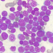
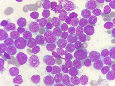
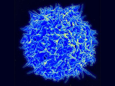
Leukemia can be a terrifying diagnosis for the more than 60,000 U.S. patients who are told they have this blood cancer every year. But the treatment for this disease can be just as frightening. For patients with certain forms of leukemia, the only chance they have for a cure is to receive a massive dose of radiation and chemotherapy that kills their hematopoietic stem cells (HSCs), the cells responsible for making new blood, and then receive new HSCs from a healthy donor.

While patients are waiting for these new cells to go to the bone marrow factory and begin churning out new blood cells, patients are left without an immune system. Devoid of working HSCs for two to four weeks – or longer, if a first transplant doesn’t take – patients are vulnerable to infections that can be just as deadly as their original cancer diagnosis.

As they wait in the protected confines of a hospital, patients who undergo HSC transplants receive blood tests every day to gauge successful engraftment, searching for the presence of immune cells called neutrophils, explains Kirsten M. Williams, M.D., blood and bone marrow transplant specialist at Children’s National Health System.

“As you head into week three post-transplant and a patient’s cell counts remain at zero, everyone starts to get nervous,” Dr. Williams says. The longer a patient goes without an immune system, the higher the chance that they’ll develop a life-threatening infection. Until recently, Dr. Williams says, there has been no way beyond those daily blood tests to assess whether the newly infused cells have survived and started to grow early healthy cells in the bone marrow, a process called engraftment.

A new study could change that paradigm. Research published online Dec. 13, 2017, by The Lancet Haematology and co-led by Dr. Williams suggests that a new imaging agent can safely show engraftment as early as days after transplant – giving a helpful and hopeful preview to patients and their doctors.

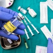
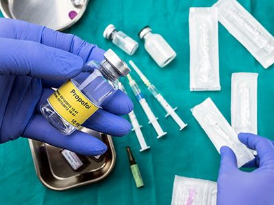
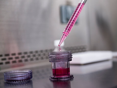
The study evaluated an investigational imaging test called 18F-fluorothymidine (18F-FLT). It’s a radio-labeled analogue of thymidine, a natural component of DNA. Studies have shown that this compound is incorporated into just three white blood cell types, including HSCs. Because it’s radioactive, it can be seen on various types of common clinical imaging exams, such as positron emission tomography (PET) and computed tomography (CT) scans. Thus, after infusion, the newly infused developing immune system and marrow is readily visible.

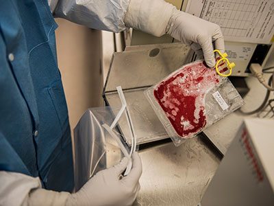
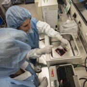
To see whether this compound can readily and safely visualize transplanted HSCs, Dr. Williams and colleagues tested it on 23 patients with various forms of high-risk leukemia.

After these patients received total-body irradiation to destroy their own HSCs, they received donor HSCs from relatives or strangers. One day before they were infused with these donor cells, and then at five or nine days, 28 days, and one year after transplantation, the patients underwent imaging with the novel PET/and CT scan imaging platform.

Each of these patients had successful engraftment, reflected in blood tests two to four weeks after their HSC transplants. However, the results of the imaging exams revealed a far more complicated and robust story.

With 18F-FLT clearly visible in the scans, the researchers saw that the cells took a complex journey as they engrafted. First, they migrated to the patients’ livers and spleens. Next, they went to the thoracic spine, the axial spine, the sternum, and the arms and legs. By one year, most of the new HSCs were concentrated in the bones that make up the trunk of the body, including the hip, where most biopsies to assess marrow function take place.

Interestingly, notes Dr. Williams, this pathway is the same one that HSCs take in the fetus when they first form. Although experimental model research had previously suggested that transplanted HSCs travel the same route, little was known about whether HSCs in human patients followed suit.

The study also demonstrated that the radiation in 18F-FLT did not adversely affect engraftment. Additionally, images could identify success of their engraftments potentially weeks faster than they would have through traditional blood tests – a definite advantage to this technique.

“Through the images we took, these patients could see the new cells growing in their bodies,” Dr. Williams says. “They loved that.”

Besides providing an early heads up about engraftment status, she adds, this technique also could help patients avoid painful bone marrow biopsies to make sure donor cells have taken residence in the bones or at the very least help target those biopsies. It also could be helpful for taking stock of HSCs in other conditions, such as aplastic anemia, in which the body’s own HSCs fade away. And importantly, if the new healthy cells don’t grow, this test could signal this failure to doctors, enabling rapid mobilization of new cells to avert life-threatening infections and help us save lives after transplants at high risk of graft failure.

“What happens with HSCs always has been a mystery,” Dr. Williams says. “Now we can start to open that black box.”

Dr. Williams’ co-authors include co-lead author Jennifer Holter-Chakrabarty, M.D., Quyen Duong, M.S., Sara K. Vesely, Ph.D., Chuong T. Nguyen, Ph.D., Joseph P. Havlicek, Ph.D., George Selby, M.D., Shibo Li, M.D., and Teresa Scordino, M.D., University of Oklahoma; Liza Lindenberg, M.D., Karen Kurdziel, M.D., Frank I. Lin, M.D., Daniele N. Avila, N.P., Christopher G. Kanakry, M.D., Stephen Adler, Ph.D., Peter Choyke, M.D., and senior author Ronald E. Gress, M.D., National Cancer Institute; Juan Gea-Banacloche, M.D., Mayo Clinic Arizona; and Catherine “Cath” M. Bollard, M.D., MB.Ch.B., Children’s National.

Research reported in this story was supported by the National Institutes of Health, Ben’s Run/Ben’s Gift, Albert and Elizabeth Tucker Foundation, Mex Frates Leukemia Fund, Jones Family fund and Oklahoma Center for Adult Stem Cell Research.

Advances in T-cell immunotherapy at ISCT

Healthy Human T Cell

T-cell immunotherapy, which has the potential to deliver safer, more effective treatments for cancer and life-threatening infections, is considered one of the most promising cell therapies today. Each year, medical experts from around the world – including leaders in the field at Children’s National Health System – gather at the International Society for Cellular Therapy (ISCT) Conference to move the needle on cell therapy through several days of innovation, collaboration and presentations.

Dr. Catherine Bollard, Children’s National chief of allergy and immunology and current president of ISCT, kicked off the week with a presentation on how specific approaches and strategies have contributed to the success of T-cell immunotherapy, a ground-breaking therapy in this fast-moving field.

Later in the week, Dr. Kirsten Williams, a blood and marrow transplant specialist, presented encouraging new findings, demonstrating that T-cell therapy could be an effective treatment for leukemia and lymphoma patients who relapse after undergoing a bone marrow transplant. Results from her phase 1 study showed that four out of nine patients achieved complete remission. Other medical options for the patients involved – those who relapsed between 2 and 12 months post-transplant – are very limited. Looking to the future, this developing therapy, while still in early stages, could be a promising solution.

Other highlights include:

  • Both Allistair Abraham, blood and marrow transplantation specialist, and Dr. Michael Keller, immunologist, presented oral abstracts, the former titled “Successful Engraftment but High Viral Reactivation After Reduced Intensity Unrelated Umbilical Cord Blood Transplantation for Sickle Cell Disease” and the latter “Adoptive T Cell Immunotherapy Restores Targeted Antiviral Immunity in Immunodeficient Patients.
  • Patrick Hanley engaged attendees with his talk, “Challenges of Incorporating T-Cell Potency Assays in Early Phase Clinical Trials,” and his poster presentation “Cost Effectiveness of Manufacturing Antigen-Specific T-Cells in an Academic GMP Facility.” He also co-chaired a session titled “Early Stage Professionals Session 1 – Advanced Strategic Innovations for Cell and Gene Therapies.”
  • To round out this impressive group, Shabnum Piyush Patel gave a talk on genetically modifying HIV-specific T-cells to enhance their anti-viral capacity; the team plans to use these HIV-specific T-cells post-transplant in HIV-positive patients with hematologic malignancies to control their viral rebound.

This exciting team is leading the way in immunology and immunotherapy, as evidenced by the work they shared at the ISCT conference and their ongoing commitment to improving treatments and outcomes for patients at Children’s National and across the country. To learn more about the team, visit the Center for Cancer and Blood Disorders site.

T-cell therapy success for relapsing blood cancer

cord blood

A unique immunotherapeutic approach that expands the pool of donor-derived lymphocytes (T-cells) that react and target three key tumor-associated antigens (TAA) is demonstrating success at reducing or eliminating acute leukemias and lymphomas when these cancers have relapsed following hematopoietic stem cell transplant (HSCT).

“There’s currently a less than 10 percent chance of survival for a child who relapses leukemia or lymphoma after a bone marrow transplant—in part because these patients are in a fragile medical condition and can’t tolerate additional intense therapy,” says Kirsten Williams, M.D., a blood and marrow transplant specialist in the Division of Hematology at Children’s National Health System, and principal investigator of the Research of Expanded multi-antigen Specifically Oriented Lymphocytes for the treatment of VEry High Risk Hematopoietic Malignancies (RESOLVE) clinical trial.

The unique manufactured donor-derived lymphocytes used in this multi-institutional Phase 1 dose-ranging study are receptive to multiple tumor-associated antigens within the cell, including WT1, PRAME, and Survivin, which have been found to be over-expressed in myelodysplastic syndromes (MDS), acute myeloid leukemia (AML), B-cell AML/MDS, B-cell acute lymphoblastic leukemia (ALL), and Hodgkins lymphoma. Modifying the lymphocytes for several antigens, rather than a single target, broadens the ability of the T-cells to accurately target and eradicate cancerous cells.

Preliminary results demonstrate a 78 percent response rate to treatment, and a 44 percent rate of total remission for participating patients. To date, nine evaluable patients with refractory and relapsed AML/MDS, B-cell ALL, or Hodgkins lymphoma have received 1-3 infusions of the expanded T-cells, and of those, seven have responded to the treatment, showing reduction in cancer cells after infusion with little or no toxicity. All of these patients had relapse of their cancer after hematopoietic cell transplantation. The study continues to recruit eligible patients, with the goal of publishing the full study results within the next 12 months.

“Our preliminary data also shows that this new approach has few if any side effects for the patient, in part because the infused T-cells target antigens that are found only in cancer cells and not found in healthy tissues,” Dr. Williams notes.

The approach used to expand existing donor-derived TAA-lymphocytes, rather than using unselected T cells or genetically modified T-cells as in other trials, also seems to reduce the incidence of post infusion graft versus host disease and other severe inflammatory side effects. Those side effects typically occur when the infused lymphocytes recognize healthy tissues as foreign and reject them or when the immune system reacts to the modified elements of the lymphocytes, she adds.

“These results are exciting because they may present a truly viable option for the 30 to 40 percent of children who will relapse post-transplant,” Dr. Williams concludes. “Many of the patients who participated were given two options: palliative care or this trial. To see significant success and fewer side effects gives us, and families with children facing relapsing leukemia, some hope for this new treatment.”

Dr. Williams discussed the early outcomes of the RESOLVE trial during an oral presentation at the American Society for Blood and Marrow Transplantation meeting on February 22, 2017.

“The early indicators are very promising for this patient population,” says Catherine Bollard, M.D., M.B.Ch.B., Chief of the Division of Allergy and Immunology, Director of the Program for Cell Enhancement and Technologies for Immunotherapy (CETI) at Children’s National, and senior author of the study. “If we can achieve this, and continue to see good responses with few side effects, it’s possible these methods could become a viable alternative to HSCT for patients with no donor match or who aren’t likely to tolerate transplant.”

This is one of the first immunotherapeutic approaches to successfully capitalize on the natural ability of human T-cells to kill cancer, though previous research has shown significant success for this approach in reducing the deadly impact of several viruses, including Epstein-Barr virus, adenovirus, and cytomegalovirus, post HSCT. These new findings have led to the development of additional clinical trials to investigate applications of this method of TAA-lymphocyte manufacture and infusion for pre-HSCT MDS/AML, B-cell ALL, Hodgkins Lymphoma, and even some solid tumors.

Training t-cells, essential players in the immune system, to fight a trio of viruses

Children's is the only U.S. pediatric hospital that manufactures specialized T-cells from native cord blood

What’s Known
Following treatment, patients with leukemia, lymphoma, and other cancers may receive a transplant in order to restore their body’s natural ability to fight infection and, sometimes, such transplants are a component of leukemia treatment. (Leukemia is the second most common blood cancer, after lymphoma, and its incidence rate has increased by 0.2 percent annually from 2002 to 2011.) A stem cell or cord blood transplant restores the body’s ability to produce infection-fighting white blood cells. After such transplants, however, patients can face heightened risk of developing a life-threatening infection with such viruses as adenovirus, cytomegalovirus, or Epstein-Barr virus.

What’s New
A head-to-head comparison of two strategies to thwart such viral infections shows that both approaches leverage the power of multivirus-specific, donor-derived T-cells (mCTL), which are highly skilled at recognizing foreign invaders. The research team, made up of nine scientists and clinicians affiliated with Children’s National Health System, grew personalized T-cells from peripheral blood (PB) of adult donors who were seropositive for CMV and also coaxed T-cells to grow from naïve cord blood (CB). PB-derived cells have long memories of past battles; naïve CB-derived cells need additional training to acquire such skills. From 35 to 384 days after their stem cell or cord blood transplant, 13 patients were infused with PB mCTL and 12 patients were infused with CB mCTL. Within four weeks, patients experienced up to a 160-fold increase in virus-specific T-cells, which coincided with their response to therapy. Overall response rate was 81 percent.

Questions for Future Research
Q: Could T-cells be personalized to attack other viruses that infect patients post-transplant, such as human parainfluenza virus and BK polyomavirus, providing the potential to target five viruses in a single infusion?
Q: Could the proteins that are used to train T-cells to attack certain viruses also be used to create a personalized approach to tumor suppression?

Source: “A Phase 1 Perspective: Multivirus-Specific T Cells From Both Cord Blood and Bone Marrow Transplant Donors.” Hanley, P., M. D. Keller, M. Martin Manso, C. Martinez, K. Leung, C.R. Cruz, C. Barese, S. McCormack, M. Luo, R.A. Krance, D. Jacobsohn, C. Rooney, H. Heslop, E.J. Shpall, and C. Bollard. Presented during the International Society for Cellular Therapy 2016 Annual Meeting, Singapore. May 26, 2016.