Tag Archive for: Hypoxic-ischemic encephalopathy

newborn in incubator

Neuroprotective effect of Src kinase in neonates affected by HIE

newborn in incubator

Hypoxic-ischemic encephalopathy (HIE) is a major cause of neonatal morbidity and mortality worldwide.

In a systematic review published by Frontiers in Neuroscience, and co-authored by Panagiotis Kratimenos, M.D., neonatologist at Children’s National Hospital, Ioannis Koutroulis, M.D., pediatric emergency medicine physician at Children’s National and Javid Ghaemmaghami, M.S., researcher with the Center for Neuroscience Research at Children’s National, it was concluded that Src kinase is an effective neuroprotective target in the setting of acute hypoxic injury.

The paper reviews hypoxic-ischemic encephalopathy (HIE), a major cause of neonatal morbidity and mortality worldwide (one in four perinatal deaths is attributed to hypoxic-ischemic). While therapeutic hypothermia has improved neurodevelopmental outcomes for some survivors of HIE, this treatment is only available to a subset of affected neonates. Src kinase, an enzyme central to the apoptotic cascade, is a potential pharmacologic target to preserve typical brain development after HIE. This paper, a product of collaboration for a Master’s Thesis with the Aristotle University School of Medicine, Thessaloniki, Greece, where Dr. Kratimenos holds the appointment of Visiting  Professor,  presents evidence of the neuroprotective effects of targeting Src kinase in preclinical models of HIE.

The systematic review shows that while heterogeneity and risk for bias were limiting factors, the overall results indicate that Src-i neuroprotective properties could be a promising therapeutic strategy for neonates after hypoxic events.

Read more about the full review.

Neuronal network with electrical activity

Neonatal hypoxia-ischemia causes damage to the cholinergic system

Neuronal network with electrical activity

Study suggests permanent injury to the cholinergic system after neonatal hypoxia-ischemia is responsible for the poor executive functions and difficulties in learning and memory.

Newborn babies who go through periods of low oxygen — also known as hypoxic-ischemic encephalopathy — during their first hours of life often experience difficulties in learning, memory and executive functions later on. Even when treated with therapeutic hypothermia, memory deficits and executive functions remain severely affected. These functions are linked to a neurotransmitter network called the cholinergic system.

“Complications from hypoxic-ischemic brain injury contribute to one-quarter of neonatal deaths worldwide and cause significant long-term neurological morbidity,” explains Panagiotis Kratimenos, M.D., Ph.D., neonatologist at Children’s National Hospital and Assistant Professor of Pediatrics at the George Washington University School of Medicine and Health Sciences.

In a study published in the Journal of Comparative Neurology led by Frances Northington, M.D., co-director of Neurosciences Intensive Care Nursery at Johns Hopkins and Professor of Pediatrics at Johns Hopkins University School of Medicine, with contributions from Dr. Kratimenos, the authors found significant injury to the neurons of the cholinergic systems in specific parts of the brain after exposure to low oxygen and restricted blood flow. These areas included the ipsilateral medial septal nucleus (MSN), the ipsilateral nucleus basalis of Meynert (nbM) and striatum. Within the injured part of the cortex at the site of injury, acetylcholine — the neurotransmitter found in cholinergic systems — was abnormally overactivated.

The authors hypothesize that permanent injury to the cholinergic system after neonatal hypoxia-ischemia is responsible for the poor executive functions and difficulties in learning and memory.

“Because cholinergic systems can easily be manipulated pharmacologically with already established treatments that have been used in other areas of medicine, they could be a good a target for therapeutic interventions for neonates with hypoxic-ischemic encephalopathy,” says Dr. Kratimenos.

Read the full article in the Journal of Comparative Neurology.

MRI of the patient's head close-up

Early versus late MRI in newborn brain injury

MRI of the patient's head close-up

A single magnetic resonance imaging (MRI) performed in the first week after birth is adequate to assess brain injury and offer prognostic information in newborn infants with hypoxic ischemic encephalopathy (HIE) treated with therapeutic hypothermia, according to a new study published in The Journal of Pediatrics.

A collaborative team of neonatology, neurology and neuroradiology experts from Children’s National Hospital that included Gilbert Vezina, M.D., Taeun Chang, M.D., and An N. Massaro, M.D., came together to evaluate the agreement in brain injury findings between early and late MRI in newborn infants with hypoxic ischemic encephalopathy (HIE) treated with therapeutic hypothermia. The team then compared the ability of early versus late MRI to predict early neurodevelopmental outcomes.

This was a prospective longitudinal study of 49 patients with HIE who underwent therapeutic hypothermia and had MRI performed at both <7 and ≥7 days of age. MRIs were reviewed by an experienced neuroradiologist and assigned brain injury severity scores according to established systems. Scores for early and late MRIs were assessed for agreement using the kappa statistic. The ability of early and late MRI scores to predict death or developmental delay at 15-30 months of age was assessed by logistic regression analyses.

The results of the study found agreement between the early and late MRI was substantial to near perfect (k>0.75, p<0.001) across MRI scoring systems. In cases of discrepant scoring, early MRI was more likely to identify severe injury when compared with late MRI. Early MRI scores were more consistently predictive of adverse outcomes compared with late MRI.

Read the full study in The Journal of Pediatrics.

newborn baby

Directly measuring function in tiny hearts

newborn baby

The amount of blood the heart pumps in one minute can be directly measured safely in newborns by monitoring changes in blood velocity after injecting saline, indicates the first clinical study of direct cardiac output measurement in newborns.

The amount of blood that the heart pumps in one minute (cardiac output) can be directly measured safely in newborns by monitoring changes in blood velocity after injecting saline, indicates a paper published online Dec. 17, 2019 in the Journal of Pediatrics and Neonatal Medicine. The research, conducted by Children’s National Hospital faculty, is believed to be the first clinical study of direct cardiac output measurement in newborns.

Right now, cardiac output is measured indirectly in the nation’s neonatal intensive care units (NICU) using newborns’ blood pressure, heart rate, urine output and other indirect measures. However, these techniques can produce imprecise readings in children. And the field lacks a feasible “gold standard” to measure cardiac output in newborns.

The COstatus monitor already uses ultrasound dilution – the expected decrease in the velocity of blood when saline is injected, producing a dilution curve. A Children’s National research team used ultrasound dilution in their small pilot study to gauge the feasibility of directly measuring cardiac output in newborns.

“Infants who stand to benefit most from directly monitoring cardiac hemodynamics are often so sick they already have central venous access,” says Khodayar Rais-Bahrami, M.D., an attending neonatologist at Children’s National and the study’s senior author. “Using the COstatus monitor in these children would enable the clinical team to personalize care based on the newborn’s current hemodynamic status, while introducing minimal fluid during measurements,” Dr. Rais-Bahrami adds.

COstatus monitor

The COstatus Monitor uses an extracorporeal loop attached to arterial and venous lines to measure cardiac output using ultrasound dilution. The research team injected 1mL/kg of body temperature saline into the loop and performed up to two measurement sessions daily.

The research team recruited 12 newborns younger than 2 weeks old who already had central venous and arterial access. The venous line of the arteriovenous AV loop is connected to the umbilical venous catheter while the COstatus monitor’s arterial line is connected to the umbilical arterial catheter. During measurement sessions, two injections of solution are injected into the venous loop, allowing for two measures of cardiac output, cardiac index, active circulating volume index, central blood volume index and systemic vascular resistance index.

Infants enrolled in the pilot study underwent up to two measurement sessions per day for up to four days, for a total of 54 cardiac hemodynamic measurements. The newborns ranged from 720 to 3,740 grams in weight and 24 to 41.3 weeks in gestational age.

The infants’ mean cardiac output was 0.43L/min and increased with gestational age. By contrast, the mean cardiac index was 197mL/kg/min and changed little with infants’ increasing maturity – either by gestational age or postnatal age. Two of the study participants were undergoing therapeutic cooling for hypoxic-ischemic encephalopathy and had their measurements taken during cooling and after rewarming.

“Although this study size is small, it demonstrates that this minimally invasive technique can safely be used in newborns to directly measure cardiac hemodynamics,” says Simranjeet S. Sran, M.D., a Children’s National neonatalogist and the study’s lead author. “This technology may allow for more precise and personalized care of critically ill newborns in a range of disease states – real-world utility in NICUs that serve some of the youngest and sickest newborns,” Dr. Sran adds.

The research team notes that direct measurement by ultrasound dilution revealed a stark increase in cardiac index as infants undergoing therapeutic hypothermia were rewarmed, raising questions about whether indirect measures using other technology, such as echocardiography, underestimate hypothermia’s effect on hemodynamics.

In addition to Drs. Rais-Bahrami and Sran, Mariam Said, M.D., also a Children’s National neonatalogist, was a study co-author.

newborn in incubator

In HIE lower heart rate variability signals stressed newborns

newborn in incubator

In newborns with hypoxic-ischemic encephalopathy (HIE), lower heart rate variability correlates with autonomic manifestations of stress shortly after birth, underscoring the value of this biomarker, according to Children’s research presented during the Pediatric Academic Societies 2019 Annual Meeting.

Tethered to an array of machines that keep their bodies nourished, warm and alive, newborns with health issues can’t speak. But Children’s research teams are tapping into what the machinery itself says, looking for insights into which vulnerable infants are most in need of earlier intervention.

Heart rate variability – or the variation between heartbeats – is a sign of health. Our autonomic nervous system constantly sends signals to adjust our heart rate under normal conditions. We can measure heart rate variability non-invasively, providing a way to detect potential problems with the autonomic nervous system as a sensitive marker of health in critically ill newborns,” says An N. Massaro, M.D., co-Director of Research for the Division of Neonatology at Children’s National, and the study’s senior author. “We’re looking for validated markers of brain injury in babies with HIE, and our study helps to support heart rate variability as one such valuable physiological biomarker.”

In most newborns, the autonomic nervous system reliably and automatically receives information about the body and the outside world and, in response, controls essential functions like blood pressure, body temperature, how quickly the baby breathes and how rapidly the newborn’s heart beats. The sympathetic part stimulates body processes, while the parasympathetic part inhibits body processes. When the nervous system’s internal auto-pilot falters, babies can suffer.

The Children’s team enrolled infants with HIE in the prospective, observational study. (HIE is brain damage that occurs with full-term babies who experience insufficient blood and oxygen flow to the brain around the time they are born.) Fifteen percent had severe encephalopathy. Mean age of babies in the observational study was 38.9 weeks gestation. Their median Apgar score at five minutes was 3; the 0-9 Apgar range indicates how ready newborns are for the rigors of life outside the womb.

The team analyzed heart rate variability metrics for three time periods:

  • The first 24 to 27 hours of life
  • The first three hours after babies undergoing therapeutic cooling were rewarmed and
  • The first three hours after babies’ body temperature had returned to normal.

They correlated the relationship between heart rate variability for 68 infants during at least one of these time periods with the stress z-score from the NICU Network Neurobehavioral Scale. The scale is a standardized assessment of newborn’s neurobehavioral integrity. The stress summary score indicates a newborn’s overall stress response, and six test items specifically relate to autonomic function.

“Alpha exponent and root mean square in short timescales, root mean square in long timescales, as well as low and high frequency powers positively correlated with stress scores and, even after adjusting for covariates, remained independently associated at 24 hours,” says Allie Townsend, the study’s lead author.

Pediatric Academic Societies 2019 Annual Meeting presentation

  • “Heart rate variability (HRV) measures of autonomic nervous system (ANS) function relates to neonatal neurobehavioral manifestations of stress in newborn with hypoxic-ischemic encephalopathy (HIE).”
    • Monday, April 29, 2019, 5:45 p.m. (EST)

Allie Townsend, lead author; Rathinaswamy B. Govindan, Ph.D., staff scientist, Advanced Physiological Signals Processing Lab and co-author; Penny Glass, Ph.D., director, Child Development Program and co-author; Judy Brown, co-author; Tareq Al-Shargabi, M.S., co-author; Taeun Chang, M.D., director, Neonatal Neurology and Neonatal Neurocritical Care Program and co-author; Adré J. du Plessis, M.B.Ch.B., MPH, chief of the Division of Fetal and Transitional Medicine and co-author; An N. Massaro, M.D., co-Director of Research for the Division of Neonatology and senior author, all of Children’s National.

An-Massaro

Looking for ‘help’ signals in the blood of newborns with HIE

An Massaro

“This data support our hypothesis that a panel of biomarkers – not a one-time test for a single biomarker – is needed to adequately determine the risk and timing of brain injury for babies with HIE,” says An N. Massaro, M.D.

Measuring a number of biomarkers over time that are produced as the body responds to inflammation and injury may help to pinpoint newborns who are more vulnerable to suffering lasting brain injury due to disrupted oxygen delivery and blood flow, according to research presented during the Pediatric Academic Societies 2019 Annual Meeting.

Hypoxic-ischemic encephalopathy (HIE) happens when blood and oxygen flow are disrupted around the time of birth and is a serious birth complication for full-term infants. To lessen the chance of these newborns suffering permanent brain injury, affected infants undergo therapeutic cooling, which temporarily lowers their body temperatures.

“Several candidate blood biomarkers have been investigated in HIE but we still don’t have one in clinical use.  We need to understand how these markers change over time before we can use them to direct care in patients,” says An N. Massaro, M.D., co-director of the Neonatal Neurocritical Care Program at Children’s National and the study’s senior author. “The newborns’ bodies sent out different ‘help’ signals that we detected in their bloodstream, and the markers had strikingly different time courses. A panel of plasma biomarkers has the potential to help us identify infants most in need of additional interventions, and to help us understand the most optimal timing for those interventions.”

Past research has keyed in on inflammatory cytokines and Tau protein as potential biomarkers of brain injury for infants with HIE who are undergoing therapeutic cooling. The research team led by Children’s faculty wanted to gauge which time periods to measure such biomarkers circulating in newborns’ bloodstreams. They enrolled 85 infants with moderate or severe HIE and tapped unused blood specimens that had been collected as cooling began, as well as 12, 24, 72 and 96 hours later. The infants’ mean gestational age was 38.7 weeks, their mean birth weight was about 7 pounds (3.2 kilograms), and 19% had severe brain disease (encephalopathy).

Cytokines – chemicals like Interleukin (IL) 6, 8 and 10 that regulate how the body responds to infection, inflammation and trauma – peaked in the first 24 hours of cooling for most of the newborns. However, the highest measure of Tau protein for the majority of newborns was during or after the baby’s temperature was restored to normal.

“After adjusting for clinical severity of encephalopathy and five-minute Apgar scores, IL-6, IL-8 and IL-10 predicted adverse outcomes, like severe brain injury or death, as therapeutic hypothermia began. By contrast, Tau protein measurements predicted adverse outcomes during and after the infants were rewarmed,” Dr. Massaro says.

IL-6 and IL-8 proteins are pro-inflammatory cytokines while IL-10 is considered anti-inflammatory.  These chemicals are released as a part of the immune response to brain injury. Tau proteins are abundant in nerve cells and stabilize microtubules.

“This data support our hypothesis that a panel of biomarkers – not a one-time test for a single biomarker – is needed to adequately determine the risk and timing of brain injury for babies with HIE,” she adds.

Pediatric Academic Societies 2019 Annual Meeting presentation

  • “Serial plasma biomarkers of brain injury in infants with hypoxic ischemic encephalopathy (HIE) treated with therapeutic hypothermia (TH).”
    • Saturday, April 27, 2019, 6 p.m. (EST)

Meaghan McGowan, lead author; Alexandra C. O’Kane, co-author; Gilbert Vezina, M.D.,  director, Neuroradiology Program and co-author; Tae Chang, M.D., director, Neonatal Neurology Program and co-author; and An N. Massaro, M.D., co-director of the Neonatal Neurocritical Care Program and senior author; all of Children’s National; and co-author Allen Everett, of Johns Hopkins School of Medicine.

An-Massaro

Keeping an eye on autonomic function for infants with HIE

An-Massaro

“By including heart rate variability measurements and other markers of autonomic function in our current predictive armamentarium,” says An Massaro, M.D., “we may be able to offer new hope for infants with HIE.”

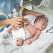
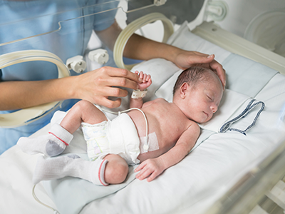
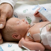
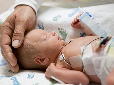
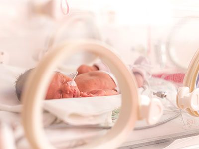
In about two to three in every 1,000 full-term births, babies develop a neurological condition called hypoxic ischemic encephalopathy (HIE) when their brains receive insufficient oxygen. HIE can be a devastating condition, leading to severe developmental or cognitive delays or motor impairments that become more evident as the child grows older. Despite improvements in care – including therapeutic hypothermia, a whole-body cooling method administered shortly after birth that can slow brain damage – about half of children with this condition die from neurological complications by age 2.

Finding ways to identify children with the most severe HIE could help researchers focus their efforts and provide even more intense neuroprotective care, explains An Massaro, M.D., a neonatologist at Children’s National Health System. But thus far, it’s been unclear which symptoms reflect the extent of HIE-induced brain damage.

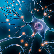
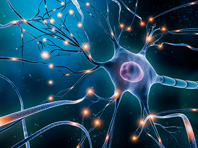
That’s why Dr. Massaro and colleagues embarked on a study published in the May 2018 issue of Journal of Pediatrics. The team sought to determine whether dysfunction of the autonomic nervous system (ANS) – the auto-pilot part of the nervous system responsible for unconscious bodily functions, such as breathing and digestion – reflected in routine care events can be used as a marker for brain injury severity.

The researchers collected data from 25 infants who were treated for HIE with therapeutic hypothermia at Children’s National. Thanks to multi-modal monitoring, these babies’ medical records hold a treasure trove of information, explains Rathinaswamy B. Govindan, Ph.D., a staff scientist in Children’s Advanced Physiological Signals Processing Lab.

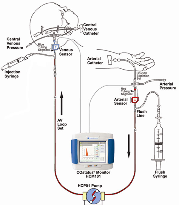
In addition to including continuous heart rate tracings and blood pressure readings that are standard for many infants in the neonatal intensive care unit (NICU), they also recorded cerebral near infrared spectroscopy, a monitor that measures brain tissue oxygen levels. The investigators performed detailed analyses to evaluate how these monitor readings change in response to a variety of routine care events, such as diaper changes, heel sticks, endotracheal tube manipulations and pupil examinations.

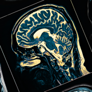
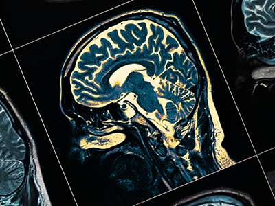
The researchers stratified these infants based on how dysfunctional their ANS behaved by using heart rate variability as a marker: The fewer natural fluctuations in heart rate, the more damaged their ANS was thought to be. And they also used non-invasive brain magnetic resonance imaging (MRI) to determine brain damage. They then compared this information with the babies’ physiological responses during each care event.

Their findings show that infants with impaired ANS, based on depressed heart rate variability before the care event, had significantly different responses to these care events compared with babies with intact ANS.

  • For stimulating interventions, such as diaper changes and heel sticks, both heart rate and blood pressure increased in babies with intact ANS but decreased in babies with impaired ones.
  • Shining a light in their pupils led to an expected decreased heart rate with stable blood pressure in ANS-intact infants, but in ANS-impaired infants, there was no responsive change in heart rate and, additionally, a decrease in blood pressure was observed.
  • Responses were similar between the two groups during breathing tube manipulations, except for a slight increase in heart rate a few minutes later in the ANS-impaired group.

These results, Govindan explains, suggest that a real-time, continuous way to assess ANS function may offer insights into the expected physiological response for a given infant during routine NICU care.

“This is exactly the type of additional information that intensivists need to pinpoint infants who may benefit from additional neuroprotective support,” he says. “Right now, it is standard practice to monitor brain activity continuously using electroencephalogram and to check the status of the brain using MRI to assess the response to therapeutic cooling. Neither of these assessments can be readily used by neonatologists at the bedside in real-time to make clinical decisions.”

Assessing ANS function in real-time can help guide neuroprotective care in high-risk newborns by providing insight into the evolving nature of brain damage in these infants, Dr. Massaro adds.

Beyond simply serving as a biomarker into brain injury, poor ANS function also could contribute to the development of secondary injury in newborns with HIE by stymieing the normal changes in heart rate and blood pressure that help oxygenate and heal injured brains. The researchers found that the cumulative duration of autonomic impairment was significantly correlated with the severity of brain injury visible by MRI in this group of infants.

“By including heart rate variability measurements and other markers of autonomic function in our current predictive armamentarium,” says Dr. Massaro, “we may be able to offer new hope for infants with HIE.”

In addition to Dr. Massaro, the Senior Author, study co-authors include Lead Author, Heather Campbell, M.D.; Rathinaswamy B. Govindan, Ph.D., Children’s Advanced Physiological Signals Processing Lab; Srinivas Kota, Ph.D.; Tareq Al-Shargabi, M.S.; Marina Metzler, B.S.; Nickie Andescavage, M.D., Children’s neonatalogist; Taeun Chang, M.D., Children’s neonatal and fetal neurologist; L. Gilbert Vezina, M.D., attending in Children’s Division of Diagnostic Imaging and Radiology; and Adré J. du Plessis, M.B.Ch.B., M.P.H., chief of Children’s Division of Fetal and Transitional Medicine.

This research was supported by the Clinical and Translational Science Institute at Children’s National under awards UL1TR000075 and 1KL2RR031987-01 and the Intellectual and Developmental Disabilities Research Consortium within the National Institutes of Health under award P30HD040677.

nurse holding newborn baby

Continuous EEG monitoring better predicts HIE outcomes

nurse holding newborn baby

For newborns who experience a serious complication that deprives their brain of oxygen, continuously monitoring brain activity and examining how the electrical signals evolve may be a more reliable way to identify infants most at risk for brain injury.

For newborns who experience a serious complication that deprives their brain of oxygen, continuously monitoring brain activity and examining how the electrical signals evolve may be a more reliable way to identify infants most at risk for brain injury, compared with doing evaluations at discreet intervals, according to a prospective cohort study led by Children’s National Health System research-clinicians.

Amplitude-integrated electroencephalogram (aEEG) is a bedside tool that permits clinicians to monitor the complex electrical activity of the child’s brain over time. It’s a positive sign when an aEEG shows babies beginning to sleep and wake normally by the time they are 3 days old. Conversely, severely abnormal aEEG readings in the first days of life predict poor outcomes.

The Children’s team used aEEG with infants born with hypoxic-ischemic encephalopathy (HIE), one of the most severe complications that can affect full-term infants. During pregnancy, birth or shortly after birth, a hypoxic-ischemic event can occur that impedes blood flow and oxygen delivery to the brain, resulting in destruction of brain tissue. Cooling (therapeutic hypothermia) is now standard for newborns with HIE in order to stave off life-long consequences, but deaths and neurodevelopmental disability still can occur.

“We know whole-body cooling – or lowering the body’s temperature by about 3 degrees Celsius – can help vulnerable newborns survive and can protect their brains from suffering profound injuries,” says An N. Massaro, M.D., a Children’s National neonatologist and senior author of the study published online Sept. 28, 2017 in the American Journal of Perinatology.  “What we were trying to determine with this study is whether evaluating the pattern of evolution of the aEEG as a whole provides more information compared with looking at snapshots in time.”

Eighty infants undergoing therapeutic cooling who met the inclusion criteria were enrolled in the five-year study, one of the largest such studies to date. The babies weighed more than 1,800 grams and were older than 35 weeks’ gestational age at birth, and either needed prolonged resuscitation after birth or had low APGAR scores – a measure of how well newborns fare outside the womb. Continuous recordings of EEG data occurred from the time of admission up to 12 hours after the infants’ temperatures were raised to normal and aEEG tracings were calculated.

After the therapeutic cooling blankets were removed, the infants underwent at least one magnetic resonance imaging (MRI) scan prior to discharge. During the routine follow-up check at about 18 months of age, the HIE survivors’ cognitive and motor skills were assessed using validated instruments.

Fifty-six of the infants in the study had favorable outcomes. Twenty-four infants had adverse outcomes, including 15 with severe brain injury detected by MRI and nine infants who died. These children had lower APGAR scores at five minutes, and were more likely to have severe HIE and to have experienced more frequent seizures.

“Infants whose aEEG abnormalities do not improve were at increased risk: Infants who do not reach a discontinuous background pattern by 15.5 hours of life, achieve cycling by 45.5 hours after birth and who fail to achieve continuous normal voltage by 78 hours after birth are most at risk for adverse outcomes,” Dr. Massaro says. “In addition to defining worrisome trends, we found that overall assessment of continuous aEEG readings through the course of hypothermia treatment provide the most meaningful predictive power. This means we can speak with families at the bedside with more confidence about their child’s outcomes after the infant undergoes cooling therapy.”

Related Resources

Patricio Ray

Toward a better definition for AKI in newborns

Patricio Ray

The National Institute of Diabetes and Digestive and Kidney Diseases convened a meeting of expert neonatologists and pediatric nephrologists, including Dr. Patricio Ray, to review state-of-the-art knowledge about acute kidney injury in neonates and to evaluate the best method to assess these patients’ kidney function.

Each year, thousands of infants in the United States end up in neonatal intensive care units (NICUs) with acute kidney injury (AKI), a condition in which the kidneys falter in performing the critical role of filtering waste products and excess fluid from the blood to produce urine. Being able to identify neonates during the early stages of AKI is critical to doctors and clinician-scientists who treat and study this condition, explains Patricio Ray, M.D., a nephrologist at Children’s National Health System.

Without an accurate definition and early identification of newborns with AKI, it is difficult for doctors to limit the use of antibiotics or other medications that can be harmful to the kidneys. Neonates who have AKI should not receive large volumes of fluids, a treatment that can cause severe complications when the kidneys do not properly function.

Until recently, there was no standard definition for AKI, leaving doctors and researchers to develop their own guidelines. Lacking set criteria led to confusion, Dr. Ray says. For example, different studies estimating the percentage of infants in NICUs with AKI ranged from 8 percent to 40 percent, depending on which definition was used. In 2012, a group known as the Kidney Disease Improved Global Outcome (KDIGO) issued practice guidelines for AKI that provide a standard for doctors and researchers to follow. They focus largely on measuring the relative levels of serum creatinine, a protein produced by muscles that is filtered by the kidneys, and the amount of urine output, which typically declines in adults and older children with failing kidneys.

The problem with these guidelines, Dr. Ray explains, is they are not sensitive enough to identify newborns experiencing the early stages of AKI during the first week of life. Newborns can have high serum creatinine levels during the first week of life due to residual levels transferred from mothers through the placenta. Also, because their kidneys are immature, failure often can mean higher – not diminished – urine production.

In 2013, the National Institute of Diabetes and Digestive and Kidney Diseases, part of the National Institutes of Health, convened a meeting of leading neonatologists and pediatric nephrologists – including Dr. Ray – to review state-of-the-art knowledge about AKI in neonates and to evaluate the best manner to assess kidney function in these patients. They published a summary of their discussion online June 12, 2017 in Pediatric Research.

Among other findings, the group concluded that the current definition of AKI lacks the sensitivity needed to identify the early stages of AKI in neonates’ first week of life. They also said that more research was needed to fill this gap.

That’s where Dr. Ray’s current research comes in. Working with fellow Children’s Nephrologist Charu Gupta, M.D., and Children’s Neonatologist An Massaro, M.D., the three clinician-scientists reviewed the medical records of 106 infants born at term with a condition known as hypoxic ischemic encephalopathy (HIE), in which the brain doesn’t receive enough oxygen. Not only does this often lead to brain injury, but it also greatly increases the risk of AKI.

Because these babies had been followed closely in the NICU to assess the possibility of AKI, their serum creatinine had been checked frequently. The researchers found that about 69 percent of the infants with HIE followed at Children’s National never developed signs of kidney failure during their first week of life. These babies’ serum creatinine concentrations dropped by 50 percent or more by the time they were 1 week old, about the same as reported previously in healthy neonates. Another 12 percent of the infants with HIE developed AKI according to the definition established by the KDIGO group in 2012. These infants:

  • Required more days of mechanical ventilation and medications to increase their blood pressure
  • Had higher levels of antibiotics in their bloodstreams
  • Retained more fluid
  • Had lower urinary levels of a molecule that their kidneys should have been cleared and
  • Had to stay in the hospital longer

A third group of the infants with HIE, about 19 percent, did not meet the standard criteria for AKI. However, these babies had a rate of decline of serum creatinine that was significantly slower than the normal newborns and the infants with HIE who had excellent outcomes. Rather, their outcomes matched those of infants with established AKI.

Dr. Ray notes that by following the rate of serum creatinine decline during the first week of life physicians could identify neonates with impaired kidney function. This approach provides a more sensitive method to identify the early stages of AKI in neonates. “By looking at how fast babies were clearing their serum creatinine compared with the day they were born, we could predict how well their kidneys were working,” he says. Dr. Ray and colleagues published these findings July 2016 in Pediatric Nephrology.

He adds that further studies will be necessary to confirm the utility of this new approach to assess the renal function of term newborns with other diseases and preterm neonates. Eventually, he hopes this new approach will become uniform clinical practice.

The search for precise blood biomarkers of neonatal brain injury

Bloodbiomarkers

PDF Version

What’s Known:
Hypoxic-ischemic encephalopathy (HIE) is characterized by reduced blood and oxygen flow to a baby’s brain around birth and may cause neurologic disability or death. It occurs most commonly after intrauterine asphyxia brought on by such difficulties as circulatory problems, placental abruption, or inflammatory processes. Newborns with HIE may suffer seizures, difficulty feeding, and disturbed control of heart rate and breathing. Cooling therapy, which is the standard of care, offers some protection to the developing brain, but up to 50 percent of HIE-affected infants still have poor outcomes.

What’s New:
Research scientists at Children’s National Health System are involved in a multi-center clinical trial to determine if erythropoietin (EPO), a hormone naturally secreted by the kidneys and commonly used to treat anemia, helps to prevent brain injury in these infants. The trial, called the HEAL Study (High Dose Erythropoietin for Asphyxia and Encephalopathy), is exploring whether EPO, given in addition to hypothermia, further lowers the risk of brain injury in HIE-affected babies. As a part of this study, researchers at Children’s National are leading the investigation to identify biomarkers of brain injury. Biomarkers are telltale chemicals in the blood and are used in tests that evaluate whether patients have suffered a heart attack. While available biomarkers warn when the heart, kidney, or liver is in trouble, there is no blood biomarker that signals ongoing brain injury. Such blood biomarkers could help to determine which infants are responding to treatment as well as to precisely identify which HIE-affected infants are still struggling and require additional treatments, such as EPO, to protect the brain and improve outcomes.

Questions for Future Research: 

  • Does EPO, in tandem with hypothermia, improve long- term neurodevelopmental outcomes in newborns with HIE?
  • Which biomarkers, or panel of biomarkers, best reflect the timing and severity of neonatal brain injury?
  • Can biomarkers direct which types of treatments are best for specific patients and when they should be used?

Source: Plasma Biomarkers of Brain Injury in Neonatal HIE (Hypoxic-Ischemic Encephalopathy).” A.N. Massaro, Y. Wu, T.K. Bammler, A. Mathur, R.C. McKinstry, T. Chang, D.E. Mayock, S. Mulkey, K. Van Meurs, L. Dong, R. Ballard, and S. Juul. Presented during the 2016 Pediatric Academic Societies Annual Meeting, Baltimore, MD. May 3, 2016.