
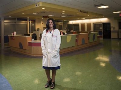
The Children’s National Heart Center is leading the way to find new and better treatments for cardiac problems as early as possible, during pregnancy and just after birth.
These critical stages are vital for a child’s lifelong heart health. “The innovations we are pursuing have the potential to transform the landscape of cardiac treatment,” says Wayne Franklin, M.D., F.A.C.C., Heart Center senior vice president.
“By focusing on the earliest stages of life, we can significantly alter the trajectory of children’s heart health, creating a lifetime of possibilities.”
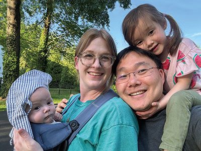
Abby with her sister and parents.
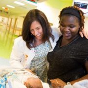
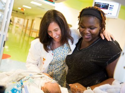
Abby, who just turned 1, is a smiley baby who loves to play peek-a-boo with her sister, Ruby. “We marvel that she is perfectly healthy,” says her father, Dan. He and wife Kelsey love to linger over ordinary moments. Her given name, Abigail, means “a father’s joy.”
When Kelsey was 18 weeks pregnant, she and Dan learned their baby had signs of heart injury, which led to a dangerous rhythm problem called “complete heart block.” The previous year, their infant son died from the same condition, which was discovered too late. The family prepared for another loss. But early detection of the problem and advanced care that started in the womb made all the difference for Abby. Children’s National prenatal cardiology experts began monitoring Abby’s development from the earliest possible moment and were able to intervene before devastating injury occurred.
Before Abby’s diagnosis, in light of the previous pregnancy, Kelsey enrolled in a clinical trial. The research sought a better way to identify and treat the heart condition Abby was at risk for. Anita Krishnan, M.D., a pediatric cardiologist and clinician scientist, met the family during their initial visit and arranged a monitoring plan that included frequent visits to make sure Abby’s heart was working normally. Soon after Kelsey’s first visit, doctors noted a problem.
Mary Donofrio, M.D., F.A.A.P., F.A.C.C., F.A.S.E., a leading pediatric and fetal cardiologist and The Van Metre Companies Professor of Fetal Cardiology, led the team that initiated lifesaving in utero therapy, followed Abby’s progress in the womb and planned for her arrival. The goal was to extend the pregnancy for as long as possible so she would survive birth and the heart surgery that would follow.
Abby’s prognosis improved as weeks passed. As a newborn, she would be a candidate for an infant pacemaker the size of a penny. It would help regulate her heartbeat and enable her to live a “normal” life.
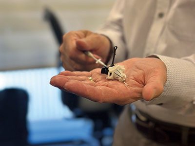
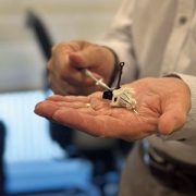
Pericardial port for pediatric pacemaker delivery, developed by Dr. Charles Berul and team.
Kelsey and Dan met with Charles Berul, M.D., emeritus chief of Cardiology and The Van Metre Companies Professor of Cardiology, and his team. There was uncertainty about whether Abby’s heart was too damaged for the pacemaker to work, but Dr. Berul, who has spent decades refining designs for this type of device, expressed confidence. Abby would be the world’s 27th infant, and the fifth at Children’s National, to have one implanted.
“To have him say, ‘We’ve developed this device, we know what we’re doing, all of the other babies who have had this are doing well and we’ll be right here in the room with you,’ was pretty incredible,” Dan says.
Kelsey’s monitoring in the clinical trial and the innovative therapy that started before birth likely helped Abby survive until she was born at 32 weeks at MedStar Washington Hospital Center. Dr. Donofrio and the Children’s National care team were in the delivery room and rushed Abby to our Cardiac Intensive Care Unit. Dr. Donofrio arranged for mom and daughter to pass in the hall on the way. “I heard her cry and felt relief for the first time,” Kelsey says.
Abby’s pacemaker enabled her heart to beat properly on its own. She soon moved to our Neonatal Intensive Care Unit. In two months, she went home with her family. Kelsey and Dan monitor her pacemaker with a handheld device that sends reports to her team at the hospital.
“We’re lucky to have doctors nearby who are at the forefront of this lifesaving research. Children’s National took care of us with a great deal of humanity. Now we can focus on being a family,” says Dan.
Read more stories like this one in the latest issue of Believe magazine.

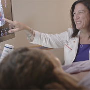
Mary Donofrio, MD, medical director of Prenatal Cardiology at Children’s National, and other dedicated pediatric cardiologists working in this evolving specialty have spent most of the last two decades defining the field.
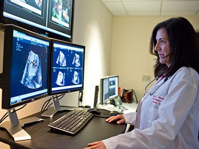
Congenital heart disease (CHD) can be detected in utero with precision and accuracy. With advanced technology, identification of a problem happens earlier than ever, including identifying details that predict whether a baby may be dangerously sick at birth. This gives fetal and pediatric cardiologists time to make plans for delivery and specialized care immediately after birth. These critical first moments can be the key to survival for infants with the most complicated defects.
Mary Donofrio, MD, medical director of Prenatal Cardiology at Children’s National Hospital, and other dedicated pediatric cardiologists working in this evolving specialty have spent most of the last two decades defining the field and demonstrating the importance of making sure every child with a congenital heart defect is diagnosed as early as possible to give them the best chance for a healthy life.
Children’s National performs more than 4,000 fetal ultrasounds each year to detect and manage the unborn child with congenital heart disease, making it one of the most experienced centers in the United States at finding these conditions and planning for their care.
For more than 20 years, every fetus diagnosed with congenital heart disease at Children’s National following an obstetrician referral has their anticipated level of delivery room care assigned by a fetal cardiologist. Protocols were created at Children’s National and validated to establish specialized delivery room management for each patient. The management plan includes specifics about the time and place of delivery and which delivery room staff members are required for stabilization and care after birth based on the severity of the condition.
The outcomes from this approach were published in a landmark 2013 study showing the impact on improving outcomes for infants with the most serious forms of congenital heart disease. Since then, these protocols have become part of more extensive fetal cardiology care guidelines that are in use both at Children’s National and around the world.
“The guidelines we wrote include recommendations about who should get a fetal echo, how to do a fetal echo, how to manage babies in utero including when a fetal intervention might be necessary, and finally how to decide the level of cardiology care that should be present in the delivery room,” according to Donofrio, who served as lead author.
In Washington, D.C., approximately 60 to 75% of congenital heart defects are diagnosed before a baby is born, giving doctors and other care providers critical days, weeks and months to plan how best to protect the fragile infant during their transition into the world from the safe haven of their mother’s body.
Fetal imaging guidelines tell obstetricians which expecting mothers should be referred for a fetal ultrasound given a higher level of risk for CHD over the population risk. However, most women do not have any risk factors that will trigger additional testing beyond obstetrical screening. Also, many families even if referred are far from a center that is qualified to perform a fetal echocardiogram to detect these conditions.
Research at Children’s National, led by Anita Krishan, MD, and Dr. Donofrio in collaboration with the Fetal Heart Society, an international research collaborative, showed that in the U.S., factors such as socio-economic status, ethnicity and geography are important barriers to detection of severe congenital heart diseases such as hypoplastic left heart syndrome and transposition of the great arteries.
In a follow-up study by Jennifer Klein, MD, and Dr. Krishnan, distance was not the only barrier to detecting CHD, however. Geo-mapping technology using zip codes allowed the team at Children’s National to pinpoint “hot spots” where detection is decreased, even in places where care should be available. The Heart Center team is hoping to work with providers in these neighborhoods to improve access to care and help educate local clinic providers about how to image and when to refer for further testing.
Donofrio and colleagues are also working to develop ways to improve the diagnosis of fetal heart disease in places that are far from the Heart Center. This includes exploring more portable diagnostic tools and applying telehealth strategies to connect fetal heart experts with local care providers to make an action plan, before a baby arrives potentially in distress. In addition, a phone-based application is under development to help sonographers to identify abnormal images in real time during routine scans in remote locations. Improved detection rates have also opened the doors to powerful new studies investigating how maternal health and stress impacts brain development in fetuses with congenital heart disease. Ongoing research looks at ways to better support expecting mothers, with the goal of helping moms cope with stress during pregnancy so her baby has the best chance possible to be born healthy and strong.
Donofrio says she won’t stop until in utero detection of congenital heart disease is 100%. “Where you live, your neighborhood, your life experience or how far you live from the Heart Center, should not decrease our ability to do everything possible to care for every baby and achieve the best outcome possible,” she says.
Extensive research has shown that children with congenital heart disease (CHD) who are born blue or who need cardiac surgery in their first year of life are at risk for developmental challenges and/or learning difficulties.
Mary Donofrio, M.D., co-director of the Cardiac Neurodevelopment Outcome (CANDO) program at Children’s National Hospital, says that we started the program — the only one of its kind in the Washington, D.C. region — to identify and manage delays in development and difficulties with learning, no matter when they arise.
“We start paying attention even before birth and then continue to evaluate neurodevelopment at key stages in a kid’s life to assure the best outcome. Our goal is for every kid born with CHD to be able to achieve their full potential, be active, make friends and succeed in school. Most important, we want each of our patients to grow up to be a happy and successful adult,” says Dr. Donofrio.
Learn more about CANDO at Children’s National Hospital and our role in developing best practices for neurodevelopmental and psychosocial services as part of the international multi-specialty Cardiac Neurodevelopmental Outcome Collaborative.
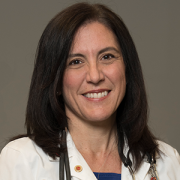
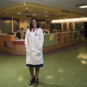
Children’s National Hospital named Mary Donofrio, M.D., FAAP, FACC, FASE, as The Van Metre Companies Professor of Fetal Cardiology at Children’s National Hospital.
Children’s National Hospital named Mary Donofrio, M.D., FAAP, FACC, FASE, as The Van Metre Companies Professor of Fetal Cardiology at Children’s National Hospital.
Dr. Donofrio serves as Medical Director of the Prenatal Cardiology Program and Critical Care Delivery Program, Director of the Advanced Cardiac Imaging Fellowship and Co-Director of the Cardiac Neurodevelopmental Outcome Program at Children’s National. She is a Professor of Pediatrics at George Washington University and is the founding and current President of the Fetal Heart Society, a non-profit organization created to advance the field of fetal cardiovascular care and science through collaborative research, education and mentorship.
Dr. Donofrio joins a distinguished group of 42 Children’s National physicians and scientists who hold an endowed chair. Professorships at Children’s National support groundbreaking work on behalf of children and their families and foster new discoveries and innovations in pediatric medicine. These appointments carry prestige and honor that reflect the recipient’s achievements and donor’s forethought to advance and sustain knowledge.
Dr. Donofrio is an international expert in fetal cardiology. She specializes in the fetal diagnosis and assessment of cardiovascular disease and the in-utero and delivery room management of newborns with complex congenital heart disease (CHD). Dr. Donofrio created an evidenced-based risk-assessment protocol for delivery room management which is now the standard of care for newborns with CHD. Dr. Donofrio has been a co-investigator on several NIH sponsored studies assessing in utero factors that influence neurodevelopmental outcome in children with CHD and more recently a study designed to minimize brain injury during heart surgery using cardiopulmonary bypass. She has published more than 130 papers, including the American Heart Association Scientific Statement on the Diagnosis and Treatment of Fetal Cardiac Disease.
The Van Metre Companies, through their vision and generosity, are ensuring that Dr. Donofrio and future holders of this professorship will launch bold, new initiatives to rapidly advance the field of fetal cardiology, elevate our leadership and improve the lifetimes of children with special hearts.
For the past 65 years, Van Metre Companies has remained one of the Greater Washington D.C. area’s most successful, private, multi-faceted real estate developers. Albert G. Van Metre, the founder of Van Metre Companies, established a tradition of philanthropy focused on local charities. As a homegrown business, perpetuating that legacy of local giving is both a responsibility and a source of pride. The Van Metre Companies Professor of Fetal Cardiology honors Albert G. Van Metre’s memory by continuing this tradition of commitment to the community they call home.
The Van Metre Companies hosts the Annual Van Metre 5K Run in support of Children’s National, a longstanding tradition that has raised nearly $3 million in the past 30 years. In 2010, Children’s National dedicated the Van Metre Companies Cardiovascular Surgery Operating Room, a state-of-the-art cardiovascular surgery suite which was funded through the Annual Van Metre 5K Run. They also established The Van Metre Companies Professorship in Cardiology held by Charles Berul, M.D., Chief of Cardiology and Co-director of Children’s National Heart Institute.


Nobuyuki Ishibashi, M.D., is the principal investigator on a $3.2 million NIH R01 to study white matter growth and repair in utero for fetal brains affected by congenital heart disease.
Many of the neurological deficits seen in children with congenital heart disease (CHD) are related to abnormal white matter development early in life caused by reduced oxygen supply to the brain while in utero. Children with immature white matter at birth also commonly sustain additional white matter injuries following cardiac surgery.
The NIH recently awarded a prestigious R01 grant totaling more than $3.2 million to a collaborative project led by the Center for Neuroscience Research, the Sheikh Zayed Institute for Pediatric Surgical Innovation and the Children’s National Heart Institute at Children’s National Hospital as well as MedStar Washington Hospital Center.
The research, titled “White matter protection in the fetus with congenital heart disease,” looks specifically at whether providing a supplemental amount of the naturally occurring tetrahydrobiopterin (BH4) for pregnant women could rescue white matter development of fetuses with congenital heart disease whose brains aren’t receiving enough oxygen – or suffering from hypoxic-ischemic events.
Previous preclinical studies have shown that this lack of oxygen depletes the brain’s natural BH4 level, and the researchers hypothesize that BH4 levels play a critical role in the growth and development of white matter in the fetal brain by triggering key cellular/molecular processes. Specifically, the study will focus on three aims:
This laboratory-based work is the first step to determining if the neurodevelopment of babies born with CHD can be preserved or recovered by addressing key brain development that occurs before the baby is even born. Findings related to congenital heart disease may also translate to other populations where white matter development is affected by hypoxia-ischemia, including premature infants.
The project is led by principal investigator Nobuyuki Ishibashi, M.D., with co-investigators Vittorio Gallo, Ph.D., Joseph Scafidi, D.O., and Mary Donofrio, M.D. as well as colleagues at MedStar Washington Hospital Center.
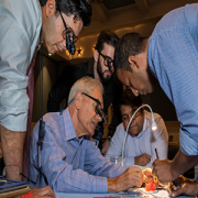
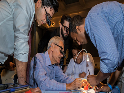
Dr. Richard Jonas shows surgical advancements using 3D heart models, which participants could bring back to their host institutions.
On July 22, 2018, more than 700 cardiac specialists met in Orlando, Fla. for the Sixth Scientific Meeting of the World Society for Pediatric and Congenital Heart Surgery (WSPCHS 2018).
The five-day conference hosted a mix of specialists, ranging from cardiothoracic surgeons, cardiologists and cardiac intensivists, to anesthesiologists, physician assistants and nurse practitioners, representing 49 countries and six continents.
To advance the vision of WSPCHS – that every child born with a congenital heart defect should have access to appropriate medical and surgical care – the conference was divided into eight tracks: cardiac surgery, cardiology, anesthesia, critical care, nursing, perfusion, administration and training.
Richard Jonas, M.D., outgoing president of WSPCHS and the division chief of cardiac surgery at Children’s National Health System, provided the outgoing presidential address, delivered the keynote lecture on Transposition of the Great Arteries (TGA) and guided a surgical skills lab with printed 3-D heart models.
Other speakers from Children’s National include:
Participants left with knowledge about how to diagnose and treat complex congenital heart disease, and an understanding of the long-term consequences of surgical management into adulthood. In addition, they received training regarding standardized practice models, new strategies in telemedicine and collaborative, multi-institutional research.
“It was an amazing experience for me to bring my expertise to a conference which historically concentrated on surgical and interventional care and long-term follow-up,” says Dr. Donofrio. “The collaboration between the fetal and postnatal care teams including surgeons, interventionalists and intensive care doctors enables new strategies to be developed to care for babies with CHD before birth. Our hope is that by intervening when possible in utero and by planning for specialized care in the delivery room, we can improve outcomes for our most complex patients”.
The Johns Hopkins University School of Medicine, Florida Board of Nursing, American Academy of Nurse Practitioners National Certification Program, American Nurses Credentialing Center and the American Board of Cardiovascular Perfusion provided continuing medical credits for eligible providers.
“I was so proud to be a member of the Children’s National team at this international conference,” notes Dr. Wernovsky. “We had to the opportunity to share our experience in fetal cardiology, outpatient cardiology, cardiac critical care, cardiac nursing and cardiac surgery with a worldwide audience, including surgical trainees, senior cardiovascular surgeons and the rest of the team members necessary to optimally care for babies and children with complex CHD. In addition, members of the nursing staff shared their research about advancements in the field. It was quite a success – both for our team and for all of the participants.”
Monitoring and managing fetuses’ heart health in the womb can greatly improve their chances of living long and productive lives
Over the 22 years that Mary T. Donofrio, M.D., has been practicing fetal cardiology, the field has changed radically. The goal once had been simply to offer parents an accurate diagnosis and prepare them for sometimes devastating outcomes. Now, Dr. Donofrio, who directs the Fetal Heart Program and Critical Care Delivery Program at Children’s National Health System, says specialists can follow fetuses throughout the pregnancy and manage many conditions in the womb, greatly improving their chances of living long and productive lives.
Case in point: Transposition of the great arteries, a congenital defect characterized by reversal of the heart’s two main arteries—the aorta, which distributes oxygenated blood throughout the body, and the pulmonary artery, which carries deoxygenated blood from the heart to the lungs. The single abnormality means that the oxygenated “red” blood flows back to the lungs while deoxygenated “blue” blood flows out to the body.
After birth, when the cord is clamped and the connection to the placenta severed, the baby’s cardiovascular system must adjust. If the fetal connections between the two sides of the heart no longer remain, the brain and other organs in infants with this defect are severely deprived of oxygen. The condition may be fatal if something is not done immediately to reopen the fetal connections to stabilize the circulation before surgery can be done. But if the fetal cardiologist can keep tabs on what’s happening to the heart over time and prepare a specialty team of cardiologists to treat the problem immediately after birth, chances of survival are significantly improved.
More than a decade ago, as a young attending physician, Dr. Donofrio witnessed a case that has stuck with her to this day. The baby’s diagnosis of transposition of the great arteries was not made until shortly before birth. In addition, the two fetal blood flow connections that allow blood to circulate had closed, causing severe heart failure. Although the care team performed an emergency delivery and immediate cardiac procedure, including initiation of a heart-lung machine in the delivery room to try to stabilize the circulation, the baby ultimately died due to complications from a very low oxygen level. “I always wonder what happened,” Dr. Donofrio says. “Was the baby’s heart always that bad and nobody noticed it, or did it change over time?”
In a paper published recently in the Journal of Neonatal-Perinatal Medicine, she and colleagues illustrate the dramatic transformation in care that has taken place in the 14 years since this unforgettable case. The new publication describes the case of a different fetus diagnosed at 22 weeks gestation with transposition of the great arteries in 2015 at Children’s National. Unlike many congenital heart disorders, the heart’s four chambers appear misleadingly normal at the typical mid-pregnancy ultrasound. Despite the challenging diagnosis for many obstetricians, this fetus’ heart condition was recognized early by looking at the arteries leaving the heart in addition to the chambers.
While such a defect is fatal if left untreated, Dr. Donofrio explains there are two pathways that can allow the blood to get to where it needs to go such that the circulation is stabilized and the damage mitigated. One is the fetal blood vessel known as the ductus arteriosus that typically stays open for a day or two after birth. The second is an opening between the heart’s two upper chambers, known as the foramen ovale, which usually closes upon delivery. By keeping those two pathways open, blood can cross from one side of the heart to the other, buying time in the delivery room so that babies can be stabilized before they receive surgery to permanently move the arteries back to their normal position.
In the 2015 case, Dr. Donofrio and colleagues had the chance to monitor the fetus and the fetal heart at follow-up appointments every four weeks after diagnosis. What they saw completely changed the course of their treatment plan and likely saved the baby’s life. With each ultrasound, they saw that the ductus arteriosus and the foramen ovale—the critical connections needed for survival—were gradually closing.
Dr. Donofrio noted at the fetal evaluation at 38 weeks that the structures had closed, and the heart was showing signs that it was not functioning well. She and her team realized that the only way to save this baby was to deliver earlier than planned and to have cardiac specialists standing by ready to perform a life-saving procedure to open the connections right after the baby was separated from the placenta. The baby was delivered by Cesarean section in the cardiac operating room at Children’s. The cardiac intervention team immediately created a hole where the foramen ovale should have been by using a balloon to open the tissue that had closed. The care team also administered a prostaglandin infusion, a drug that can keep the ductus arteriosis open. This time, however, the medicine did not work. The baby was stabilized with several cardiac medications and, with little time to spare, the cardiac surgeons operated on the one-day-old baby to switch his great arteries back to the normal position, saving his life.
The baby is now 1-year-old, Dr. Donofrio says, and is healthy—a scenario that likely wouldn’t have happened had the fetal team not made the diagnosis and continually monitored the condition in the womb.
“I remember back to that first case when we were really scrambling to do everything we could at the last minute because we didn’t have the information we needed until the very end,” Dr. Donofrio says. “Now, we can spot problems early and do something about it. For me, that’s amazing. We’re making a difference, and that’s a really great thing.”
 Mary T. Donofrio, M.D., Director of the Fetal Heart Program and Critical Care Delivery Program at Children’s National Health System In a small percentage of pregnancies, the fetuses’ hearts develop in the wrong place. In the congenital anomaly known as heterotaxy syndrome that often includes a severe heart defect, the heart is often displaced from its usual position in the left chest. In other instances, the heart starts out in a normal position; however, it is pushed out of its normal position by a mass that grows in the chest cavity, by abnormal development of the lungs, or due to other causes. Although rare, babies born with cardiac malpositions associated with other congenital defects can be the most serious of all possible birth defects. Sometimes, fetuses with these congenital problems die in the womb. Others do not survive long after birth. In some pregnancies, surgery is performed shortly after childbirth to stabilize the circulation so newborns even have a chance at life. Correctly diagnosing these cardiac conditions during pregnancy can help doctors and parents alike make the most informed decisions and plan ahead. However, the tools now used most often to reveal the overall anatomic details of cardiac malpositions — obstetrical ultrasound and fetal echocardiography — often don’t give a full picture. A clear view of the fetus can be obscured by the position of the fetus, insufficient amniotic fluid, or even a mother’s body habitus. Imaging techniques sometimes also have a hard time distinguishing between liver, bowel, and lung because the echogenicity of these tissues — the signature that sound waves make as they bounce back from their targets — is so similar. “To be able to offer parents the best and most comprehensive counseling, and to begin planning for the type of intensive and multidisciplinary care that many of these babies will require, we need to have access to as much information as we can about each baby, not only relating to the heart but all the other organs as well,” says Mary T. Donofrio, M.D., a pediatric cardiologist who directs the Children’s National Health System Fetal Heart Program and Critical Care Delivery Program. “Unfortunately in some instances, obstetrical ultrasound and fetal echocardiography, the two diagnostic tools used most often in these cases, can be limited in what they tell us.” What fetal MRI can showAn underutilized technique that gathers more details about the associated abnormalities that often accompany cardiac malposition during pregnancy is fetal magnetic resonance imaging, or fetal MRI, says Dr. Donofrio. Even though this technique is widely used to diagnose other fetal conditions, such as brain anomalies, it’s rarely used to better define the overall anatomy in cardiac malposition. To determine whether fetal MRI is effective in complementing obstetrical ultrasound and fetal echocardiography, the current standard of care for this condition, Dr. Donofrio and colleagues took a retrospective look at all cases of cardiac malposition in which fetuses were evaluated using MRI between 2008 to 2013 at Children’s National. Their search turned up 42 cases. Twenty-three cases had been diagnosed with obstetrical ultrasound and fetal echocardiography as having additional abnormalities beyond the heart’s changed position, and 19 had been given the diagnosis of heterotaxy syndrome. Each patient had been assigned to various known subtypes of these conditions, with some classified as having an unknown etiology for the findings. After fetal MRI, the diagnoses of nearly one-third changed or were better delineated. Seven of the 23 cases of cardiac malposition attributed to an extra cardiac anomaly were reassigned to a cause different from the original diagnosis based on the new, more detailed information provided by fetal MRI, including three in which a complete diagnosis could not be made due to poor visualization by ultrasound. Five of the 19 cases attributed of heterotaxy were reassigned to different subgroups within this disorder or were given a different diagnosis completely after fetal MRI. In eight of these 12 diagnoses that changed after fetal MRI, doctors were able to confirm these findings postnatally. Other cases were either lost to follow-up, pregnancy termination, or fetal demise. The research team led by Dr. Donofrio published these results in the August 2016 issue of Prenatal Diagnosis. Overall, she says the findings demonstrated the benefits of using fetal MRI as an adjunct to obstetrical ultrasound and fetal echocardiography. MRI offers advantages over ultrasound, she explains, including better spatial resolution, a wider field of view, and a way to see through or around maternal body fat, overlying fetal bone, or a fetus whose position is not optimal. “Determining the etiology of cardiac malposition remains a challenging diagnosis, and the value of accurate prenatal diagnosis has been long recognized,” Donofrio and colleagues write in the study. “Ultimately, fetal MRI can assist with identifying the etiology of cardiac malposition for informative prenatal counseling and multidisciplinary planning.” |
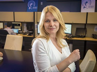
A team of researchers used 3-D volumetric magnetic resonance imaging (MRI) in an innovative study that reported that when the placenta fails to grow adequately in a fetus with congenital heart disease (CHD), it contributes to impaired fetal growth and premature birth. Fetal CHD involves an abnormality of the heart and is associated with increased risk for neurodevelopmental morbidity.Until now, CHD in the fetus and its relationship to placental function has been unknown. But the advanced fetal imaging study has shown for the first time that abnormal growth in the fetus with CHD relates to impaired placental growth over the third trimester of pregnancy. Catherine Limperopoulos, PhD, Director of Children’s National Developing Brain Research Laboratory in the Division of Diagnostic Imaging and Radiology, is the senior author of the study published in the September 2015 issue of the journal Placenta, “3-D Volumetric MRI Evaluation of the Placenta in Fetuses With Complex Heart Disease.”
Specifically, the decreased 3-D volumetric MRI measurements of pregnant women reported in this study suggest placental insufficiency related to CHD. The placenta nourishes and maintains the fetus, through the delivery of food and oxygen. Its volume and weight can determine fetal growth and birth weight.
Abnormality in placental development may contribute to significant morbidity in this high risk-population. This study shows impaired placental growth in CHD fetuses is associated with the length of the pregnancy and weight at birth. Nearly 1 in every 100 babies is born in the United States with a congenital heart defect.
Developing the capacity to examine the placenta non-invasively using advanced MRI is needed to identify early markers of impaired placental structure and function in the high-risk pregnancy. This is a critical first step towards developing strategies for improved fetal monitoring and management, Dr. Limperopoulos says.
“We are trying to develop the earliest and most reliable indicators of placental health and disease in high-risk pregnancies. Our goal is to bring these early biomarkers into clinical practice and improve our ability to identify placental dysfunction,” Dr. Limperopoulos says. “If we can develop the capacity to reliably identify when things begin to veer off course, we then have a window of opportunity to develop therapies to restore function.”
The study used in-vivo 3-D MRI studies and explored placental development and its relationship to neonatal outcomes by comparing placental volumetric growth in healthy pregnancies and pregnancies complicated by CHD.
While mortality rates continue to decrease steadily in newborns diagnosed with complex CHD, long-term neurodevelopmental impairments are recognized with increasing frequency in surviving infants, Dr. Limperopoulos says.
“Our goal is to better support the developing fetus with CHD. We can best accomplish this if we develop technology that can allow us to safely and effectively monitor the fetal-placental unit as a whole throughout pregnancy,” Dr. Limperopoulos says.
“This is the new frontier, not only to ensure survival but to safeguard the fetus and to ensure the best possible quality of life,” she says.
