Addressing long-term brain effects of congenital heart disease

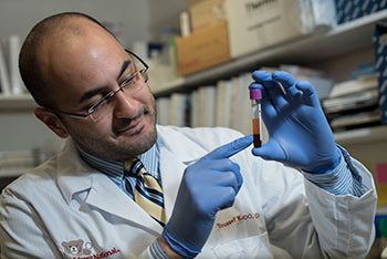
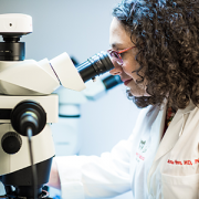
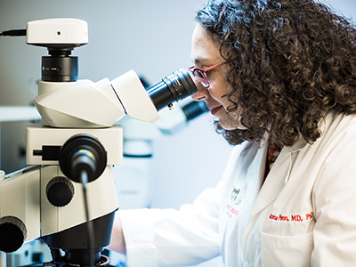
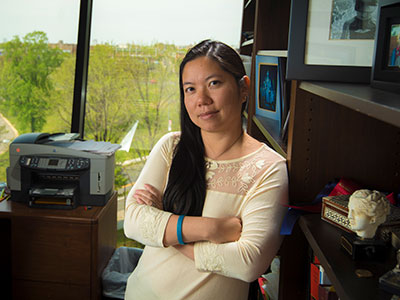
Dr. Anitha John, medical director of the Washington Adult Congenital Heart Program at Children’s National Hospital, presenting on the lifelong effects of congenital heart disease on brain health at a recent symposium.
About 81% of the 40,000 babies born in the United States with congenital heart disease (CHD) are expected to survive to at least age 35, according to the Centers for Disease Control and Prevention. As survival rates have increased in recent decades, clinicians treating CHD patients are seeking to improve outcomes by understanding the long-term health effects and complications that arise for them.
Anitha John, M.D., Ph.D., medical director of the Washington Adult Congenital Heart Program at Children’s National Hospital, presented an overview of what researchers currently know about the lifelong effects of CHD on brain health at a symposium focused on the heart-brain continuum presented by Children’s National Innovation Ventures, CobiCure and JLABS @ Washington, D.C. She also discussed critically needed advancements in monitoring technology to help clinicians better understand and address how CHD affects the brain.
Why it matters
Based on data collected from adults and children with the condition, Dr. John shared that people with CHD face many potential lifelong challenges and risks, which vary based on disease severity:
- About one-third report a mood disorder, either anxiety or depression
- 25% higher risk of substandard academic outcomes
- 50% more likely to require special education services
- Higher incidence of motor skills impairment
- Higher lifetime prevalence of ADHD
- Generally lower educational attainment at adulthood
- Higher risk of autism spectrum disorders
- Higher rate of dementia before the age of 65
Why do some people with CHD experience profound, lifelong brain effects? Dr. John notes that clinicians and researchers are seeking those answers, recognizing that they likely involve various factors and accumulating issues that occur over the entire lifespan, from fetal life onward.
Because the heart supplies the brain with oxygen through circulated blood, the diagnostic tool clinicians most want for patients of all ages is a technology that enables noninvasive monitoring of central venous pressure, an indicator of the volume of blood returning to the heart and the pressure within the heart. Currently, the most reliable way to measure this pressure is by an invasive procedure in which a catheter is inserted into the patient’s subclavian or internal jugular vein or by placing a device into the patient’s pulmonary artery. These procedures have limitations and cannot be used for routine surveillance.
What’s next
Dr. John says noninvasive central venous pressure monitoring is important to understanding and addressing what is causing brain injury in CHD patients. She says the challenges in developing this monitoring solution include the need for an individualized approach, a design that accommodates multidisciplinary use, sizing for patients from infants to adulthood, usability for all age groups and avoiding stigma for wearers.
To address this need, the Alliance for Pediatric Device Development – a consortium funded by the Food and Drug Administration and led by Children’s National – is partnering with CobiCure to issue a request for proposals for direct device funding. The goal is to provide funding to innovators who offer solutions to the dire unmet need for pediatric devices that provide noninvasive monitoring of the circulatory system and heart performance. Details will be announced in June 2024.