Graph showing magnesium reduces arrhythmia risk

Magnesium helps prevent postsurgical arrhythmias in pediatric patients

Graph showing magnesium reduces arrhythmia risk

Magnesium (Mg) helps reduce arrhythmias, irregular heart rhythms, in adults. It also helps alleviate the symptoms of postoperative atrial fibrillation, or AFib, which can lead to blood clots, stroke and heart failure. Can it help prevent postsurgical arrhythmias in pediatric patients with congenital heart disease?

New research from Children’s National Health System finds a 25- or 50-mg dose of Mg used during congenital heart surgery (CHS) helps prevent arrhythmias, especially junctional ectopic tachycardia (JET) and atrial tachycardia (AT), common arrhythmias following CHS, according to a study published in the August 2018 edition of The Journal of Thoracic and Cardiovascular Surgery.

To reach this conclusion, the researchers separated 1,871 CHS patients from Children’s National into three groups: a control group of 750 patients who had surgery without Mg, a group of 338 patients receiving a 25-mg /kg dose of Mg during surgery and a group of 783 patients receiving a 50-mg/kg dose of Mg during surgery. The data looked at CHS cases over eight years, from 2005 to 2013, to determine if Mg administration during surgery alleviates postoperative arrhythmias and if the amount, measured by a 25- or 50-mg/kg dose, makes a difference.

“This study, the first conducted in pediatric patients, finds administering magnesium during congenital heart surgery reduces the likelihood of postsurgical arrhythmias,” says Charles Berul, M.D., a study author and the chief of cardiology at Children’s National. “We don’t detect a dose-dependent relationship, which means a small or larger amount of magnesium is equally effective at preventing arrhythmias following surgery.”

The researchers found that up to one-third of CHS patients experience postoperative arrhythmias, with JET and AT accounting for more than two-thirds of arrhythmias following CHS. They note that despite the administration of Mg during surgery, there continues to be a high incidence of postoperative arrhythmias – affecting 18 percent or about one in five CHS patients.

“We hope this study guides future research to see if adding new or additional agents to magnesium eliminates, or further reduces, postoperative arrhythmias,” notes Dr. Berul. “For now, we’re happy to find an algorithm to put into practice and to share with other medical centers, as a way to help pediatric patients recover from congenital heart surgery—stronger, faster and with a reduced risk of complications.”

The researchers note that postoperative arrhythmias impact the recovery period of CHS, increase the duration of intubation and CICU stay and prolong hospital stay.

Making the grade: Children’s National is nation’s Top 5 children’s hospital

Children’s National rose in rankings to become the nation’s Top 5 children’s hospital according to the 2018-19 Best Children’s Hospitals Honor Roll released June 26, 2018, by U.S. News & World Report. Additionally, for the second straight year, Children’s Neonatology division led by Billie Lou Short, M.D., ranked No. 1 among 50 neonatal intensive care units ranked across the nation.

Children’s National also ranked in the Top 10 in six additional services:

For the eighth year running, Children’s National ranked in all 10 specialty services, which underscores its unwavering commitment to excellence, continuous quality improvement and unmatched pediatric expertise throughout the organization.

“It’s a distinct honor for Children’s physicians, nurses and employees to be recognized as the nation’s Top 5 pediatric hospital. Children’s National provides the nation’s best care for kids and our dedicated physicians, neonatologists, surgeons, neuroscientists and other specialists, nurses and other clinical support teams are the reason why,” says Kurt Newman, M.D., Children’s President and CEO. “All of the Children’s staff is committed to ensuring that our kids and families enjoy the very best health outcomes today and for the rest of their lives.”

The excellence of Children’s care is made possible by our research insights and clinical innovations. In addition to being named to the U.S. News Honor Roll, a distinction awarded to just 10 children’s centers around the nation, Children’s National is a two-time Magnet® designated hospital for excellence in nursing and is a Leapfrog Group Top Hospital. Children’s ranks seventh among pediatric hospitals in funding from the National Institutes of Health, with a combined $40 million in direct and indirect funding, and transfers the latest research insights from the bench to patients’ bedsides.

“The 10 pediatric centers on this year’s Best Children’s Hospitals Honor Roll deliver exceptional care across a range of specialties and deserve to be highlighted,” says Ben Harder, chief of health analysis at U.S. News. “Day after day, these hospitals provide state-of-the-art medical expertise to children with complex conditions. Their U.S. News’ rankings reflect their commitment to providing high-quality care.”

The 12th annual rankings recognize the top 50 pediatric facilities across the U.S. in 10 pediatric specialties: cancer, cardiology and heart surgery, diabetes and endocrinology, gastroenterology and gastrointestinal surgery, neonatology, nephrology, neurology and neurosurgery, orthopedics, pulmonology and urology. Hospitals received points for being ranked in a specialty, and higher-ranking hospitals receive more points. The Best Children’s Hospitals Honor Roll recognizes the 10 hospitals that received the most points overall.

This year’s rankings will be published in the U.S. News & World Report’s “Best Hospitals 2019” guidebook, available for purchase in late September.

Research and Education Week awardees embody the diverse power of innovation

cnmc-research-education-week

“Diversity powers innovation” was brought to life at Children’s National April 16 to 20, 2018, during the eighth annual Research and Education Week. Children’s faculty were honored as President’s Award winners and for exhibiting outstanding mentorship, while more than 360 scientific poster presentations were displayed throughout the Main Atrium.

Two clinical researchers received Mentorship Awards for excellence in fostering the development of junior faculty. Lauren Kenworthy, Ph.D received the award for Translational Science and Murray M. Pollack, M.D., M.B.A., was recognized in the Clinical Science category as part of Children’s National Health System’s Research and Education Week 2018.

Dr. Kenworthy has devoted her career to improving the lives of people on the autism spectrum and was cited by former mentees as an inspirational and tireless counselor. Her mentorship led to promising new lines of research investigating methods for engaging culturally diverse families in autism studies, as well as the impact of dual language exposure on cognition in autism.

Meanwhile, Dr. Pollack was honored for his enduring focus on motivating early-career professionals to investigate outcomes in pediatric critical care, emergency medicine and neonatology. Dr. Pollack is one of the founders of the Collaborative Pediatric Critical Care Research Network. He developed PRISM 1 and 2, which has revolutionized pediatric intensive care by providing a methodology to predict mortality and outcome using standardly collected clinical data. Mentees credit Dr. Pollack with helping them develop critical thinking skills and encouraging them to address creativity and focus in their research agenda.

In addition to the Mentorship and President’s Awards, 34 other Children’s National faculty, residents, interns and research staff were among the winners of Poster Presentation awards. The event is a celebration of the commitment to improving pediatric health in the form of education, research, scholarship and innovation that occurs every day at Children’s National.

Children’s Research Institute (CRI) served as host for the week’s events to showcase the breadth of research and education programs occurring within the entire health system, along with the rich demographic and cultural origins of the teams that make up Children’s National. The lineup of events included scientific poster presentations, as well as a full slate of guest lectures, educational workshops and panel discussions.

“It’s critical that we provide pathways for young people of all backgrounds to pursue careers in science and medicine,” says Vittorio Gallo, Ph.D., Children’s chief research officer and CRI’s scientific director. “In an accelerated global research and health care environment, internationalization of innovation requires an understanding of cultural diversity and inclusion of different mindsets and broader spectrums of perspectives and expertise from a wide range of networks,” Gallo adds.

“Here at Children’s National we want our current and future clinician-researchers to reflect the patients we serve, which is why our emphasis this year was on harnessing diversity and inclusion as tools to power innovation,” says Mark L. Batshaw, M.D., physician-in-chief and chief academic officer of Children’s National.

“Research and Education Week 2018 presented a perfect opportunity to celebrate the work of our diverse research, education and care teams, who have come together to find innovative solutions by working with local, national and international partners. This event highlights the ingenuity and inspiration that our researchers contribute to our mission of healing children,” Dr. Batshaw concludes.

Awards for the best posters were distributed according to the following categories:

  • Basic and translational science
  • Quality and performance improvement
  • Clinical research
  • Community-based research and
  • Education, training and program development.

Each winner illustrated promising advances in the development of new therapies, diagnostics and medical devices.

Diversity powers innovation: Denice Cora-Bramble, M.D., MBA
Diversity powers innovation: Vittorio Gallo, Ph.D.
Diversity powers innovation: Mark L. Batshaw, M.D.

child measuring belly with tape measure

Children’s obesity research team presents compelling new findings

child measuring belly with tape measure

Faculty from Children’s National Health System’s Department of Psychology & Behavioral Health set out to learn if any demographic, psychiatric, or cognitive factors play a role in determining if an adolescent should be eligible for bariatric surgery, and what their weight loss outcomes might be. Presenting at the Society for Pediatric Psychology Annual Conference earlier this month, a group of researchers, fellows and clinicians, including surgeons from Children’s National showcased their findings. One of the posters developed by Meredith Rose, LGSW, ML, who works as an interventionist on a Children’s National clinical research team, received special recognition in the Obesity Special Interest Group category.

One presentation reported on a total of 222 pediatric patients with severe obesity, which is defined as 120 percent of the 95th percentile for Body Mass Index. Mean age of the participants was 16 years of age, 71 percent were female and 80 percent where Hispanic or non-White. As part of their preparation for surgery, all patients were required to complete a pre-bariatric surgery psychological evaluation, including a clinical interview and Schedule for Affective Disorders and Schizophrenia (KSADS-PL) screening. The studies by the Children’s teams were based on a medical record review of the pre-screening information. Adolescents being evaluated for surgery had high rates of mental health diagnoses, particularly anxiety and depression, but also included Attention Deficit Hyperactivity Disorder, eating disorders, and intellectual disability.

Another Children’s presentation at the conference looked at weight loss outcomes for adolescents based on IQ and intellectual disability. Overall, neither Full Scale IQ from the Wechsler Abbreviated Intelligence Scale – 2nd edition, nor the presence of an intellectual disability predicted weight loss following surgery.

“The sum of our research found that kids do really well with surgery,” said Eleanor Mackey, PhD, assistant professor of psychology and behavioral health. “Adolescents, regardless of the presence of intellectual disability areas are likely to lose a significant amount of weight following surgery,” added Dr. Mackey.

“This is a particularly important fact to note because many programs and insurers restrict weight loss surgery to ‘perfect’ candidates, while these data points demonstrate that our institution does not offer or deny surgery on the basis of any cognitive characteristics,” says Evan P. Nadler, M.D., associate professor of surgery and pediatrics. “Without giving these kids a chance with surgery, we know they face a lifetime of obesity, as no other intervention has shown to work long-term in this patient population. Our research should empower psychologists and physicians to feel more confident recommending bariatric surgery for children who have exhausted all other weight loss options.”

The research team concluded that examining how individual factors, such as intellectual disability, psychiatric diagnoses, and demographic factors are associated with the surgery process is essential to ensuring adequate and empirically supported guidelines for referral for, and provision of bariatric surgery in adolescents. Next steps by the team will include looking into additional indicators of health improvement, like glucose tolerance, quality of life, or other lab values, to continue evaluating the benefits of surgery for this population.

Sean Donahue

Pediatric ophthalmology celebrates 75th anniversary in Washington, D.C.

Sean Donahue

Angeline M. Parks Visiting Professor Sean P. Donahue, M.D., Ph.D., (front left) enjoys a light moment during the celebration of the 75th anniversary while Anthony Sandler, M.D., Children’s surgeon in chief, senior vice president of the Joseph E. Robert Jr. Center for Surgical Care and director of the Sheikh Zayed Institute, speaks to the group.

After 75 years dedicated to the eyes of children, the world’s pediatric ophthalmologists gathered in Washington, D.C., the specialty’s birthplace, to share the latest research and innovation in the field. The group gathered for a joint meeting of the International Strabismological Association (ISA) and the American Association for Pediatric Ophthalmology and Strabismus (AAPOS), which was held March 18-22, 2018.

“This year marks the 75th anniversary of our specialty, which was founded right here, at Children’s National, in Washington, D.C., when Dr. Frank Costenbader restricted his practice exclusively to children and began to train residents in the nuance of treating children’s eyes,” says Mohamad S. Jaafar, M.D., chief of the Division of Ophthalmology at Children’s National Health Center. “It is a tremendous honor to welcome my colleagues back to the birthplace of pediatric ophthalmology on this grand occasion.”

In advance of the larger meeting, Children’s Division of Ophthalmology welcomed some of the international attendees to Children’s National for a special gathering on Saturday, March 17, 2018.

The event at Children’s featured a special lecture by this year’s Angeline M. Parks Visiting Professor, Sean P. Donahue, M.D., Ph.D. Dr. Donahue is the Sam and Darthea Coleman Chair in Pediatric Ophthalmology and Chief of Pediatric Ophthalmology at the Children’s Hospital at Vanderbilt. This Annual Visiting Professorship was established by the members of the Costenbader Society (The Children’s National Pediatric Ophthalmology Alumni Society) in memory of Angeline M. Parks, the wife of pediatric ophthalmologist Marshall M. Parks, M.D., to carry on her legacy of establishing a warm and supportive environment between physician and spouse, which benefits the physicians and their young patients.

Three former division chiefs of Ophthalmology at Children’s National, Drs. Costenbader, Parks and Friendly, have national lectureships established in their names to reflect their contributions to the field. Dr. Frank Costenbader, the society’s namesake, established the sub-specialty of pediatric ophthalmology. Dr. Parks founded the Children’s Eye Foundation and the AAPOS, and David S. Friendly, M.D., codified pediatric ophthalmology fellowship training across the United States.

Honor Awards for Children’s pediatric ophthalmologists at ISA-AAPOS

During the ISA-AAPOS meeting, two current Children’s National pediatric ophthalmologists were recognized with Honor Awards for their long-term dedication to pediatric ophthalmology, their patients, and their engagement in the AAPOS to advance the field.

William Madigan, M.D., vice chief of Ophthalmology at Children’s, a professor of surgery at the Uniformed Services University of the Health Sciences, and a clinical professor of Ophthalmology and Pediatrics at the George Washington University School of Medicine and Health Sciences. He was recognized by AAPOS for his long-time service, including:

  • Chair of the organization’s audit committee and the Costenbader Lecture selection committee.
  • Membership on the fellowship directors’ committee that developed nationwide requirements for pediatric ophthalmology fellowships and established the certification process to insure high quality and uniform education in the specialty.
  • Invited lectures in Shanghai, China; Geneva, Switzerland; and Sao Paolo, Brazil, among others.
  • Many posters and presentations about clinical and research topics of importance for members of the AAPOS and other distinguished professional societies.

Marijean Miller, M.D., director of Neonatal Ophthalmology, division research director at Children’s National and clinical professor of Ophthalmology and Pediatrics at the George Washington University School of Medicine and Health Sciences, was recognized by AAPOS for her cumulative contributions to the society, including:

  • Multiple memberships on vital committees, including AAPOS’s training and accreditation committee and audit committee.
  • Presentation of original research via posters and oral presentations on topics including best practices in neonatal clinical care, innovative tools and applications and advocacy for patients and their families.

“We are so grateful to have a team that continues the tradition of excellence in pediatric ophthalmology here at Children’s National,” Dr. Jaafar says. “Drs. Madigan and Miller exemplify the dedication of our division to caring for the children we serve, and to advancing our field. Congratulations to both!”

banner year

2017: A banner year for innovation at Children’s National

banner year

In 2017, clinicians and research faculty working at Children’s National Health System published more than 850 research articles about a wide array of topics. A multidisciplinary Children’s Research Institute review group selected the top 10 articles for the calendar year considering, among other factors, work published in high-impact academic journals.

“This year’s honorees showcase how our multidisciplinary institutes serve as vehicles to bring together Children’s specialists in cross-cutting research and clinical collaborations,” says Mark L. Batshaw, M.D., Physician-in-Chief and Chief Academic Officer at Children’s National. “We’re honored that the National Institutes of Health and other funders have provided millions in awards that help to ensure that these important research projects continue.”

The published papers explain research that includes using imaging to describe the topography of the developing brains of infants with congenital heart disease, how high levels of iron may contribute to neural tube defects and using an incisionless surgery method to successfully treat osteoid osteoma. The top 10 Children’s papers:

Read the complete list.

Dr. Batshaw’s announcement comes on the eve of Research and Education Week 2018 at Children’s National, a weeklong event that begins April 16, 2018. This year’s theme, “Diversity powers innovation,” underscores the cross-cutting nature of Children’s research that aims to transform pediatric care.

Matthew Oetgen

3D printed implant used to repair knee cartilage

Matthew Oetgen

“Our preliminary study shows this novel 3D printed material is able to allow ingrowth from the bone, so the body started to grow into the material to help fix it in place,” says Matthew Oetgen, M.D., M.B.A. “These are the first step requirements for an implant like this to be acceptable for treating lesions.”

Every year, an estimated 1 million children tear the articulate cartilage that lines their knees. Unfortunately, these types of injuries are extremely hard to repair because of the cartilage’s poor healing qualities and unique physiochemical properties.

Now, a new study by Children’s National Health System researchers has found that a three dimensional (3D) printed synthetic implant can be successfully used as a scaffold to encourage the healing and repair of articulate cartilage lesions.

Three bones meet in the knee joint: the femur, the tibia and the patella. The surface of these bones is covered with articulate cartilage, which can be damaged by injury or by normal wear and tear. Because articulate cartilage has poor healing qualities, these injuries will rarely heal or regenerate on their own, especially in younger and more active patients.

“These are active 12 to 19 year olds, so it can really affect relatively normal kids,” says Matthew Oetgen, M.D., M.B.A., Division Chief of Orthopaedic Surgery and Sports Medicine at Children’s National. “While there are many ways to repair these lesions — from implanting autogenous cells to using grafts to fill the defect — none of these options are perfect, and they all have some down sides.”

To facilitate repair of these injuries, a team of researchers led by Dr. Oetgen received a grant from the Pediatric Orthopaedic Society of North America (POSNA) to design a 3D printed implant that promotes bone and cartilage growth.

To make the implant, the team used nanoporous thermoplastic polyurethane (TPU), a biodegradable material that is highly elastic and yet strong, very much like the native cartilage in the osteochondral region. TPU is also porous, which allows blood and nutrient flow through the implant.

“The implant is designed to allow native cells to repair the lesions with normal articular cartilage and not scar tissues like some repairs,” says Dr. Oetgen.

The implant itself has a stratified structure: an upper region that contains micro channels to allow for increased perfusion; a middle zone with a nanoporous structure that mimics porous cartilage and encourages stem cell recruitment, growth and differentiation; and a lower region, or articular surface, that allows for smooth transition from the articulating surface to the implant surface and minimizes adverse interactions between the articulate cartilage and the meniscus.

When tested in vitro, the implant was able to support the growth of stem cells and vascular cells, and structurally mature vascularized bone was formed around the implant after 10 days. In animal models with full thickness osteochondral lesions the implant did just as well: The scaffold was able to promote bone, soft tissue and vascular growth without eliciting an immune response.

“Our preliminary study shows this novel 3D printed material is able to allow ingrowth from the bone, so the body started to grow into the material to help fix it in place,” says Dr. Oetgen. “These are the first step requirements for an implant like this to be acceptable for treating lesions.”

Because of the ease with which 3D printing can be scaled up, Dr. Oetgen is hopeful that the implant will one day become a viable option for repairing articulate cartilage injuries. He plans on trying the implants in a larger animal model and on larger lesions, and is also looking at custom printing for the implants to match natural lesion shapes and sizes.

William Gaillard

Putting childhood epilepsy in the spotlight at American Epilepsy Society Meeting

William Gaillard

“We aim to build the evidence base for treatments that are effective specifically for children with epilepsy,” says William D. Gaillard, M.D., chief of Child Neurology, Epilepsy and Neurophysiology, and director of the Comprehensive Pediatric Epilepsy Program.

While epilepsy affects people of all ages, the unique way it manifests in infants, children and adolescents can be attributed in part to the complexities of the growing and developing brain. Researchers from the Children’s National Comprehensive Pediatric Epilepsy Program brought their expertise on the challenges of understanding and treating epilepsy in children to the recent American Epilepsy Society Annual Meeting, the largest professional gathering on epilepsy in the world.

“We aim to build the evidence base for treatments that are effective specifically for children with epilepsy,” says William D. Gaillard, M.D., chief of Child Neurology, Epilepsy and Neurophysiology, and director of the Comprehensive Pediatric Epilepsy Program. “We have learned much from studies in adult populations but technologies like functional MRI allow us to get in-depth understanding, often in non-invasive ways, of precisely how epilepsy is impacting a child.”

Dr. Gaillard was also recently elected to serve as the Second Vice President of the American Epilepsy Society. “The AES is the largest multidisciplinary professional and scientific society dedicated to the understanding, treatment and eradication of epilepsy and associated disorders, and I am honored to serve as the new Second Vice President,” he said.

The team’s presentations and poster sessions focused on several key areas in pediatric epilepsy:

Better ways to see, measure and quantify activity and changes in the brain for children with epilepsy before, during and after surgery

  • Novel applications of fMRI for children with epilepsy
    • Evaluation of an fMRI tool that tracks verbal and visual memory in children with epilepsy – one of the first to capture memory functions in this population of children using noninvasive fMRI;
    • Early study of the use of “resting-state” fMRI to map language skills before epilepsy surgery – an important first step toward noninvasively evaluating children who are too young or neurologically impaired to follow tasks in traditional MRI studies;
  • A study of whether intraoperative MRI, i.e. imaging during neurosurgery, allows for more complete removal of abnormal brain tissue associated with focal cortical dysplasia in children, which is a common cause of intractable epilepsy;
  • A preliminary case review of existing data to see if arterial spin labeling MRI, which measures blood flow to the brain, has potential to identify blood flow changes in specific locations of the brain where seizures occur;
  • An analysis of language laterality – the dominant side of the brain controlling language –  questioning the true reasons that the brains of children with epilepsy have differences in the hemisphere that predominantly controls language;
  • A review of some common assessments of language and working memory that are used pre- and post-operatively to gauge the impacts of pediatric epilepsy surgery. The study found that using multiple assessments, and studying results individually rather than as a group average, resulted in a more complete picture of the outcomes of surgery on these areas of brain function;
  • A preliminary study examining whether continuous EEG monitoring of neonates with hypoxic ischemic encephalopathy, or lack of oxygen to the brain, can be a reliable predictor of neurodevelopmental outcomes while the infant is undergoing therapeutic hypothermia.

“In order to expand our understanding of causes, impacts and outcomes, the range of research is broad given the complexity of epilepsy,” says Madison M. Berl, Ph.D. “This is the only way we can contribute to the goal of providing our colleagues and the families they serve with better resources to make informed decisions about how best to assess and treat pediatric epilepsy.”

The molecular, genetic and biological factors that contribute to onset and severity of pediatric epilepsy

  • A retrospective study of young patients with malformations in cortical development that are important causes of childhood epilepsy;
  • Investigation of a simple saliva test to effectively identify the presence of two common viral infections, human herpesvirus-6B and Epstein-Barr virus, that may be contributors to onset of epilepsy in otherwise normally functioning brains;
  • A preliminary review of the possible relationship between febrile infection-related epilepsy syndrome and the co-occurrence of another neuro-inflammatory condition – hemophagocytic lymphohistiocytosis.

Madison Berl, Ph.D., director of research in the Division of Pediatric Neuropsychology, and a pediatric neuropsychologist in the Comprehensive Pediatric Epilepsy Program, adds, “In order to expand our understanding of causes, impacts and outcomes, the range of research is broad given the complexity of epilepsy. This is the only way we can contribute to the goal of providing our colleagues and the families they serve with better resources to make informed decisions about how best to assess and treat pediatric epilepsy.”

Exchanging ideas

Exchanging ideas, best practices in China

Exchanging ideas

Physicians from the Children’s National delegation attended the Shanghai Pediatric Innovation Forum in June 2017. Pictured (left to right): Roberta DeBiasi, M.D., Michael Mintz, M.D., Robert Keating, M.D., Lawrence Jung, M.D., Peter Kim, M.D., and Sarah Birch, D.N.P., A.P.R.N.

In late June, a delegation of international pediatric experts from Children’s National Health System journeyed across the world to learn about the practice of pediatric medicine in China and to exchange ideas with colleagues there. Leaders from several of Children’s key specialties joined the delegation, including:

The group, led by Drs. Keating and Gaillard, traveled to China with Children’s Outreach Coordinator John Walsh, whose longtime connections and close familiarity with the pediatric medical community in Hangzhou and Shanghai made the collaboration possible. The team toured several of the largest children’s hospitals in country, including The Children’s Hospital of Zhejiang University School of Medicine in Hangzhou and Shanghai Children’s Medical Center, connecting with pediatric specialists there.

“Some of the most important parts of this trip were the opportunities to exchange ideas and solidify long term relationships that will allow us to work closely with our peers in China as they develop their pediatric programs. The potential is tremendous for unique collaborations between our teams and theirs for research and the development of clinical care improvements for children,” said Roger Packer, M.D., senior vice president of the Center for Neuroscience and Behavioral Medicine, who joined the delegation in Beijing.

A keynote lecture and more at the 3rd China International Forum on Pediatric Development

The delegation also was honored with an invitation to participate in the 3rd China International Forum on Pediatric Development. The forum is one of the largest pediatric focused meetings in the country and is led by all the major children’s hospitals in China, including those in Beijing and Shanghai. Close to 4,000 pediatricians attended the meeting, and presenters included esteemed international leaders in pediatric medicine from around the world.

Dr. Packer delivered one of the opening keynote lectures, entitled, “Translation of molecular advances into care: the challenge ahead for children’s hospitals.” His talk focused on the tremendous promise and significant challenges posed by the latest scientific advances, through the lens of a neurologist.

“Across the world, we are looking at the same challenges: How can we use scientific advances to find better outcomes? How can we financially support the new types of interventions made possible by these molecular biologics insights when they can cost millions of dollars for one patient?”

“There’s palpable excitement that these new developments will give us potential therapies we never dreamed about before, ways to reverse what we initially thought was irreversible brain damage, ways to prevent severe illnesses including brain tumors, but the issue is how to turn this promise into reality. That’s a worldwide issue, not simply a single country’s issue,” he continued.

He also flagged mental health and behavioral health as a crucial, universal challenge in need of addressing on both sides of the Pacific.

The Children’s National delegation, including Drs. DeBiasi, Song, Keating, Gaillard and Packer were also honored to share their insight in a series of specialty-specific breakout sessions at the Forum.

Overall, the long journey opened a dialogue between Children’s National and pediatric care providers in China, paving the way for future discussion about how to learn from each other and collaborate to enhance all institutions involved.

two doctors perform surgery

Working miracles to control seizures and preserve brain power in newborns

Oluigbo and Myseros neurosurgery

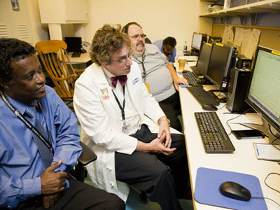
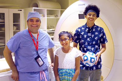
In the spring of 2017, a multidisciplinary team applied an innovative approach to help preserve function in the working right hemisphere of a baby who experienced her first seizure hours after birth.

When orderly early fetal brain development is disturbed in one half of the brain, infants can be born with hemimegalencephaly—a rare occurrence—that results in one of the brain’s two hemispheres being oversized, heavy and malformed. This brain malformation arises early in the fetal period of life, is not inherited and is associated with seizures early in life.

Children with hemimegalencephaly can develop horrible seizures within the first hours or days of life. According to published research, every month these infants experience uncontrolled seizures correlates to a steep decline in IQ.

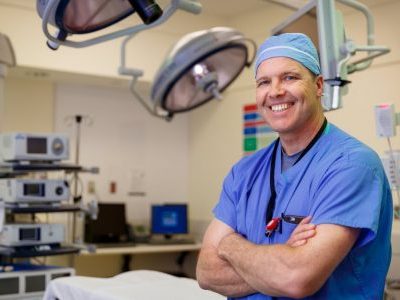
Because these types of seizures do not respond to multiple anti-seizure medications—medicines which may also cause worrisome side effects of their own in neonates—care teams attempt to schedule surgery as soon as feasible to remove or disconnect the hemisphere triggering the damaging seizures. “The ‘bad’ brain does not sustain any function and it interferes with the ‘good’ brain doing what it needs to do,” says William D. Gaillard, M.D., chief of Children’s division of Epilepsy and Neurophysiology and chief of Neurology.

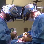
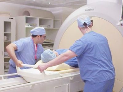
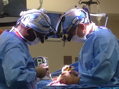
Hemispherectomy is intricate surgery on an organ that is softer than normal and crisscrossed with a tangle of blood vessels that supply the damaged hemisphere with blood. Because of the risks of life-threatening blood loss in very young infants, the dramatic surgery is usually not performed until babies are at least 3 months old and weigh at least 10 pounds.

The challenge: The vulnerable babies who most need relief, infants who have been seizing since early life, are too young for the operation.

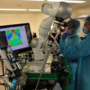
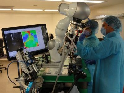
Neurosurgeons have clamped the carotid artery that supplies blood to the brain to minimize blood loss when the hemisphere is surgically removed. Dr. Gaillard says knowledge of that approach led the team to think: What if we use embolization—blocking blood supply to targeted locations in the brain—to achieve the same effect?  The plan effectively destroys the malformed brain from within, neutralizing its ability to cause the seizures.

“It was eye-opening for us to think about actually inflicting brain injury as a way of treating something in the brain that was causing seizures. That is really novel in itself: We’re thinking out of the box in applying existing techniques in a different age group. The conventional thinking with newborns is to let them be; their seizures don’t look that bad,” says Taeun Chang, M.D., director of Children’s Neonatal Neurology and Neonatal Neurocritical Care Program.

“We have evidence to suggest this is a safe and effective way of avoiding recurrent seizures and minimizing the need to give these infants potentially toxic medications so early in life. Ultimately, this helps a select group of babies who need the surgery to get to the point of being old enough to have it—all the while, sparing the healthy part of their brain,” Dr. Gaillard adds.

Darcy hemimegalencephaly

Once the embolization ended Darcy’s most severe seizures, the little girl could make eye contact, started smiling, and then graduated from smiling to full laughs. In weekly physical therapy, the infant works on tummy time, head control and ensuring her eyes track.

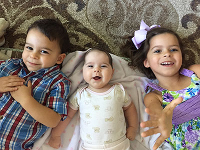
In the spring of 2017, the multidisciplinary team applied the innovative approach to help preserve function in the working right hemisphere of a baby named Darcy Murphy. Darcy experienced her first seizure hours after she was born, and when she arrived at Children’s National had been in and out of two different emergency rooms in another state for the first few weeks of her life.

The team explained to the Murphy family that Darcy was on multiple medications, but her seizures continued unabated. The options included inducing a coma, sending Darcy home despite ongoing seizures or minimally invasive embolization.

“We would not have even posed this if we were not confident in our ability to do the procedure and deal with potential complications,” Dr. Chang says.

“Oh my gosh, as a parent you know what you’re doing is permanent,” says Rachel Murphy, 29, Darcy’s mom said of the decisions that she and husband Ryan, 33, faced for the youngest of their three children. “What if it’s not the right decision? What if in a week they come out with a new procedure you could have done? We were horrified all the time. The nice part with this procedure is the reward is apparent very quickly, and it just gets better. You don’t have to wait two years to know you made the right decision. You can see half a brain is better than the whole thing for this specific child.”

Once the embolization ended Darcy’s most severe seizures, the little girl could initiate and maintain eye contact with family members, started smiling and then graduated from smiling to full laughs. In weekly physical therapy, the infant works on tummy time, head control and ensuring her eyes track.

Children’s multidisciplinary care team includes experts in newborn intensive care (neonatologists) to aggressively manage seizures in the traditional fashion as they occur and to monitor vital signs; a neonatal neurologist/neurointensivist at the bedside and in the Angio suite monitoring Darcy’s brain activity; a neonatal epileptologist; a surgical epilepsy team; an interventional neuroradiologist; neurosurgeons to perform the delicate functional hemispherectomy to remove any residual brain tissue from the bad hemisphere; and physical therapists working to help Darcy achieve maximum function after surgery.

“We were just like one unit in the sense of being able to provide coherent, comprehensive care. It’s about blood pressure management, breathing, electrolytes, making sure everything is right for going to the operating room,” Dr. Chang explains. “Darcy’s case highlights the ways in which Children’s National is different and offers personalized care that is superior to other centers.”

The team, which recently published a case report of two previous serial embolizations followed by hemispherectomy, plans follow-up papers describing EEG manifestations during an acute stroke in a newborn, advice to the field on best practices for the embolization and using cooling to control the planned brain injury during embolization hemispherectomy.

Revised Nov. 7, 2017

Related resources

Karun Sharma, M.D., poses with two patients

Treating osteoid osteoma with MR-HIFU

Karun Sharma, M.D., poses with two patients

Karun Sharma, M.D., poses with two patients who participated in the MR-HIFU trial for pediatric osteoid osteoma.

Doctors from the Sheikh Zayed Institute for Pediatric Surgical Innovation and surgeons from Children’s National are the first in the U.S. to use Magnetic Resonance-Guided High-intensity Focused Ultrasound (MR-HIFU) to treat pediatric osteoid osteoma.

The trial, led by Principal Investigator Karun Sharma, M.D., Ph.D., Director of Interventional Radiology at Children’s National, began in 2015 and is demonstrating early success in establishing the safety and feasibility of noninvasive MR-HIFU as an alternative to the current, more invasive approaches to remove tumor tissue.

Osteoid osteoma is a painful, but benign, bone tumor that commonly occurs in children and young adults. Removal generally requires orthopaedic surgery to scrape the tumor from the bone or CT (computerized tomography) image-guided radiofrequency ablation (RFA), which is less invasive than surgery but is associated with ionizing radiation exposure and requires drilling through muscle and soft tissue into bone.

MR-HIFU, on the other hand, is a precise and controlled method that does not require a scalpel or needle, greatly reducing the risk of complications, including infections and bone fractures. Even better, it promises reduced procedure time, typically an hour or less.

“Our team set out to provide a noninvasive and radiation free treatment option for children with osteoid osteoma and our pilot feasibility and safety trial is almost completed. We have treated 9 patients and we’re very pleased with the success of the treatments so far. Although follow up will continue for another year, results to date that show that MR-HIFU may be a completely non-invasive and radiation free treatment for osteoid osteoma,” Dr. Sharma says. “Several of the children we treated were very active prior to the onset of their tumor, one a soccer player and the other a swimmer, but because of pain from the tumor, they were unable to enjoy their favorite activities, until now.”

“The use of MR-HIFU ablation of osteoid osteoma is a perfect example of our mission in the Sheikh Zayed Institute to make pediatric surgery more precise and less invasive,” adds Peter Kim, M.D., C.M., Ph.D., Vice President of the Sheikh Zayed Institute, who leads the Image Guided Non-Invasive Therapeutic Energy (IGNITE) program.

IGNITE is a joint clinical and research collaboration between the Sheikh Zayed Institute and the Divisions of Radiology, Oncology, Surgery, and Anesthesiology at Children’s National. MR-HIFU is also being used to treat pediatric refractory soft tissue tumors, a first-in-the-world clinical trial that is a collaboration between Children’s National and the NIH Center for Interventional Oncology directed by Bradford Wood, MD. Additionally, the IGNITE team has started preliminary work to explore applications of MR-HIFU for noninvasive ablation of growth plates and pediatric solid tumors.

In addition to Drs. Sharma and Kim, the team for the ablation of osteoid osteoma clinical trial includes: AeRang Kim, MD, PhD, pediatric oncologist; Matthew Oetgen, M.D., Division Chief of Orthopaedic Surgery and Sports Medicine; Kaleb Friend, M.D., pediatric orthopedic surgeon; Pavel Yarmolenko, Ph.D., Haydar Celik, Ph.D., and Avinash Eranki, biomedical engineers; Viktoriya Beskin, MR technologist; and Janish Patel, M.D., and Domiciano Santos, M.D., pediatric anesthesiologists.

MAGEC Rod Tool

MAGEC growing rod improves orthopaedics

MAGEC Spinal Growing Rod Inside Boy

After implanting a MAGEC Spinal Growing Rod, doctors use an external remote control to lengthen the magnetically controlled rod as the child grows.

Children’s National Health System was among the first in the country to offer a novel spinal growing rod for children with scoliosis after it was approved by the FDA just three years ago – and has now treated 30 patients with this innovative technique. The MAGEC™ (MAGnetic Expansion Control) Spinal Growing Rod is a non-invasive treatment for children with early onset scoliosis.

After the initial procedure to implant the rod, doctors use an external remote control outside of the body to lengthen the magnetically controlled rod as the child grows. The adjustments are non-invasive, reducing the number of surgeries required during the course of treatment.

Growing rods have become effective tools for children whose spinal curvature is too significant to control with bracing or casting. The rods—which are surgically attached to the spine above and below the curve and then traditionally lengthened during follow-up surgical procedures—allow the spine to continue growing while managing the curve until the child is old enough for spinal fusion.

The problem: Children must bear the physical and psychological burden of undergoing lengthening procedures every six to 12 months until they are skeletally mature enough to have spinal fusion—typically around age 10 for girls and age 12 or 13 for boys.

Now, instead of returning to the hospital for a major surgery to adjust growth rods twice a year, children with the MAGEC rod have adjustments in just a few minutes four times a year – minus invasive surgery and recovery time, says Matthew Oetgen, M.D., Division Chief of Orthopaedic Surgery and Sports Medicine and Director of Orthopaedic Research at Children’s National.

“Traditional growing rods work, but they require multiple surgeries that increase complication rates and time spent in the hospital,” he says. “We treat many children each year who have or are candidates for growing rods, so it’s important for us to embrace new technology to make the lengthening process easier and less painful for children while decreasing morbidity.”

Children’s National orthopaedic surgeons lengthen the MAGEC rod every three to four months in the office using the electronic remote control. They then monitor the scoliosis and treatment progress with radiographs. Like traditional growing rods, MAGEC is a means, not an end—the system provides a bridge treatment spanning the years between the initial lengthening surgery and spinal fusion.

Dr. Oetgen says this game-changing technology may not be the right solution for every patient, but is the preferred choice because the patients can avoid some additional surgeries down the line. Patients in the 5-7 year age range at the time the rod is in place would potentially face 10 years of surgeries every six months with traditional growing rods.

“We’ve eliminated these regularly scheduled procedures, which is great if you’re a healthy kid,” Oetgen says. “But if you are a kid with other health challenges, such as a neuromuscular disease – it’s really life changing not to have to go into surgery every six months. It saves them a tremendous amount of intervention.”

Following MAGEC’s approval by the U.S. Food and Drug Administration in February 2014, surgeons at Children’s National performed two of the first 15 MAGEC implantations in the country, and the first in the greater Washington, DC, area. MAGEC rods are approved for children with scoliosis greater than 50 degrees in magnitude and under 10 years of age.

On the horizon for this new technology are some improvements Oetgen says would allow physicians using MAGEC rods to improve the patient experience even more. “Smarter” devices could potentially tell doctors how much lengthening has actually occurred after they’ve pushed the buttons on the remote control – instead of having to follow up the procedure with an x-ray to see how the rod interacted with soft tissue around the spine, he says.

And the next generations of MAGEC rods may be smaller devices, allowing younger, smaller kids to reap the benefits.

“In the future these improvements will allow us to treat more patients, and allow us to know what we’re doing and what kind of feedback we’re getting,” Oetgen says.

Study to evaluate heat-activated chemotherapy drug

Children’s National Health System and Celsion Corp., a leading oncology drug-development company, will be the first to launch a clinical study in the U.S. that evaluates the use of ThermoDox®, a heat-activated chemotherapy drug, in combination with noninvasive magnetic resonance-guided high-intensity focused ultrasound (MR-HIFU) to treat refractory or relapsed solid tumors in children and young adults.

The investigator-sponsored Phase I study, which is partially funded by an NIH R01 grant, will determine a safe and tolerable dose of ThermoDox, a lyso-thermosensitive liposomal doxorubicin (LTLD), which can be administered in combination with MR-HIFU. Under the guidance of an MRI, the high-intensity focused ultrasound directs soundwave energy to heat the tumor and the area around the tumor. When heated, the liposome rapidly changes structure and releases doxorubicin directly into and around the targeted tumor.

“There is currently no known cure for many patients with refractory recurring solid tumors, despite the use of intensive therapy, so we need to identify new, smarter therapies that can improve outcomes,” said AeRang Kim, M.D., Ph.D., oncologist and member of the Sheikh Zayed Institute for Pediatric Surgical Innovation at Children’s National, who is also principal investigator for the study. “Recent advances in the use of noninvasive MR-HIFU coupled with novel therapies, such as LTLD, may provide us with a mechanism to noninvasively administer high concentrations of the drug directly to the site where it is most needed and avoid toxicity to other areas of the body.”

A First to Treat Childhood Cancer

This is the first time LTLD is being combined with MR-HIFU and the first time it is being evaluated in children.

“Celsion’s experience in combining ThermoDox with HIFU, a noninvasive next generation heating technology, supports this very important research in childhood cancers. From a safe dose, ThermoDox’s proven ability to deliver high concentrations of an effective chemotherapy directly to a heated tumor makes it an ideal candidate for a trial involving children and young adults,” said Michael H. Tardugno, Celsion’s chairman, president and CEO. “This study will further elucidate ThermoDox’s potential in combination with ultrasound-induced hyperthermia, and highlight potential applications of ThermoDox in combination with a broad range of heating technologies that could address an even larger population of patients.”

A Multidisciplinary Approach

The study targeting the treatment of childhood sarcomas will be carried out as a multidisciplinary collaboration between Children’s National, Celsion, and Dr. Bradford Wood’s team at the National Institutes of Health.

This is the latest study from the Image-Guided Non-Invasive Therapeutic Energy (IGNITE) program, a collaboration of the Sheikh Zayed Institute for Pediatric Surgical Innovation at Children’s National and the pediatric health system’s Divisions of Radiology, Oncology, Surgery, and Anesthesiology. The goal of the IGNITE program is to improve the quality of life and outcomes for pediatric patients through the development and clinical introduction of novel minimally invasive and noninvasive surgery technologies and combination therapy approaches. In 2015, doctors from Children’s National were the first in the U.S. to treat osteoid osteoma, a benign and painful bone tumor, using MR-HIFU.

ThermoDox is currently in late-stage clinical trials in primary liver cancer and recurrent chest wall breast cancer. It is positioned for use with multiple heating technologies, and has the potential for applications in the treatment of other forms of cancer including metastatic liver and nonmuscle invading bladder cancers.

Minimally invasive surgery brings lasting relief to pediatric achalasia patients

tkane_atmospheric_2015

Achalasia affects only a small number of people around the world, estimated at 1.6 per 100,000, and children make up fewer than 5 percent of that total. In most cases, the causes are unknown, but it is attributed to a combination of heredity and autoimmune or nerve cell disorders. For adults, treatment might include oral medication to prevent narrowing, balloon dilation, or botulinum toxin injections to relax the muscle at the end of the esophagus. For a growing child, who faces not just months but a lifetime of injections and potential repeat procedures, these methods aren’t viable. Instead, surgical correction is the standard of care. In the past 10 years, the surgical option evolved from a traditional open procedure with weeks of recovery and pain to less-invasive approaches.

“The total number of children with achalasia is small,” says Timothy D. Kane, M.D., Division Chief of General and Thoracic Surgery at Children’s National Health System. “But Children’s National treats more of these cases than most other children’s hospitals around the world, and that gives us the ability to look at a larger population and see what works.”

Dr. Kane is senior author of a study recently published in the Journal of Pediatric Surgery that analyzed the outcomes from nearly a decade’s worth of these cases to gauge the effectiveness of two different minimally invasive surgical approaches for children with achalasia.

A look at the two surgical options

The most common surgical intervention is laparoscopic Heller myotomy, performed through small incisions in the belly. Additionally, Dr. Kane and the Children’s surgical team are one of only two teams in the country who perform a different procedure called peroral endoscopic myotomy (POEM) on children. The POEM procedure is completed entirely through the mouth using an endoscope, with no additional incision needed. The procedure is commonly used for adult achalasia cases, but is not widely available for children elsewhere as it requires specialized training and practice to perform.

“Heller myotomy works very well for most kids — that’s why it’s the standard of care,” Dr. Kane says. “Our study found that patients who underwent the POEM procedure experienced the same successful outcomes as Heller patients, and we already knew from adult data that POEM patients reported less pain following surgery — a win-win for children.”

The retrospective study included all children who had undergone surgical treatment for achalasia at Children’s from 2006 to 2015. Since achalasia cases are few and far between, with most children’s hospitals seeing maybe one to five cases over 10 years, collecting reliable data on outcomes is challenging. This study provides a large enough sample to allow doctors to use the findings as a guide to find the interventions that are the best fit for each patient.

“Now we’re very comfortable presenting families with two really good options and letting them choose the one that works best for them,” he concludes.

Imagine the feeling of food stuck in your throat. For children with esophageal achalasia, that feeling is a constant truth: The muscles in the esophagus fail to function properly and the lower valve, or sphincter, of the esophagus controlling the flow of food into the stomach doesn’t relax enough to allow in food — causing a backup, heartburn, chest pain, and many other painful symptoms. For children, surgery is the best hope for permanent relief.

Smart Tissue Autonomous Robot (STAR)

Popular Science awards smart tissue autonomous robot

stm-star01rescaled

Technology developed in the Sheikh Zayed Institute for Pediatric Surgical Innovation at Children’s National has been named one of the 12 Most Important Health Innovations of the Year in the November/December 2016 issue of Popular Science. Smart Tissue Autonomous Robot (STAR), a technology that performed the first supervised, autonomous robotic soft tissue surgery on a live subject (in vivo) this year, has been awarded a 2016 Popular Science Best of What’s New Award in the Health category.

How the smart tissue autonomous robot works

STAR removes the surgeon’s hands from the procedure, instead utilizing the surgeon as supervisor, with soft tissue suturing autonomously planned and performed by the STAR robotic system.  The system integrates near infrared florescent (NIRF) markers and 3-D plenoptic vision to provide uninhibited tracking of tissue motion. This tracking is combined with an intelligent algorithm that autonomously adjusts the surgical plan in real time as tissue movements occur.

About Popular Science health innovations of the year

Each year, the editors of Popular Science review thousands of products in search of the top 100 tech innovations of the year—breakthrough products and technologies that represent a significant leap in their categories.

The Best of What’s New awards honor the innovations that shape the future,” says Kevin Gray, Executive Editor, Popular Science. “From lifesaving technology to incredible space engineering to gadgets that are just breathtakingly cool, this is the best of what’s new.”

pediatric medical device competition winners

Winning innovators of pediatric medical device competition announced

_ksc2940-2

The Sheikh Zayed Institute for Pediatric Surgical Innovation at Children’s National Health System hosted the 4th annual Pediatric Surgical Innovation Symposium on Oct. 8. One of the highlights: Six pediatric medical device innovations that address a significant unmet need were awarded a total of $250,000 in grant money by the National Capital Consortium for Pediatric Device Innovation (NCC-PDI).

Kurt Newman, M.D., President and CEO of Children’s National said: “Even though they are a small portion of the patient population, it’s critical for children to have medical devices that are built specifically for them. Children’s National is committed to bringing together the key stakeholders including innovators, clinicians, policy makers, and investors, to support advancements in the care of children.”

“We are honored to recognize these exciting innovations with this funding,” said Kolaleh Eskandanian, Ph.D., Executive Director of NCC-PDI and the Sheikh Zayed Institute for Pediatric Surgical Innovation at Children’s National. “It takes millions of dollars to bring a device to market and our program provides the funding needed to bridge the critical gap that often follows the prototyping phase in life cycle of the device.”

       Winning innovations receiving $50,000 awards are:

  • Maternal Life, Palo Alto, Calif. – low-cost closed system that captures and administers colostrum to newborns with zero percent loss
  • JustRight Surgical, Louisville, Colo. – second generation surgical 5mm stapler scaled for a wider range of pediatric surgical procedures and bringing the benefits of laparoscopy to patients
  • Lully, San Francisco – moisture sensor and Smart Pod monitor wirelessly connected to a smartphone app to prevent bedwetting episodes
  • Center for Advanced Sensor Technology, University of Maryland Baltimore County, Baltimore – low-cost, disposable multifunctional incubator for at-risk, low-birth-weight babies

Winning innovations receiving $25,000 awards are:

  • Nebula Industries, Melrose, Mass. – quick-release medical tape to prevent neonatal and pediatric skin injuries
  • May & Meadow, Inc., Redwood City, Calif. – low-cost, mobile medical device for assessing feeding ability in infants at risk for feeding problems

11 Children’s National surgeons and physicians to participate at WOFAPS 2016

a90dbd49-0ca5-42b7-b6e5-a4f1be664809-large

Pediatric surgeons, physicians, and scientists from around the world are meeting in the nation’s capital Oct. 8 to 11 , for the 5th World Congress of the World Federation of Associations of Pediatric Surgeons (WOFAPS) hosted by The Sheikh Zayed Institute for Pediatric Surgical Innovation at Children’s National Health System. It’s the first time that the meeting will be in North America. This year’s theme is “re-imagining children’s surgery through global innovation and integration.”

The 5th WOFAPS congress includes many scientific and research plenary sessions by pediatric surgical experts from around the world. Eleven Children’s National and Sheikh Zayed Institute surgeons and physicians are participating in panels covering different topics and areas of expertise including:

  • Minimally Invasive Surgery: Current State of Endoscopic & Minimally Invasive Bariatric Surgery
  • The Current Standards of Management & Controversies in Pediatric Tumors: Neuroblastoma & Wilms Tumor
  • Per Oral Endoscopic Myotomy (POEM) Techniques for Pediatric Achalasia: Approach, Techniques & Setting up Program
  • Hot Topics in Pediatric Urology: Controversies & Advances