Matthew Oetgen

Matthew Oetgen, M.D., M.B.A, lauded for paper on treating Compartment Syndrome

Matthew Oetgen

For his work in advancing the care for children with complex orthopaedic needs, Matthew Oetgen, M.D., M.B.A., chief of Orthopaedics at Children’s National Hospital, recently accepted the award for Best Basic Science Paper at the 2021 Pediatric Orthopaedic Society of North America (POSNA) annual meeting. Dr. Oetgen co-authored the paper titled “Activation of A Central Immunosuppressive Cascade Prevents Ischemia Reperfusion Injury after Acute Compartment Syndrome in a Murine Model.”

Compartment Syndrome is rare and often difficult to diagnose. It occurs when interstitial pressure exceeds perfusion pressure and results in warm ischemia and cell death due to impaired aerobic metabolism. Following surgical decompression and reperfusion of the extremity, a robust innate inflammatory response results in further tissue injury due to the production of reactive oxygen species and local capillary dysfunction.

The authors described using varenicline, an FDA-approved medication for smoking cessation, to mitigate inflammation after ischemia reperfusion injury in murine models. Twenty-four hours after reperfusion, the treatment reduced acute leukocyte infiltrate, 7 days following reperfusion, the expression of pro-fibrotic genes was reduced and 14 days following treatment, histologic evidence of collagen deposition was also significantly reduced.

“The promising results of this study show that this medication may have the potential to blunt the immune response resulting in better outcomes for children with compartment syndrome,” says Dr. Oetgen.

Watch Dr. Oetgen’s presentation here.

Drs. Wernovsky and Martin

Cardiac care leaders recognized for mentorship and innovation at AAP

Two Children’s National Hospital cardiac care leaders received prestigious recognition awards from the American Academy of Pediatrics (AAP) during that organization’s virtual National Conference and Exhibition in October 2021.

  • Gil Wernovsky, M.D., cardiac critical care specialist at Children’s National Hospital, received the 2021 Maria Serratto Master Educator Award from AAP Section on Pediatric Cardiology and Cardiac Surgery, celebrating his 30-plus-years as a clinician, educator, mentor and leader in the field.
  • Gerard Martin, M.D., FAAP, FACC, FAHA, C. Richard Beyda Professor of Cardiology, Children’s National Hospital, received the AAP Section on Advances in Therapeutics and Technology (SOATT) Achievement Award, in recognition of his work to establish the use of pulse oximetry to screen newborn infants for critical congenital heart disease in the first 24 hours of life.

Dr. Wernovsky: 2021 Maria Serratto Master Educator Award, AAP Section on Pediatric Cardiology and Cardiac Surgery

Gil Wernovsky

Gil Wernovsky, M.D., received the 2021 Maria Serratto Master Educator Award from AAP Section on Pediatric Cardiology and Cardiac Surgery.

The Master Educator Award is presented each year to a pediatric cardiologist or cardiothoracic surgeon who exemplifies excellence as an educator, mentor and/or leader in the field.

A practicing cardiac critical care specialist with more than 30 years’ experience in pediatric cardiology, Dr. Wernovsky trained and mentored more than 300 fellows in pediatric cardiology, cardiac surgery, neonatology, critical care medicine and cardiac anesthesia, in addition to countless residents and fellows. He also organizes national and international symposia to share expertise around the world. During the COVID-19 public health emergency, for example, he co-founded the Congenital Heart Academy (CHA). The CHA provides content from an international faculty of cardiac care to more than 26,000 practitioners in 112 countries and includes a thriving YouTube channel.

Dr. Wernovsky is also a founding member of several international societies focused on bringing together clinicians, researchers and students across sub-specialties of pediatric cardiology and cardiac surgery for knowledge exchange and best practice sharing. These include: the Pediatric Cardiac Intensive Care Society, World Society for Pediatric and Congenital Heart Surgery, the International Society of Pediatric Mechanical Circulatory Support and the Cardiac Neurodevelopmental Outcome Collaborative.

Dr. Wernovsky received the award on October 10 at the virtual Scientific Sessions of the 2021 American Academy of Pediatrics National Conference and Exhibition.

Dr. Martin: AAP Section on Advances in Therapeutics and Technology (SOATT) Achievement Award

Gerard Martin

Gerard Martin, M.D., FAAP, FACC, FAHA, C. Richard Beyda Professor of Cardiology, Children’s National Hospital, received the AAP Section on Advances in Therapeutics and Technology (SOATT) Achievement Award.

The Section on Advances in Therapeutics and Technology (SOATT) educates physicians, stimulates research and development and consults on therapeutics and technology-related matters for the AAP. The Achievement Award recognizes someone who has shown leadership in applying innovative approaches to solve pressing problems.

Dr. Martin is the C. Richard Beyda Professor of Cardiology and has cared for children at Children’s National for more than 30 years. As an advocate for congenital heart disease efforts nationally and internationally, he played an integral role in the development of an innovative use of existing hospital technology—the pulse oximeter—to detect critical congenital heart disease in newborn babies.

Today, Dr. Martin and colleagues across the United States and around the world have worked to make this screening method a standard of care for newborns everywhere. It is a part of the Health Resources and Services Administration (HRSA) Recommended Uniform Screening Panel and has become law in every state. They continue to conduct research to refine the recommendations and hone-in on the most effective ways to harness these tools.

Dr. Martin was selected for this award in 2020. He accepted it and offered remarks during the 2021 virtual AAP National Conference and Exhibition on Monday, October 11, 2021.

doctor looking at brain MRIs

NINDS awards $10 million for pediatric concussion research

doctor looking at brain MRIs

Researchers will use advanced brain imaging and blood tests to explore biological markers—changes in blood pressure, heart rate and pupil reactivity—that could predict which children will develop persistent symptoms after concussion.

The National Institute of Neurological Disorders and Stroke has awarded a $10-million grant to the Four Corners Youth Consortium, a group of academic medical centers studying concussions in school-aged children. Led in part by the Safe Concussion Outcome Recovery and Education (SCORE) program at Children’s National Hospital, the project is named Concussion Assessment, Research and Education for Kids, or CARE4Kids.

Researchers will use advanced brain imaging and blood tests to explore biological markers—changes in blood pressure, heart rate and pupil reactivity—that could predict which children will develop persistent symptoms after concussion. The five-year CARE4Kids study will enroll more than 1,300 children ages 11-18 nationwide.

The five-year study will be led by Gerard Gioia, Ph.D., division chief of Neuropsychology at Children’s National Hospital, Frederick Rivara, M.D., M.P.H., at Seattle Children’s Center for Child Health, Behavior and Development and University of Washington’s Medicine’s Department of Pediatrics, and Dr. Chris Giza at University of California, Los Angeles (UCLA).

“We will be gathering innovative data to help answer the critical question asked by every patient: ‘When can I expect to recover from this concussion?’” said Dr. Gioia. “We have a great team and are excited to have been selected to study this important issue.”

Christopher G. Vaughan, Psy.D., neuropsychologist, and Raquel Langdon, M.D., neurologist, both at Children’s National, will join Dr. Gioia as principal investigators of the study at this site.

Every year, more than 3 million Americans are diagnosed with concussions. Symptoms continue to plague 30 percent of patients three months after injury—adolescents face an even higher risk of delayed recovery. Chronic migraine headaches, learning and memory problems, exercise intolerance, sleep disturbances, anxiety and depressed mood are common.

“Providing individualized symptom-specific treatments for youth with a concussion has been a longstanding aim of the SCORE program,”Dr. Vaughan said. “This project will lead to a better understanding of the specific markers for which children may have a longer recovery. With this knowledge, we can start individualized treatments earlier in the process and ultimately help to reduce the number of children who experienced prolonged effects after concussion.”

The grant was announced on September 9, 2021.

In Washington, D.C., an estimated 240 children ages 11 to 18, will participate in the study.

The study will unfold in two phases. The first part will evaluate children with concussion to identify a set of biomarkers predictive of persistent post-concussion symptoms. To validate the findings, the next stage will confirm that these biomarkers accurately predict prolonged symptoms in a second group of children who have been diagnosed with concussion. The goal is to develop a practical algorithm for use in general clinical practice for doctors and other health professionals caring for pediatric patients.

Institutions currently recruiting patients for the study include Children’s National Hospital, UCLA Mattel Children’s Hospital, Seattle Children’s, the University of Washington, University of Rochester, University of Texas Southwestern Medical Center and Wake Forest School of Medicine. Indiana University, the National Institute of Nursing Research, University of Arkansas, University of Southern California and the data coordinating center at the University of Utah are also involved in the project.

Earlier research conducted by the Four Corners Youth Consortium that led to this project was funded by private donations from Stan and Patti Silver, the UCLA Steve Tisch BrainSPORT Program and the UCLA Easton Clinic for Brain Health; Children’s National Research Institute; as well as from the Satterberg Foundation to Seattle Children’s Research Institute; and an investment from the Sports Institute at UW Medicine.

smiling baby sleeping

Link between early lower respiratory tract infections and obstructive sleep apnea

smiling baby sleeping

For the first time, researchers at Children’s National Hospital have identified the association between early LRTI and the development of OSA in children.

Several birth cohorts have defined the pivotal role of early lower respiratory tract infections (LRTI) in the inception of pediatric respiratory conditions. However, the association between early LRTI and the development of obstructive sleep apnea (OSA) in children had not previously been made.

Now, for the first time, researchers at Children’s National Hospital have identified the association between early LRTI and the development of OSA in children, according to a study published in the journal SLEEP.

“These results suggest that respiratory syncytial virus LRTI may contribute to the pathophysiology of OSA in children,” said Gustavo Nino, M.D., director of sleep medicine at Children’s National.

The study also demonstrated that children with a history of severe respiratory syncytial virus (RSV) bronchiolitis during early infancy had more than double the odds of developing OSA during the first five years of life independently of other risk factors.

“The results suggest that RSV LRTI may contribute to the pathophysiology of OSA in children, raising concern for the possibility that primary prevention strategies can hinder the initial establishment of OSA following early viral LRTIs,” said Dr. Nino. “Primary prevention of OSA in children would have a dramatic effect in reducing the increasing incidence of this condition and in preventing its detrimental effects on childhood health and beyond.”

The novel findings also raise the possibility that anticipatory strategies and interventions can be developed to identify and prevent the initial establishment of OSA following viral respiratory infections during early infancy. This could provide a dramatic effect in reducing the increasing incidence of this condition and its multiple detrimental effects on childhood health and beyond.

“Our study offers a new paradigm for investigating mechanisms implicated in the early pathogenesis of OSA in the pediatric population,” said Dr. Nino.

Marishka Brown, Ph.D., director of the National Center on Sleep Disorders Research at the National Heart, Lung, and Blood Institute (NHLBI), part of the National Institutes of Health (NIH), agreed.

“The findings from this study suggest that viral lower respiratory tract infections could predispose to the development of sleep-disordered breathing in later childhood,” Brown said. “More research to determine how these infections affect airway function could lead to a better understanding of how sleep apnea develops in pediatric patients.”

This study includes funding support from the NIH, including the NHLBI.

The Pulmonary Division at Children’s National has been ranked as one of the top ten programs in the nation by U.S. News & World Report.

child being bullied

Food allergy-related bullying assessment methods don’t fully capture hurdles

child being bullied

When asked a simple “yes” or “no” question about food allergy-related bullying, 17% of kids said they’d been bullied, teased or harassed about their food allergy. But when asked to reply to a multi-item list of victimization behaviors, that number jumped to 31%.

Living with a food allergy can greatly impact a child’s everyday life – from limiting participation in social activities to being treated differently by peers. While previous research indicates many kids experience food allergy-related bullying, a new study in the Journal of Pediatric Psychology found that offering kids with food allergies a multi-question assessment gives a more accurate picture of the size and scope of the problem.

When asked a simple “yes” or “no” question about food allergy-related bullying, 17% of kids said they’d been bullied, teased or harassed about their food allergy. But when asked to reply to a multi-item list of victimization behaviors, that number jumped to 31%. Furthermore, Children’s National Hospital researchers found that only 12% of parents reported being aware of it.

The reported bullying ranged from verbal teasing or criticism to more overt acts such as an allergen being waved in their face or intentionally put in their food. Researchers say identifying accurate assessment methods for this problem are critical so children can get the help they need.

“Food allergy-related bullying can have a negative impact on a child’s quality of life. By using a more comprehensive assessment, we found that children with food allergies were bullied more than originally reported and parents may be in the dark about it,” says Linda Herbert, Ph.D., director of the Psychosocial Clinical and Research Program in the Division of Allergy and Immunology at Children’s National and one of the study’s researcher.

“The results of this study demonstrate a need for greater food allergy education and awareness of food allergy-related bullying among communities and schools where food allergy-related bullying is most likely to occur,” Herbert adds.

The study looked at food allergy-related bullying among a diverse patient population and evaluated parent-child disagreement and bullying assessment methods. It included 121 children and 121 primary caregivers who completed questionnaires. The children ranged in age from 9 to 15-years-old and were diagnosed by an allergist with at least one of the top eight IgE-mediated food allergies – peanut, tree nut, cow’s milk, egg, wheat, soy, shellfish and fish.

Of the 41 youth who reported food allergy-related bullying:

  • 51% reported experiencing overt physical acts such as an allergen being waved in their face, thrown at them or intentionally put in their food.
  • 66% reported bullying experiences that are categorized as non-physical overt victimization acts including verbal teasing, remarks or criticisms about their allergy and verbal threats or intimidation.
  • Eight reported relational bullying, such as rumors being spread, people speaking behind their back and being intentionally ignored or excluded due to their food allergy.

The researchers also note that food allergy bullying perpetrators included, but were not limited to, classmates and other students, and bullying most commonly occurred at school.

The authors found that only 12% of parents reported that their child had been bullied because of their food allergy and of those, 93% said their child had reported the bullying to them. Some parents reported they had been made fun of or teased themselves because of concerns about their child’s food allergy.

“It’s important to find ways for children to open up about food allergy-related bullying,” Herbert says. “Asking additional specific questions about peer experiences during clinic appointments will hopefully get children and caregivers the help and support they need.”

Research reported in this publication was supported by the National Institute of Allergy and Infectious Diseases, part of the National Institutes of Health, under Award Number K23AI130184 and National Institute on Minority Health and Health Disparities, part of the National Institutes of Health, under Award Number P20MD000198. The content is solely the responsibility of the authors and does not necessarily represent the official views of the National Institutes of Health.

x-ray of human skull

Researchers awarded $3.5 million to study brain and cranium development in children

x-ray of human skull

Currently, studies on typical brain and cranium development are limited. One reason for this is that imaging techniques are optimized to best visualize either bone or soft tissue, but not both.

With prevalence of developmental disorders on the rise, the need to understand brain development has never been more critical. Development of the brain is strongly influenced by the cranium, but this relationship has not been adequately studied because of limitations in imaging technology. Now, researchers from Children’s Hospital Los Angeles and Children’s National Hospital are working together to develop techniques that will provide greater insight into this relationship. Their studies will be funded by The National Institute of Dental and Craniofacial Research, which has awarded them $3.5 million.

Natasha Leporé, Ph.D., of Children’s Hospital Los Angeles, studies methods to interpret brain imaging data. “There’s a lot of interaction between the skull and the brain,” she says, “and we want to better understand how they grow together.”

Currently, studies on typical brain and cranium development are limited. One reason for this is that imaging techniques are optimized to best visualize either bone or soft tissue, but not both.

The brain — mostly composed of water, protein and fat — doesn’t show up well on computerized tomography (CT) scans, which use X-ray images. In addition, radiation exposure limits the amount of CT scan data available in children. On the other hand, magnetic resonance imaging (MRI) scans are excellent for brain images but are not optimal for surrounding bone.

This presents researchers with a dilemma if they want to see the brain and the skull together in one image. Fortunately, research barriers like these are often overcome by collaboration.

Leporé will work with Marius George Linguraru, D.Phil, M.A., MS.c., principal investigator in the Sheikh Zayed Institute for Pediatric Surgical Innovation at Children’s National Hospital.

Linguraru works on a set of tools for cranial phenotyping, using existing CT images from typically developing children. In their collaboration, Leporé and Linguraru will extend the tools to MRI scans, allowing the team to analyze the brain and cranium simultaneously. The pair has received a $3.5 million award over 5 years.

“The tools we develop together will help us to better understand the healthy growth of children,” says Linguraru. “We will have the ability to analyze the joint cranial and brain development from large medical image datasets of pediatric patients.”

This, the team says, will be invaluable to the medical community.

“These tools will help clinicians to better assess, diagnose and plan treatment for infants with cranial deformities,” says Linguraru.

Collaborations like this allow expertise to be shared across specialties, ultimately benefiting children in need. Exceptional pediatric care is a result of teamwork; not only doctors, nurses and clinical staff, but also biomedical research, which arms clinicians with the information they depend on.

“We need to have a clear idea of what is expected in normal development,” says Leporé. “This allows doctors to detect and better understand differences in development.”

Other members of the research team include: Vidya Rajagopalan, Ph.D.; Marvin Nelson, M.D.; Alexis Johns, Ph.D.; Niharika Gajawelli, Ph.D. (from Children’s Hospital Los Angeles and University of Southern California); Robert Keating, M.D. (Children’s National Hospital); Yalin Wang, Ph.D. (Arizona State University); Antonio Porras, Ph.D. (University of Colorado); Sean Deoni, Ph.D. (Rhode Island Hospital and Brown University).

A version of this story appeared on the Children’s Hospital Los Angeles newsroom.

x-ray of child's chest with COVID

Chest X-rays help distinguish COVID-19 from other types of viral respiratory infections

x-ray of child's chest with COVID

COVID-19 in a 9-month infant demonstrating a GGO/consolidation pattern.

Increased infections of COVID-19 and other respiratory viruses in kids are filling up children’s hospitals, pushing them to critical occupancy nationwide. As schools open, the community spread of viral infections has become more common, and the rapid differentiation of pediatric COVID-19 from other viruses is — more than ever — relevant to pediatric clinicians.

“Pediatric cases have increased exponentially and currently represent over 15% of the total cases, and about 26% of the new infections in the U.S. Chest imaging is a powerful tool for determining their status.” said Marius George Linguraru, D.Phil., M.A., M.Sc., principal investigator in the Sheikh Zayed Institute for Pediatric Surgical Innovation at Children’s National Hospital.

In a new peer-reviewed study, researchers from Children’s National found novel and clinically relevant data regarding the specific lung imaging patterns of pediatric COVID-19 on chest radiographs (CXR), their relationship to clinical outcomes and the possible differences from infections caused by other respiratory viruses in children.

“While most studies have focused on clinical manifestations and lung imaging of COVID-19 in adults, this study is the first to define specific patterns of clinical disease and imaging signatures in CXR in different age groups of children infected with COVID-19,” said Gustavo Nino, M.D., director of sleep medicine at Children’s National.

Lung imaging has become critically important for the early identification and treatment of pediatric patients affected by COVID-19 and may play an important role in distinguishing COVID-19 infection from viral bronchiolitis.

“The old perception that COVID-19 only affects older patients is no longer true,” said Dr. Nino. “Pediatric intensive care units and emergency departments are overwhelmed with COVID-19 cases, and now hospitals are admitting more children with COVID-19 than ever.”

As next steps, Nino et al. will develop pediatric-centered technology for early identification, risk stratification, and outcome prediction of COVID-19 in children, similar to what the scientific community has done for adults.

RSV infected infant cells

$2.13M grant accelerates treatments for kids with Down syndrome experiencing respiratory viruses

RSV infected infant cells

Children’s National Hospital received a combined $2.13 million award from the National Institutes of Health’s (NIH) National Heart, Lung and Blood Institute to better understand the mechanisms of severe viral respiratory infections in patients with Down syndrome and to develop new diagnostic tools and innovative precision medicine approaches for this vulnerable population.

“We have a unique opportunity to discover novel targets that can treat severe viral respiratory infections, including SARS-CoV-2,” said Gustavo Nino, M.D., M.S.H.S., D’A.B.S.M., principal investigator in the Center for Genetic Medicine at Children’s National. “Part of the award will help us accelerate the development of these novel approaches to prevent severe respiratory infections caused by SARS-CoV-2 and other viruses like respiratory syncytial virus infection (RSV) in children and adults with Down syndrome.”

Lower respiratory tract infections are a leading cause of hospitalization and death in children with Down syndrome. Those children have a nine times higher risk for hospitalization and mortality due to respiratory viruses that cause lower respiratory tract infections.

Chromosome 21, which is an extra chromosome copy found in patients with Down syndrome, encodes four of the six known interferon receptors, leading to hyperactivation of interferon response in Down syndrome. With the central role of interferons focused on antiviral defense, it remains puzzling how interferon hyperactivation contributes to severe viral lower respiratory tract infections in children with Down syndrome. This is an area that the researchers will explore to better manage and treat viral lower respiratory tract infections in these patients, with the support of NIH’s INCLUDE initiative. INCLUDE provides institutions with grants to help clinical research and therapeutics to understand and diminish risk factors that influence the overall health, longevity, and quality of life for people with Down syndrome related to respiratory viruses.

“While many of the other studies focus on intellectual and other disabilities, we are exploring a novel viral respiratory infectious disease mechanism and are doing so by working directly with patients and patient-derived samples,” said Jyoti Jaiswal, M.Sc., Ph.D., senior investigator in the Center for Genetic Medicine Research at Children’s National.

Children with Down syndrome have historically been excluded in research related to airway antiviral immunity, which is a focus of this human-based transformative study to improve the health and survival of patients with Down syndrome. There is a critical need for studies that define targetable molecular and cellular mechanisms to address dysregulated antiviral responses in this patient population.

“The clinical expertise at Children’s National in studying Down syndrome and the work of our team in caring for these patients with respiratory and sleep disorders positions us well to pursue this work,” said Jaiswal. “This is further supplemented by our initial studies that have identified a novel mechanism of impaired airway antiviral responses in these patients.”

Congresswoman Eleanor Holmes Norton (D-DC) also celebrated Children’s National and its NIH research funding benefitting people with Down syndrome.

“I am pleased to congratulate Dr. Nino and staff on being the recipients of the National Heart, Lung, & Blood Institute grant. You were chosen from a competitive group of applicants and should be proud of this notable achievement,” said Norton in a letter. “By receiving this grant, you have demonstrated outstanding promise in your field. It is my hope that this grant will enable you to better the local and global community.”

3d render of brain form

LEND program to support physicians with interdisciplinary training for NDD and ASD

3d render of brain form

In a time with dearth of specialties, LEND will train allied health professionals, parent advocates and self-advocates, provide continuing education and technical assistance, research and consultation while preparing professionals for leadership roles in the provision of health and related care.

A new program at Children’s National Hospital, known as The Leadership Education in Neurodevelopmental and Other Related Disabilities (LEND CN), will provide interdisciplinary training to enhance clinical expertise and leadership skills while reducing the shortage of medical specialists — a hurdle also present nationwide. Participating institutions such as Children’s National Hospital, Howard University and University of the District of Columbia will enhance the care for children and families with neurodevelopmental disorders (NDD), including autism spectrum disorder (ASD).

The program seeks to improve the health of infants, children and adolescents with or at risk for NDD and related disabilities. LEND CN will also prepare future leaders in this space that offer a comprehensive support tailored to a child’s specific condition.

“There are very few opportunities for training a broad multidisciplinary team to work with and provide leadership in the neurodevelopmental and autism space,” said Andrea Gropman, M.D., neurodevelopmental pediatrics and neurogenetics division chief at Children’s National Hospital and principal investigator of the LEND CN program. “This grant funding will allow the LEND CN leadership and curriculum team to develop innovative training and leverage community resources, universities and institutions to provide a broad, diverse and inclusive training.”

The Health Resources and Services Administration (HRSA) awarded the program with $2,200,000. The funding will help develop, implement, evaluate and innovate the curriculum and experiential activities of LEND CN. These efforts will be led by Dr. Gropman and Anne Pradella Inge, Ph.D., clinical director of the Center for Autism Spectrum Disorders at Children’s National Hospital and LEND educational content director.

In a time with dearth of specialties, LEND will train allied health professionals, parent advocates and self-advocates, provide continuing education and technical assistance, research and consultation while preparing professionals for leadership roles in the provision of health and related care.

“We have a broad multidisciplinary team of specialists in developmental pediatrics, neuropsychology, speech and hearing, and other allied health specialists,” Dr. Gropman said, adding that Children’s National is uniquely positioned to participate in this grant opportunity. “This grant is exciting because it allows us to take advantage of the full potential the D.C. area has to offer to establish comprehensive and individualized training.”

Many of the trainees of this program remain local and in the field of developmental disabilities and autism, while many others also have risen to leadership positions. Some who have completed the program return as LEND educators to the next generation of trainees, proving the many doors this program can open for those seeking a career in neurodevelopmental pediatrics and work that intersects with developmental disabilities and their families.

illustration of Research & Innovation Campus

NIH awards $6.7M to build additional lab space at Children’s National Research & Innovation Campus

Children’s National Hospital today announced a $6.7 million award from the National Institute of Health (NIH) for the new Children’s National Research & Innovation Campus (RIC). The funds will help transform a historic building on the former site of Walter Reed Army Medical Center into new research labs. The NIH construction grant marks the first secured grant funding for Phase II of the campus project, signaling continued momentum for the first-of-its-kind pediatric research and innovation hub.

The funding was announced as D.C. Mayor Muriel Bowser, D.C. Deputy Mayor for Planning and Economic Development John Falcicchio and D.C. Council Chair Phil Mendelson took their first tour of the already-renovated Phase I of the RIC. The campus began opening in early 2021 and brings together Children’s National with top-tier research and innovation partners: Johnson & Johnson Innovation – JLABS @ Washington, DC and Virginia Tech. They come together with a focus on driving discoveries and innovation that will save and improve the lives of children.

“This NIH award is the latest confirmation that we are creating something very special at the Children’s National Research & Innovation Campus,” said Kurt Newman, M.D., president and CEO of Children’s National. “Only the D.C. region can offer this proximity to federal science agencies and policy makers. When you pair our location with these incredible campus partners, I know the RIC will be a truly transformational space where we develop new and better ways to care for kids everywhere.”

The campus is an enormous addition to the BioHealth Capital Region, the fourth largest research and biotech cluster in the U.S., with the goal of becoming a top-three hub by 2023. The RIC exemplifies the city’s commitment to building the partnerships necessary to drive discoveries, create jobs, promote economic growth, treat underserved populations, improve health outcomes, and keep D.C. at the forefront of innovation and change.

“We are proud to officially welcome the Children’s National Research & Innovation Campus to the District and to the Ward 4 community,” said Mayor Bowser, after touring the campus. “This partnership pairs a world-class hospital with a top university and a premier business incubator – right here in the capital of inclusive innovation. Not only will our community benefit from the jobs and opportunities on this campus, but the ideas and innovation that are born here will benefit children and families right here in D.C. and all around the world.”

The NIH grant funding announced today will go toward the expansion and relocation of the DC Intellectual and Developmental Disabilities Research Center (DC-IDDRC). This research center will increase the efforts to improve the understanding and treatment of children with developmental disabilities, including autism, cerebral palsy, epilepsy, inherited metabolic disorders and intellectual disability.

The space where the new lab will be built used to be the Armed Forces Institute of Pathology Building, a portion of the Walter Reed Army Medical Center. The site closed and Children’s National secured 12 acres in 2016, breaking ground on Phase I construction in 2018.

The new space will offer highly cost-effective services and unique state-of-the-art research cores that are not available at other institutions, boosting the interdisciplinary and inter-institutional collaboration between Children’s National, George Washington University, Georgetown University and Howard University. Investigators from the four institutions will access the center, which includes hoteling laboratory space for investigators whose laboratories are not on-site but are utilizing the core facilities — Cell and Tissue Microscopy, Genomics and Bioinformatics, and Inducible Pluripotent Stem Cells.

“While we have explored outsourcing some of these cores, especially genomics, we found that expertise, management, training and technical support needed for pediatric research requires on-site cores,” said Vittorio Gallo, Ph.D., interim chief academic officer, interim director of the Children’s National Research Institute, and principal investigator for the DC-IDDRC. “The facility is designed to support pediatric studies that are intimately connected with our community. We operate in a highly diverse environment, addressing issues of health equity through research.”

The RIC provides graduate students, postdocs and trainees with unique training opportunities, expanding the workforce and talent of new investigators in the D.C. area. Young investigators will have job opportunities as research assistants and facility managers as well. The new labs will support these researchers so they can tackle pressing questions in pediatric research by integrating pre-clinical and clinical models.

Phase II will place genetic and neuroscience research initiatives of the DC-IDDRC at the forefront to treat a variety of pediatric developmental disorders. Other Children’s National research centers will also benefit from this additional space. The clinical and research campuses will be physically and electronically integrated with new informatics and video-communication systems.

The total projected cost of Phase II is $180 million, with design and construction to take up to three years to complete once started.

illustration of Research & Innovation Campus

Phase II will place genetic and neuroscience research initiatives of the DC-IDDRC at the forefront to treat a variety of pediatric developmental disorders. Other Children’s National research centers will also benefit from this additional space. The clinical and research campuses will be physically and electronically integrated with new informatics and video-communication systems.

Dr. Matthew Bramble, Vincent Kambale, and Neerja Vashist

Gut microbiome may impact susceptibility to konzo

Dr. Matthew Bramble, Vincent Kambale, and Neerja Vashist

From left to right: Dr. Matthew Bramble, Vincent Kambale, and Neerja Vashist. Here, the team is processing samples in the field collected from the study cohort prior to storage in liquid nitrogen. Bramble et al. Nature Communications (2021).

Differences between gut flora and genes from konzo-prone regions of the Democratic Republic of Congo (DRC) may affect the release of cyanide after poorly processed cassava is consumed, according to a study with 180 children. Cassava is a food security crop for over half a billion people in the developing world. Children living in high-risk konzo areas have high glucosidase (linamarase) microbes and low rhodanese microbes in their gut, which could mean more susceptibility and less protection against the disease, suggest Children’s National Hospital researchers who led the study published in Nature Communications.

Konzo is a severe, irreversible neurologic disease that results in paralysis. It occurs after consuming poorly processed cassava — a manioc root and essential crop for DRC and other low-income nations. Poorly processed cassava contains linamarin, a cyanogenic compound. While enzymes with glucosidase activity convert starch to simple sugars, they also break down linamarin, which then releases cyanide into the body.

Neerja Vashist learning how to make fufu

Neerja Vashist is learning how to make fufu. Fufu is a traditional food made from cassava flour, and the cassava flour used in the making of the fufu here has gone through the wetting method to further remove toxins from the cassava flour prior to consumption. Bramble et al. Nature Communications (2021).

“Knowing who is more at risk could result in targeted interventions to process cassava better or try to diversify the diet,” said Eric Vilain, M.D., Ph.D., director of the Center for Genetic Medicine Research at Children’s National. “An alternative intervention is to modify the microbiome to increase the level of protection. This is, however, a difficult task which may have unintended consequences and other side effects.”

The exact biological mechanisms underlying konzo disease susceptibility and severity remained poorly understood until now. This is the first study to shed light on the gut microbiome of populations that rely on toxic cassava as their primary food source.

“While the gut microbiome is not the sole cause of disease given that environment and malnourishment play a role, it is a required modulator,” said Matthew S. Bramble, Ph.D., staff scientist at Children’s National. “Simply stated, without gut microbes, linamarin and other cyanogenic glucosides would pose little to no risk to humans.”

To understand the influence of a detrimental subsistence on the gut flora and its relationship to this debilitating multifactorial neurological disease, the researchers compared the gut microbiome profiles in 180 children from the DRC using shotgun metagenomic sequencing. This approach evaluates bacterial diversity and detects the abundance of microbes and microbial genes in various environments.

The samples were collected in Kinshasa, an urban area with diversified diet and without konzo; Masi-Manimba, a rural area with predominant cassava diet and low prevalence of konzo; and Kahemba, a region with predominant cassava diet and high prevalence of konzo.

Dr. Nicole Mashukano and Dr. Matthew Bramble wetting cassava flour

From left to right: Dr. Nicole Mashukano and Dr. Matthew Bramble. Dr. Mashukano leads the efforts in Kahemba to teach the wetting method to individuals in different health zones. The wetting method is used as an additional step to further detoxify toxins from cassava flour prior to consumption. Here, Dr. Mashukano and Dr. Bramble are spreading out the wet mixture of cassava flour and water into a thin layer on a tarp for drying in the sun, which allows cyanogen breakdown and release in the form of hydrogen cyanide gas. Bramble et al. Nature Communications (2021).

“This study overcame many challenges of doing research in low-resource settings,” said Desire Tshala-Katumbay, M.D., M.P.H., Ph.D., FANA, co-senior author and expert scientist at Institut National de Recherche Biomédicale in Kinshasa, DRC, and professor of neurology at Oregon Health & Science University. “It will open novel avenues to prevent konzo, a devastating disease for many children in Sub-Saharan Africa.”

For next steps, the researchers will study sibling pairs from konzo-prone regions of Kahemba where only one sibling is affected with the disease.

“Studying siblings will help us control for factors that cannot be controlled otherwise, such as the cassava preparation in the household,” said Neerja Vashist, Ph.D. candidate and research trainee at Children’s National. “In this work, each sample had approximately 5 million DNA reads each, so for our follow-up, we plan to increase that to greater than 40 million reads per sample and the overall study cohort size. This study design will allow us to confirm that the trends we observed hold on a larger scale, while enhancing our ability to comprehensively characterize the gut microbiome.”

Hodgkin lymphoma cells

T-cell therapy alone or combined with nivolumab is safe and persistent in attacking Hodgkin’s lymphoma cells

Hodgkin lymphoma cells

Hodgkin’s lymphoma is a type of cancer that attacks part of the immune system and expresses tumor-associated antigens (TAA) that are potential targets for cellular therapies.

It is safe for patients with relapsed or refractory Hodgkin’s lymphoma (HL) to receive a novel tumor-associated antigen specific T-cell therapy (TAA-T) either alone or combined with a checkpoint inhibitor, nivolumab — a medication used to treat several types of cancer. The study, published in Blood Advances, further suggests that nivolumab aids in T-cell persistence and expansion to ultimately enhance anti-tumor activity. This offers a potential option for patients who do not have a durable remission with checkpoint inhibitors alone or are at a high risk of relapse after a transplant.

“The fact that this combination therapy is so safe was very encouraging for the treatment of patients with lymphomas,” said Catherine Bollard, M.D., M.B.Ch.B., director of the Center for Cancer and Immunology Research at Children’s National Hospital. “In addition, this data allows us to consider this combination immunotherapy for other patients, including those with solid tumors.”

HL is a type of cancer that attacks part of the immune system and expresses tumor-associated antigens (TAA) that are potential targets for cellular therapies. While it may affect children and adults, it is most common in those who are between 20 and 40 years old. The survival rate for this condition has improved due to scientific advances.

A new approach in cancer therapy is the use of “checkpoint inhibitors,” which are a class of drugs that block some of the inhibitory pathways of the immune system to unleash a powerful tumor killing immune response. Similarly, T-cell therapies have also shown to enhance anti-tumor immune response. Therefore, combining these novel immune therapies is an attractive and targeted alternative to conventional untargeted therapies – such as chemotherapy and radiation – which not only kill the tumor cells but also can kill healthy cells and tissues.

“In five to 10 years we can get rid of chemotherapy and radiation therapy and have an immunotherapy focused treatment for this disease,” said Dr. Bollard.

To determine the safety of infusing TAA-T with and without checkpoint inhibitors, eight patients were infused with TAA-specific T-cell products manufactured from their own blood. Two other patients received TAA-T generated from matched healthy donors as adjuvant therapy after hematopoietic stem cell transplant. According to Dave et al., the TAA-T infusions were safe and patients who received TAA-T as adjuvant therapy after transplant remained in continued remission for over two years.

Of the eight patients with active disease, one patient had a complete response, and seven had stable disease at three months, three of whom remained with stable disease during the first year.

“Treating Hodgkin’s lymphoma with cellular therapy has not yet achieved the same success that we have seen for other lymphoma subtypes,” said Keri Toner, M.D., attending physician at Children’s National. “This study brings us closer to overcoming some of the current barriers by developing methods to improve the persistence and function of the tumor-specific T-cells.”

This study builds upon the researchers’ latest findings in another study, which demonstrated that TAA-T manufactured from patients were safe and associated with prolonged time to progression in solid tumors.

“The addition of a checkpoint inhibitor like Nivolumab to the TAA-T treatment is a powerful next step, but previously, the safety of this combination was unknown,” said Patrick Hanley, Ph.D., chief and director of the Cellular Therapy Program at Children’s National, leader of the GMP laboratory and co-author of the study. “Now that we have demonstrated a safety profile, the next step will be to evaluate the efficacy of this combination in a larger subset of patients.”

boy with a chromosomal developmental disability.

NIH award will support intellectual and developmental disabilities research at Children’s National

boy with a chromosomal developmental disability.

Children’s National Hospital announces a $7 million award from the National Institutes of Health’s Eunice Kennedy Shriver National Institute of Child Health and Human Development (NICHD) to support the DC Intellectual and Developmental Disabilities Research Center (DC-IDDRC).

Children’s National Hospital announces a $7 million award from the National Institutes of Health’s Eunice Kennedy Shriver National Institute of Child Health and Human Development (NICHD) to support the DC Intellectual and Developmental Disabilities Research Center (DC-IDDRC). Through this award, the DC-IDDRC will enhance the recruitment and training of investigators, generate innovation and promote transdisciplinary research to facilitate the development, implementation and dissemination of new diagnostic and therapeutic advances for the care of individuals with intellectual and developmental disabilities.

The DC-IDDRC, led by Children’s National in partnership with George Washington University, Howard University and Georgetown University, is one of only 14 IDDRCs in the United States funded by NICHD. This long standing NICHD program supports researchers whose goals are to advance understanding of a variety of conditions and topics related to intellectual and developmental disabilities.

“Children’s National cares for one of the largest cohorts of children with developmental disabilities in the U.S. — which uniquely positions us to lead the way in both care and research of developmental disabilities in young children,” said Vittorio Gallo, Ph.D., interim chief academic officer and interim director of the Children’s National Research Institute, and principal investigator for the DC-IDDRC.

The research strategy for this period will address three key areas: neural development and neurodevelopmental disorders, fetal and neonatal brain injury and genetic disorders by leveraging the core facilities and core innovation — including the Genomics and Bioinformatics Core, Cell and Tissue Microscopy Core, Neuroimaging Core, Clinical Translational Core and Neurobehavioral Evaluation Core.

“In spite of tremendous advances in our understanding of how abnormalities in brain development cause neurodevelopmental disorders and developmental disabilities, integrated knowledge in all these areas of research is still lacking. In particular, it is still unknown how specific genetic defects and cellular abnormalities result in behavioral phenotypes,” said Gallo.

One in six children suffers from a chronic, complex neurodevelopmental disability — conditions such as intellectual disability, learning disability, attention deficit hyperactivity disorder, autism spectrum disorder, cerebral palsy and Down syndrome. For 20 years, the DC-IDDRC has been a home for researchers from different specialties and different institutions to discover new therapies and treatments for children with these types of neurodevelopmental disabilities.

“The DC-IDDRC promises to be a great vehicle to spawn new research and collaborative networks for D.C. area investigators,” said Chandan Vaidya, Ph.D., vice provost for faculty and professor at Georgetown University. “We will be examining whether a behavioral intervention to enhance self-regulation in adolescents with Autism changes how they learn and use computational modeling to understand learning strategy and identify associated changes in the brain using functional magnetic resonance imaging.”

The robust relationships and spirit of cooperation built over two decades of collaboration have laid a strong groundwork for the establishment of the expansive post-doctoral training program and continuous growth of the research programs within the DC-IDDRC. Gallo continues his efforts in expanding access to these programs and building a sustainable pipeline of young scholars from diverse backgrounds. The partnership between Children’s National and Howard University continues to play a crucial role in these goals.

The DC-IDDRC continues to work toward translating research findings into novel approaches and personalized treatments for people with developmental disabilities and their caregivers. This work will be amplified when the DC-IDDRC moves into the expanded facility at the Children’s National Research & Innovation Campus, which houses startup incubator programs and other support for device innovation.

control population and population with Williams-Beuren syndrome.

Machine learning tool detects the risk of genetic syndromes

control population and population with Williams-Beuren syndrome.

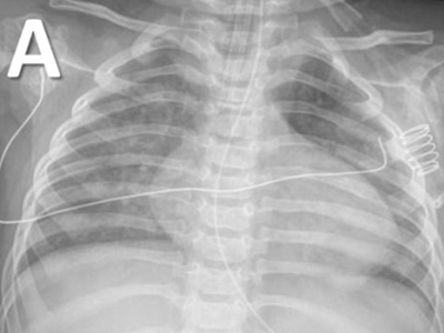
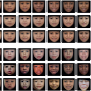
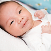
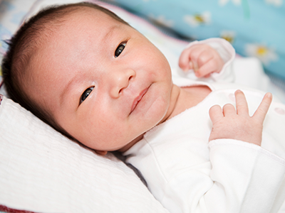
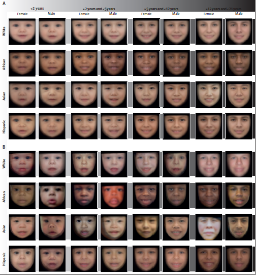
(A) Control population. (B) Population with Williams-Beuren syndrome. Average faces were generated for each demographic group after automatic face pose correction.

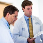
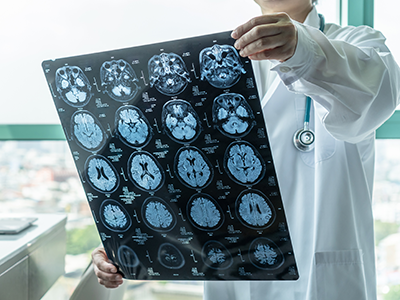
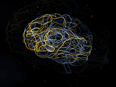
With an average accuracy of 88%, a deep learning technology offers rapid genetic screening that could accelerate the diagnosis of genetic syndromes, recommending further investigation or referral to a specialist in seconds, according to a study published in The Lancet Digital Health. Trained with data from 2,800 pediatric patients from 28 countries, the technology also considers the face variability related to sex, age, racial and ethnic background, according to the study led by Children’s National Hospital researchers.

“We built a software device to increase access to care and a machine learning technology to identify the disease patterns not immediately obvious to the human eye or intuition, and to help physicians non-specialized in genetics,” said Marius George Linguraru, D.Phil., M.A., M.Sc., principal investigator in the Sheikh Zayed Institute for Pediatric Surgical Innovation at Children’s National Hospital and senior author of the study. “This technological innovation can help children without access to specialized clinics, which are unavailable in most of the world. Ultimately, it can help reduce health inequality in under-resourced societies.”

This machine learning technology indicates the presence of a genetic syndrome from a facial photograph captured at the point-of-care, such as in pediatrician offices, maternity wards and general practitioner clinics.

“Unlike other technologies, the strength of this program is distinguishing ‘normal’ from ‘not-normal,’ which makes it an effective screening tool in the hands of community caregivers,” said Marshall L. Summar, M.D., director of the Rare Disease Institute at Children’s National. “This can substantially accelerate the time to diagnosis by providing a robust indicator for patients that need further workup. This first step is often the greatest barrier to moving towards a diagnosis. Once a patient is in the workup system, then the likelihood of diagnosis (by many means) is significantly increased.”

Every year, millions of children are born with genetic disorders — including Down syndrome, a condition in which a child is born with an extra copy of their 21st chromosome causing developmental delays and disabilities, Williams-Beuren syndrome, a rare multisystem condition caused by a submicroscopic deletion from a region of chromosome 7, and Noonan syndrome, a genetic disorder caused by a faulty gene that prevents normal development in various parts of the body.

Most children with genetic syndromes live in regions with limited resources and access to genetic services. The genetic screening may come with a hefty price tag. There are also insufficient specialists to help identify genetic syndromes early in life when preventive care can save lives, especially in areas of low income, limited resources and isolated communities.

“The presented technology can assist pediatricians, neonatologists and family physicians in the routine or remote evaluation of pediatric patients, especially in areas with limited access to specialized care,” said Porras et al. “Our technology may be a step forward for the democratization of health resources for genetic screening.”

The researchers trained the technology using 2,800 retrospective facial photographs of children, with or without a genetic syndrome, from 28 countries, such as Argentina, Australia, Brazil, China, France, Morocco, Nigeria, Paraguay, Thailand and the U.S. The deep learning architecture was designed to account for the normal variations in the face appearance among populations from diverse demographic groups.

“Facial appearance is influenced by the race and ethnicity of the patients. The large variety of conditions and the diversity of populations are impacting the early identification of these conditions due to the lack of data that can serve as a point of reference,” said Linguraru. “Racial and ethnic disparities still exist in genetic syndrome survival even in some of the most common and best-studied conditions.”

Like all machine learning tools, they are trained with the available dataset. The researchers expect that as more data from underrepresented groups becomes available, they will adapt the model to localize phenotypical variations within more specific demographic groups.

In addition to being an accessible tool that could be used in telehealth services to assess genetic risk, there are other potentials for this technology.

“I am also excited about the potential of the technology in newborn screening,” said Linguraru. “There are approximately 140 million newborns every year worldwide of which eight million are born with a serious birth defect of genetic or partially genetic origin, many of which are discovered late.”

Children’s National as well recently announced that it has entered into a licensing agreement with MGeneRx Inc. for its patented pediatric medical device technology. MGeneRx is a spinoff from BreakThrough BioAssets LLC, a life sciences technology operating company focused on accelerating and commercializing new innovations, such as this technology, with an emphasis on positive social impact.

“The social impact of this technology cannot be underestimated,” said Nasser Hassan, acting chief executive officer of MGeneRx Inc. “We are excited about this licensing agreement with Children’s National Hospital and the opportunity to enhance this technology and expand its application to populations where precision medicine and the earliest possible interventions are sorely needed in order to save and improve children’s lives.”

Sickle-Cell-Blood-Cells

Children’s National joins ASH RC Sickle Cell Disease Clinical Trials Network

Sickle-Cell-Blood-Cells

The American Society of Hematology Research Collaborative (ASH RC) has announced the first 10 clinical research consortia to join the ASH RC Sickle Cell Disease Clinical Trials Network. Children’s National Hospital will be one of the clinical trials units to serve in the DMV Sickle Cell Disease Consortium (DMVSCDC).

The sites will be able to enroll children and adults living with sickle cell disease (SCD) within their patient populations in clinical trials as part of an unprecedented national effort to streamline operations and facilitate data sharing to expedite the development of new treatments for this disease.

“As part of the ASH RC SCD clinical trials network, we will learn regionally and nationally how sickle cell patients respond differently to therapies, hopefully giving us clues to provide more successful targeted and individualized treatments that will improve the morbidity and mortality in sickle cell disease patients,” said Andrew Campbell, M.D., director of Comprehensive Sickle Cell Disease Program at Children’s National.

SCD is a chronic, progressive, life-threatening, inherited blood disorder that affects more than 100,000 Americans and an estimated 100 million persons worldwide. Clinical trials hold incredible promise for the development of much-needed new treatments, and possibly even a cure. While there are currently only four U.S. Food and Drug Administration (FDA)-approved drugs to treat the disease, there is a robust SCD drug development pipeline that will drive demand for clinical trials to a new level, providing a prime opportunity to advance treatment and care of those affected by SCD.

“We are proud that the DMV Sickle Cell Disease Consortium will contribute regionally, allowing our patients and families to benefit from new clinical trials investigating new therapies that may improve the clinical course and quality of life of patients living with sickle cell disease in the DMV region,” Dr. Campbell added. “We will also have an integrated Community Advisory Board who will continue to provide guidance and expertise for our consortium including patients, families and caregivers.”

Read the full list of other hospitals joining the network.

flow chart of pulse ox study

Newborn screening for critical congenital heart disease serves as vital safety net

One of the nation’s longest-running newborn screening programs for critical congenital heart disease (CCHD) finds that screening continues to serve as a necessary tool to help identify every child with CCHD — even in states where the majority of babies are diagnosed before birth.

The screening program study findings were published in Pediatrics. The data is some of the first to provide long-term evidence for using pulse oximetry to screen newborns for critical congenital heart disease 24 hours after birth. This screening test was added to the Department of Health and Human Services Recommended Uniform Screening Panel in 2011 and is now required in all 50 states.

“This study reinforces why pulse oximetry screening for CCHD is an important tool in our arsenal to identify and treat critical congenital heart disease, and other conditions that affect the flow of oxygen throughout the body, as soon as possible,” says Bryanna Schwarz, M.D., a cardiology fellow at Children’s National Hospital and lead author. “We know that prompt, early detection and swift intervention is crucial to positive long-term outcomes for these kids.”

The team looked at the data and outcomes for all babies born throughout eight years at Holy Cross Hospital in suburban Maryland, one of the first community birthing hospitals in the country to routinely perform the screening. Over the eight-year period, 64,780 newborns were screened at the site. Of those:

  • Thirty-one failed the screening, and every baby who failed was found to have congenital heart disease or another important medical condition.
  • Twelve of the failures (38.7%) were babies with critical congenital heart disease who were not previously identified by prenatal detection.
  • Nine others (29%) had a non-critical congenital heart condition.
  • Ten additional babies (32%) had a non-cardiac condition.

The authors note that the 12 newborns with CCHD identified through pulse oximetry screening are noteworthy because they represent critical congenital heart disease cases that are not found before birth in the state of Maryland, where rates of prenatal diagnosis are relatively high. The finding indicates that screening after birth continues to play a critical role in ensuring every baby with critical congenital heart disease is identified and treated as quickly as possible.

“Holy Cross Health and Children’s National have had a decades-long relationship, as we mutually care for women and infants throughout the region. With Children’s National having the U.S. News & World Report #1 ranking Neonatology service in the nation and Holy Cross Hospital being among the top 10 hospitals for the number of babies delivered each year, we are honored to be leading together the great work that is being done to serve our health care community,” says Ann Burke, M.D., vice president of Medical Affairs at Holy Cross Hospital. “We are committed to continuing to do our part to care for women and infants, as well as contribute to the national landscape for neonatal care. We are delighted in the outcomes we have seen and look forward to continued advancement.”

In this study, infants who did not have critical congenital heart disease were considered “false positives” for CCHD. Still, every one of them was found to have another underlying condition, including non-critical congenital heart disease or non-cardiac conditions (such as sepsis and pneumonia) that would also require monitoring and treatment.

The researchers also ran a projection of recently recommended updates to the screening protocol, which include removing a second re-screen after a newborn fails the initial test, to look at whether removing the second rescreen to verify results would decrease accuracy. While the false positive rate did increase slightly from .03% to .04%, eliminating a second re-screen allowed the newborns who were identified to receive crucial care sooner without having to wait an additional hour for one more test to verify their condition.

“It’s time to stop asking if pulse oximetry is a necessary tool to detect critical heart disease in babies,” says Gerard Martin, M.D., M.A.C.C., senior author of the study and C.R. Beyda Professor of Cardiology at Children’s National Hospital. “Our focus now should be on making evidence-based refinements to the screening protocol based on collected data to ensure the process is simple, can be performed consistently and provides as accurate results as possible.”

A transient low-dose MEKi treatment in a pre-clinical model prevents NF1-OPG formation

Using targeted signaling pathway therapy to prevent pediatric glioma formation

Researchers at Children’s National Hospital identified a vulnerability in a developmental signaling pathway that can be hijacked to drive pediatric low-grade glioma (pLGG) formation, according to a pre-clinical study published in Developmental Cell. The study demonstrated that targeted treatment prevents tumor formation, long before irreversible damage to the optic nerve can cause permanent loss of vision. This finding will inform chemo-prevention therapeutic trials in the future.

Brain tumors are the most common solid tumors in children, the most prevalent of which are pLGGs. Approximately 10% to 15% of pLGGs arise in patients with the familial cancer predisposition syndrome known as neurofibromatosis type 1 (NF1). This is a genetic condition that increases risks of developing tumors along the nerves and in the brain.

Nearly 20% of children with NF1 develop pLGGs along the optic pathway, also known as NF1-associated optic pathway glioma (NF1-OPG). Despite many advances in cancer therapy, there are no definitive therapies available that prevent or alleviate the neurological deficits (i.e. vision loss) and that could improve the quality of life.

“The evidence presented can inform chemoprevention therapeutic trials for children with NF1-OPG,” said Yuan Zhu, Ph.D., scientific director and Gilbert Family Endowed professor at the Gilbert Family Neurofibromatosis Institute and associate director of the Center for Cancer and Immunology Research, both part of Children’s National. “This therapeutic strategy may also be applicable to children with the developmental disorders that are at high risk of developing pediatric tumors, such as other RASopathies.”

The mechanism of vulnerability to pLGGs during development is not fully understood. It has been implied that the cell population of origin for this debilitating tumor is transiently proliferative during development. The NF1 gene produces a protein that helps regulate normal cell proliferation, survival and differentiation by inhibiting MEK/ERK signaling. When there is loss of function in NF1, it abnormally activates the MEK/ERK signaling pathway and leads to tumor formation.

Certain cells that exist transiently during the normal development of the brain and optic nerve are vulnerable to tumor formation because they depend on the MEK/ERK signaling. In this study, researchers in Zhu’s lab identified cells that were MEK/ERK pathway dependent and grew during a transient developmental window as the lineage-of-origin for NF1-OPG in the optic nerve. The researchers used a genetically engineered pre-clinical model to design a transient, low-dose chemo-preventative strategy, which prevented these tumors entirely.

“When we provided a dose-dependent inhibition of MEK/ERK signaling, it rescued the emergence and increase of brain lipid binding protein-expressing (BLBP+) migrating GPs glial progenitors, preventing NF1-OPG formation,” wrote Jecrois et al. “Equally importantly, the degree of ERK inhibition required for preventing NF1-OPG formation also greatly improved the health and survival of the NF1-deficient model.”

Ongoing clinical trials using MEK inhibitors (MEKi) are being performed for children as young as 1 month old. Thus, it becomes increasingly feasible to design a chemo-preventative trial using a MEKi to treat children with NF1. These treatment paradigms may have the potential to not only prevent OPG formation, but also other NF1-associated and RASopathies-associated developmental defects and tumors.

A transient low-dose MEKi treatment in a pre-clinical model prevents NF1-OPG formation

A transient low-dose MEKi treatment in a pre-clinical model prevents NF1-OPG formation. The middle panels highlighted by a red dashed box show an OPG in the optic nerve (arrows, top), exhibiting abnormal triply-labeled tumor cells, inflammation and nerve damage (the bottom three panels), which are absent in the normal (left panels) or MEKi-treated Nf1-deficient optic nerves (right panels). [Credit: Jecrois et al., Developmental Cell, (2021)]

Could whole-exome sequencing become a standard part of state newborn screening?

smiling baby boy

There are concerns about implementing whole-exome sequencing since it takes away the child’s right to decide if they want to know — or not — about their specific inherited disease.

It is still premature to standardize an innovative methodology known as whole-exome sequencing (WES) as part of state newborn screening programs, argues Beth A. Tarini, M.D., M.S., associate director for the Center of Translational Research at Children’s National Hospital, in a new editorial published in JAMA Pediatrics.

About 4 million infants are born annually in the United States. Newborn screening is a mandatory state-run public health program that screens infants for inherited diseases in the first days of life so they can receive treatment before irreversible damage occurs. Several of these screening tests are done on blood drawn from an infant’s heel.

WES holds the potential to screen infants for thousands of disorders and traits, including those that appear in adulthood. But there are concerns about implementing WES since it takes away the child’s right to decide if they want to know — or not — about their specific inherited disease. There is also the unknown effect that it could have on their ability to obtain health insurance.

“As caretakers for their children, parents have the challenge of deciding what kind of information, including genetic, will be valuable for their child,” says Dr. Tarini. “As a society, we have the responsibility of deciding where the healthcare dollars get the best return – especially when it comes to children. We need to start that conversation for universal genomic sequencing of newborns sooner rather than later.”

The Pereira et al. study, appearing in the new edition of JAMA Pediatrics and referenced in Dr. Tarini’s editorial, is the first to demonstrate no significant harm in the initial 10 months of life after performing WES under the best conditions of access to resources and a controlled environment.

While the Pereira et al. study has limited data on the effects of WES on families from underrepresented backgrounds, Dr. Tarini notes that it does provide a critical first step in this area of pediatric genomic research and for policy decision-making about the widespread implementation of WES in newborns.

“Moving forward, the U.S. will have to make a collective decision about the value of WES for newborns,” says Dr. Tarini. That value calculus cannot be made without consideration of the general state of healthcare for infants. As she points out, “This is not an easy question to answer in a country whose infant mortality ranks 34th according to the Organization for Economic Co-operation and Development (OECD).”

Dr. Tarini’s research identifies ways to optimize the delivery of genetic services to families and children, particularly newborn screening. She has also chaired state newborn screening committees and served on several federal newborn screening committees.

Ugandan boy in hospital bed

Acute rheumatic fever often goes undiagnosed in sub-Saharan Africa

Ugandan boy in hospital bed

Despite low numbers of documented acute rheumatic fever cases in sub-Saharan Africa, the region continues to show some of the highest numbers of people with, and dying from, rheumatic heart disease, the serious heart damage caused by repeat instances of rheumatic fever.

Despite low numbers of documented acute rheumatic fever cases in sub-Saharan Africa, the region continues to show some of the highest numbers of people with, and dying from, rheumatic heart disease, the serious heart damage caused by repeat instances of rheumatic fever. A population-based study in the Lancet Global Health collected evidence of acute rheumatic fever in two areas of Uganda, providing the first quantifiable evidence in decades that the disease continues to take a deadly toll on the region’s people.

“These findings matter. Access to life-saving heart surgery is only available to a very small fraction of the hundreds of thousands of patients in Africa who have irreversible heart damage from rheumatic heart disease,” says Craig Sable, M.D., associate chief of Cardiology at Children’s National Hospital and one of the senior authors of the study. “It’s time to focus upstream on capturing these conditions sooner, even in low-resource settings, so we can implement life-sustaining and cost-saving preventive treatments that can prevent further heart damage.”

The authors, who hail from Uganda and several institutions around the United States, including Children’s National and Cincinnati Children’s Hospital Medical Center, note this is the first study to use an active case-finding strategy for diagnosing acute rheumatic fever. They also note that raising awareness in the community and among its healthcare workers while also finding new ways to overcome some of the diagnostic challenges in these low-resource settings greatly improved diagnosis and treatment of the condition.

The study also described clinical characteristics of children ages 5 to 14 presenting with both definitive and possible acute rheumatic fever, providing further clinical data points to help healthcare workers in these communities differentiate between this common infection and some of the other frequently diagnosed conditions in the region.

“With this study, we can now confidently dismiss the myth that acute rheumatic fever is rare in Africa,” the authors write. “It exists at elevated rates in low-resource settings such as Uganda, even though routine diagnosis remains uncommon. While these incidence data have likely underestimated the cases of acute rheumatic fever in two districts in Uganda, they show that opportunity exists to improve community sensitization and healthcare worker training to increase awareness of acute rheumatic fever. Ultimately this leads to diagnosing more children with the condition before they develop rheumatic heart disease, so that they can be offered secondary prophylaxis with penicillin.”

Children with suspected acute rheumatic fever participated in this population-based study. Data was collected over 12 months in Lira district (January 2018 to December 2018) and over nine months (June 2019 to February 2020) in Mbarara district.

Follow-up of children diagnosed in this study will provide more data on the outcomes of acute rheumatic fever, including a better understanding of the risk for a child to develop rheumatic heart disease.

This work was funded by the American Heart Association Children’s Strategically Focused Research Network Grant #17SFRN33670607 and by DEL‐15‐011 to THRiVE‐2 and General Electric.

Learn more about the challenges of rheumatic heart disease in sub-Saharan Africa and other developing parts of the world through the Rheumatic Heart Disease microdocumentary series:


coronavirus

One-half of MIS-C patients at a single center experienced heart complications

coronavirus

A single center study of patients with multisystem inflammatory disease in children (MIS-C) found that half of children diagnosed with MIS-C had a heart complication as part of the disease. The study collected and analyzed data from 39 cases of MIS-C at Children’s National Hospital in 2020. MIS-C is a pediatric disease that has been linked to SARS-CoV-2, the virus that causes COVID-19.

The study’s findings appear in the journal Cardiology of the Young. The authors aimed to describe the type and frequency of cardiac complications in children with MIS-C while also outlining the disease’s short-term progression. They also hoped to better understand the demographics, clinical and laboratory findings, as well as the therapeutic successes for children with cardiac complications from MIS-C.

“While half of all children at our hospital diagnosed with MIS-C did experience a cardiac complication, it’s important to note that almost all of them (84%) also fully recovered from that cardiac complication within 50 days of diagnosis,” says Ashraf Harahsheh, M.D., director of Quality Outcomes in Cardiology at Children’s National Hospital, who led the study. “We were also able to identify a few common factors among those with cardiac complications that, with further research, may help us identify earlier the children with MIS-C who are at greater risk for heart problems.”

The study found that children with cardiac complications had higher levels of natriuretic peptides, which appear in greater numbers when the heart isn’t pumping enough blood to the rest of the body. Additionally, children who developed heart complications also had higher initial white blood cell counts. MIS-C cardiac complications ranged from mild systolic dysfunction to coronary artery abnormalities and/or artery dilation.

This was a retrospective, observational study of 39 patients admitted to Children’s National Hospital from March 2020 to September 2020 who met the Centers for Disease Control and Prevention MIS-C case definition. Patient demographics, clinical features, laboratory values, diagnostic investigations, including echocardiograms, and therapies were extracted from the electronic medical records.

“This syndrome has some similarities to Kawasaki disease, another inflammatory syndrome that is known to cause cardiac complications,” says Dr. Harahsheh. “Thankfully what we’ve learned from studying and treating Kawasaki disease in children has helped us collaborate with partners around the world to find treatments for MIS-C that seem to minimize the impact of these complications, at least in the short term.”