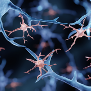
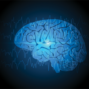
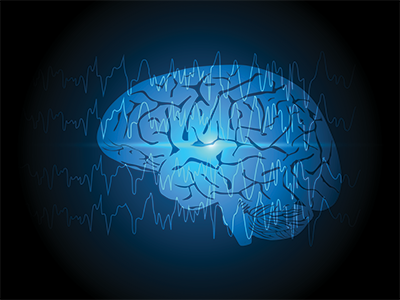
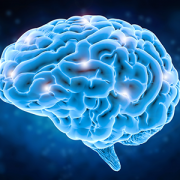
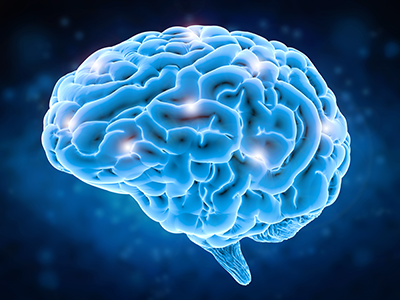
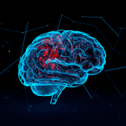
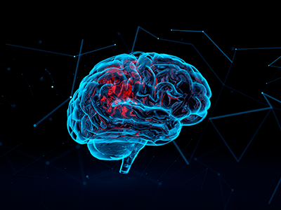
Future TBI treatments may hinge on understanding a new cell type
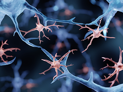
Only recently have investigators begun to understand how a cell type – the NG2-glia – may respond to injuries, offering clues into the brain’s healing and regeneration.
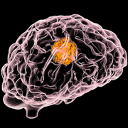
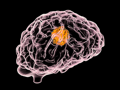
Traumatic brain injury (TBI) afflicts 69 million people, including 630,000 children, worldwide each year. Yet only recently have investigators begun to understand how a cell type – the NG2-glia – may respond to injuries, offering clues into the brain’s healing and regeneration.
In a new paper published in GLIA, investigators from Children’s National Hospital reviewed 25 years of neuroscience research to lay out what’s known about the molecular response of these NG2-glia cells after TBI. Researchers said they see “a seductive possibility” that tapping into the regenerative potential of NG2-glia cells after neurotrauma could lead to therapies in the future. The impact could be profound, given that TBI is the leading cause of death among all people ages 1-44 and the global cost of this ‘silent epidemic’ is estimated to top $102 billion annually.
What they’re saying
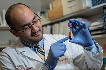
“Our review lays out what’s known about these fascinating cells,” said Terry Dean, M.D., Ph.D., critical care specialist at Children’s National and investigator at the Center for Neuroscience Research (CNR). “NG2-glia are found throughout the brain, and we know that these cells undergo several dynamic changes in the hours, days and weeks after TBI. They are unique, and we want to understand their molecular characteristics to eventually enhance patients’ cellular recovery after TBI.”
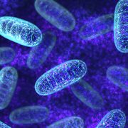
Although only encompassing 4% to 8% of brain cells, these NG2-glia cells make up the largest population of regenerative cells in the adult central nervous system. In their article, Dean and Vittorio Gallo, Ph.D., Children’s National Research Institute interim director, lay out a number of unique features of these cells:
- They proliferate, or multiply, and can form different cell types, especially after brain injuries.
- They are structurally dynamic and can move and migrate throughout the cortex, including toward injury sites.
- They appear to play a role in cell-to-cell signaling, which may prove vital after injuries.
The big picture
“As we study the brain after injuries, we hope our work will reveal the role these NG2-glia cells play in recovery, driving us to possible therapies,” Gallo said. “We believe the big answers will come through understanding the brain on a molecular level. This type of deep investigation is the foundation of our bench-to-bedside approach and positions researchers like Dr. Dean to find answers for our patients.”
Moving the field forward
Researchers have only begun to unlock how NG2-glia respond to injury, making this a fruitful area for research. Gallo, Dean and others at CNR hope to build on their knowledge about what happens to the brain immediately after an injury to learn more about what happens months after a debilitating impact. They are also considering new types of research models to expand their knowledge about cellular destruction, immune interaction and blood vessel compromise after different types of brain injuries.
“We look forward to the day when we have a truly targeted therapy for TBI patients,” Dean said. “Imagine the relief this could provide patients suffering from the persistent physical, cognitive and psychological disabilities that often accompany these brain injuries.”


 The choice between stereoelectroencephalography (SEEG) and subdural evaluation is not mutually exclusive, according to a new