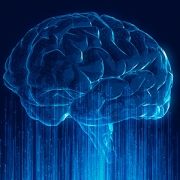
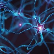
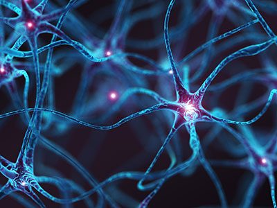
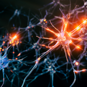
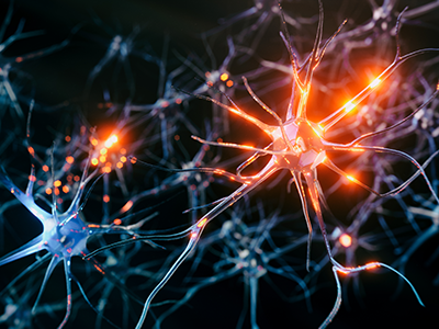
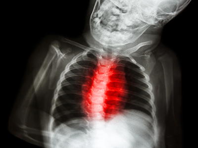
A new AI tool developed by Children’s National and Howard University analyzes brain immune cells 10,000x faster than manual methods.
A new Machine Learning and Artificial Intelligence tool from researchers at Children’s National Hospital (CNH) and Howard University (HU) accelerates discoveries in brain inflammation. Called StainAI, it rapidly and accurately analyzes microglia, the brain’s immune cells. Scientists currently analyze microglia slowly by hand. StainAI automates this process and speeds it up 10,000-fold. Its use will aid discovery of new treatments for inflammatory brain conditions such as infection, autoimmunity, and aging.
Solving a problem
Traditionally, scientists study microglia one cell at a time. They reconstruct each cell’s shape by hand under a microscope. The shape helps classify microglia as “resting” (normal) or “activated” (inflamed). The manual process is tedious and slow. It limits analyses to a few microglia in small brain areas.
StainAI changes that. It uses deep machine learning and artificial intelligence to overcome and exceed the manual method’s limitations. It correctly classifies millions of microglia from standard microscopic images. StainAI also localizes each microglia to its brain region in 3D. These features enable single-cell analyses of immune activity at a scale not feasible before – the entire brain.
A tool with broad impact
The team applied StainAI to two models of brain injury and inflammation to show its utility. In a rodent model of pediatric cardiac arrest, StainAI identified new brain regions susceptible to injury. In a simian model of viral infection, StainAI localized rod-shaped microglia normally found in white matter to an unexpected brain region – the hippocampus. These findings point towards new treatments and highlight StainAI’s value across diseases and species.
StainAI is fast, accurate and adaptable. It uses common laboratory equipment. Its creators, Michael Shoykhet, MD, PhD, at CNH and Dr. Tsang-Wei Tu at HU, are making StainAI available to other researchers. They hope StainAI will help labs worldwide discover new ways to protect children’s brains from inflammation and injury.
Children’s National Hospital hosted its fifteenth annual Research, Education and Innovation Week from March 31–April 4, 2025, bringing together clinicians, scientists, educators and innovators from across the institution to celebrate discovery and collaboration. This year’s theme, “Empowering the Future in Pediatric Research and Innovation with Equity, Technology and a Global Reach,” served as a call to action for advancing science that improves child health both locally and around the world.
Each day of the week-long event featured thought-provoking lectures — now available to watch — dynamic panel discussions, interactive workshops and vibrant poster sessions, all highlighting the diverse and interdisciplinary work taking place across Children’s National.
Centering the patient and the planet
REI Week began on Monday with a powerful keynote lecture from Lynn R. Goldman, MD, MS, MPH, Michael and Lori Milken dean of the Milken Institute School of Public Health at the George Washington University. In her talk, “Children: Uniquely vulnerable to climate-related threats,” Dr. Goldman underscored the urgent need to protect children from the environmental hazards of a changing climate and to integrate climate science into pediatric care and advocacy.
At mid-morning, Mary-Anne “Annie” Hartley, MD, PhD, MPH, director of the LiGHT Laboratory at École Polytechnique Fédérale de Lausanne, introduced the “MOOVE” platform — Massive Open Online Validation and Evaluation of clinical LLMs. Her talk demonstrated how artificial intelligence, when rigorously validated, has the potential to transform clinical decision-making and global health equity.
Monday’s final keynote, “Zinc and childhood diarrhea,” was presented by Christopher Duggan, MD, MPH, director of the Division of Nutrition at Harvard Medical School. Dr. Duggan highlighted the global health impact of zinc supplementation in reducing childhood mortality — a reminder that simple, evidence-based interventions can save millions of lives.
In that first day, the first poster session of the week showcased projects in adolescent medicine, global health, infectious diseases, oncology and more. The session reflected the full breadth of research taking place across Children’s National.
Ambroise Wonkam, MD, PhD, professor of genetic medicine at Johns Hopkins University, then delivered Tuesday’s Global Health Keynote Lecture, “Harnessing our common African genomes to improve health and equity globally.” His work affirmed that inclusive genomics is key to building a healthier world.
Later, the Global Health Initiative event and GCAF Faculty Seminar encouraged attendees to pursue collaborative opportunities at home and abroad, reflecting the growing global footprint of Children’s National research programs.
Transforming education and care delivery
On Wednesday, Larrie Greenberg, MD, professor emeritus of pediatrics, kicked off the day with a Grand Rounds keynote on educational transformation: “Shouldn’t teachers be more collaborative with their learners?” He followed with a CAPE workshop exploring the effectiveness of case-based learning.
In the Jill Joseph Grand Rounds Lecture, Deena J. Chisolm, PhD, director of the Center for Child Health Equity at Nationwide Children’s Hospital, challenged attendees to move beyond dialogue into action in her talk, “Health equity: A scream to a whisper?,” reminding researchers and clinicians that advocacy and equity must be foundational to care.
The day continued with a poster session spotlighting medical education, neonatology, urology and neuroscience, among other fields.
Posters and pathways to progress
Throughout the week, poster sessions highlighted cutting-edge work across dozens of pediatric disciplines. These sessions gave attendees the opportunity to engage directly with investigators and reflect on the shared mission of discovery across multiple disciplines, including:
The REI Week 2025 Awards Ceremony celebrated outstanding contributions in research, mentorship, education and innovation. The winners in each category were:
POSTER SESSION AWARDS
Basic & Translational Research
Faculty: Benjamin Liu, PhD
“Genetic Conservation and Diversity of SARS-CoV-2 Envelope Gene Across Variants of Concern”
Faculty: Steve Hui, PhD
“Brain Metabolites in Neonates of Mothers with COVID-19 Infection During Pregnancy”
Faculty: Raj Shekhar, PhD
“StrepApp: Deep Learning-Based Identification of Group A Streptococcal (GAS) Pharyngitis”
Post docs/Fellows/Residents: Dae-young Kim, PhD
“mhGPT: A Lightweight Domain-Specific Language Model for Mental Health Analysis”
Post docs/Fellows/Residents: Leandros Boukas, MD, PhD
“De Novo Variant Identification From Duo Long-Read Sequencing: Improving Equitable Variant Interpretation for Diverse Family Structures”
Staff: Naseem Maghzian
“Adoptive T Lymphocyte Administration for Chronic Norovirus Treatment in Immunocompromised Hosts (ATLANTIC)”
Graduate Students: Abigail Haffey
“Synergistic Integration of TCR and CAR T Cell Platforms for Enhanced Adoptive Immunotherapy in Brain Tumors”
High School/Undergraduate Students: Medha Pappula
“An ADHD Diagnostic Interface Based on EEG Spectrograms and Deep Learning Techniques”
Clinical Research
Faculty: Folasade Ogunlesi, MD
“Poor Air Quality in Sub-Saharan Africa is Associated with Increase Health Care Utilization for Pain in Sickle Cell Disease Patients”
Faculty: Ayman Saleh, MD
“Growth Parameters and Treatment Approaches in Pediatric ADHD: Examining Differences Across Race”
Post docs/Fellows/Residents: Nicholas Dimenstein, MD, MPH
“Pre-Exposure Prophylaxis (PrEP) Eligibility in the Pediatric Emergency Department”
Staff: Tayla Smith, MPH
“The Public Health Impact of State-Level Abortion and Firearm Laws on Health Outcomes”
Graduate Students: Natalie Ewing
“Patterns of Bacteriuria and Antimicrobial Resistance in Patients Presenting for Primary Cloacal Repair: Is Assisted Bladder Emptying Associated with Bacteriuria?”
Graduate Students: Manuela Iglesias, MS
“Exploring the Relationship Between Child Opportunity Index and Bayley-III Scores in Young Children”
High School/Undergraduate Students: Nicholas Lohman
“Preliminary Findings: The Efficacy, Feasibility and Acceptability of Group Videoconference Cognitive Behavioral Therapy with Exposure and Response Prevention for Treating Obsessive-Compulsive Disorder Among Children and Young People”
Community-Based Research
Faculty: Sharon Shih, PhD “Assessing Pediatric Behavioral Health Access in DC using Secret Shopper Methodology”
Post docs/Fellows/Residents: Georgios Sanidas, MD “Arrested Neuronal Maturation and Development in the Cerebellum of Preterm Infants”
Staff: Sanam Parwani
“Intersectionality of Gender and Sexuality Diversity in Autistic and Non-Autistic Individuals”
Graduate Student: Margaret Dearey “Assessing the Burden of Period Poverty for Youth and Adolescents in Washington, DC: A Pilot Study”
Quality and Performance Improvement
Faculty: Nichole L. McCollum, MD
“A Quality Improvement Study to Increase Nurse Initiated Care from Triage and Improve Timeliness to Care”
Post docs/Fellows/Residents: Hannah Rodriguez, MD
“Reducing Unnecessary Antibiotic Use in a Level IV NICU”
Staff: Amber K. Shojaie, OTD, OTR/L
“Implementing Dynamic Axilla Splints in a Large Burn Patient”
Meleah Boyle, PhD, MPH
“Understanding and Addressing Environmental Sustainability to Protect the Health of the Children’s National and Global Communities”
Eiman Abdulrahman, MD
“Research Capacity Building to Improve Pediatric Emergency and Critical Care in Ethiopia”
Pilot Awards
Alexander Andrews, MD
“EEG as a Diagnostic and Prognostic Marker in Severe Pediatric Malaria, Blantyre Malawi”
Daniel Donoho, MD & Timothy Singer, MD
“Feasibility Study of a Novel Artificial Intelligence-Based Educational Platform to Improve Neurosurgical Operative Skills in Tanzania”
Hasan Syed, MD
“Bridging the Gap an Educational Needs Assessment for Pediatric Neurosurgery Training in Pakistan”
Sofia Perazzo, MD & Lamia Soghier, MD, MEd, MBA
“QI Mentorship to Improve Pediatric Screening and Follow-up in Rural Argentina”
Benjamin Liu, PhD
“AI-Empowered Real-Time Sequencing Assay for Rapid Detection of Schistosomiasis in Senegal”
Rae Mittal, MD
“Assessment and Enhancement of Proficiency in Emergency Child Neurology Topics for Post-Graduate Emergency Medicine Trainees in India”
Innovation Day ignites bold thinking
Thursday, REI Week shifted to the Children’s National Research & Innovation Campus for Innovation Day, a celebration of how bold ideas and collaborative culture can accelerate progress in pediatric medicine.
REI Week 2025 reaffirmed the values that define Children’s National: a commitment to excellence, collaboration and equity in pediatric research and care. As discoveries continue to emerge from our hospital and our research campuses, the connections built and ideas sparked during this week will help shape the future of pediatric health — locally and globally.
By elevating voices from the bedside to the bench, with the support of the executive sponsors Nathan Kuppermann, MD, MBChB, Catherine Bollard, MBChB, MD, Kerstin Hildebrandt, MSHS, Linda Talley, MS, RN, NE-BC and David Wessel, MD, REI Week demonstrated that we must embrace the community in all aspects of our work. Because we know that there are answers we can only get from the patients that we serve—and we need to be their voice.
Research, Education & Innovation Week will be back next year on April 13-17, 2026.
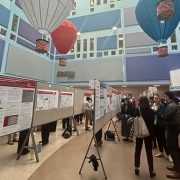
Posters at the REI Week 2025 Monday, March 31 poster session.
Panelists discuss innovation during REI Week 2025.
Global Health Initiative community engagement event during REI Week 2025.
Chris Rees presents his REI Week 2025 lecture.
Nathan Kuppermann listens to a presenter during the REI Week 2025 Tuesday, April 1, poster session.
Michelle Riley-Brown, Nathan Kuppermann, Catherine Bollard and Naomi Luban on stage during the REI Week 2025 awards ceremony.
Brandy Salmon presents on innovation programs at Virginia Tech during the REI Week 2025 Innovation Day.
Catherine Bollard listens to a presenter during the REI Week 2025 Monday, March 21 poster session.
Ambroise Wonkman poses for a picture with Children’s National staff.
Tanzeem Choudhury presenting during REI Week 2025.
https://innovationdistrict.childrensnational.org/wp-content/uploads/2025/04/REI-Week-2025-Monday-Poster-Session-CNRI.jpg385685Innovation Districthttps://innovationdistrict.childrensnational.org/wp-content/uploads/2023/12/innovationdistrict_logo-1-1030x165.pngInnovation District2025-04-22 10:31:052025-06-10 12:20:52REI Week 2025 empowers the future in pediatric research and innovation
The course attracts a national audience and brings together neuroscience clinicians and pediatricians in the Washington, D.C. and Mid-Atlantic region.
Guest speakers include Annapurna Poduri, MD, MPH, Deputy Director for NINDS, Emily Freilich, MD, from the FDA and Conor Mallucci, MBBS, Chief of Neurosurgery at Alder Hays, England.
This year’s course highlights 3 major areas:
Updates in Epilepsy
Innovations in Vascular Neurosurgery and Neurointerventional Radiology
Addressing Mental and Behavioral Health in Neurological Conditions
We invite you to join us for presentations from experts in the field during this full-day, CME accredited event on April 10, 2025. This is a hybrid event that will be held virtually or in-person at the Children’s National Research & Innovation Campus.
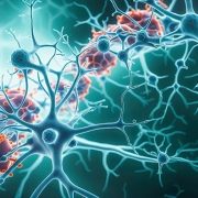
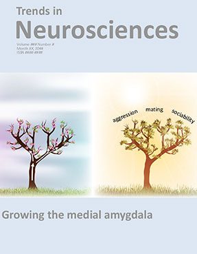
A recent review by researchers at Children’s National, published in Trends in Neurosciences, offers a new and in-depth understanding of how the amygdala is formed during fetal developmental.
The medial amygdala (MeA) is a central structure of the brain for regulation of social and emotional behaviors. Amygdala dysfunction is associated with a host of developmental conditions including autism spectrum disorders (ASD), post-traumatic stress disorder (PTSD) and the consequences of early life stress. To date, there has been a lack of comprehensive understanding of how the amygdala forms developmentally.
A recent review by researchers at Children’s National Hospital, published in Trends in Neurosciences, offers a new and in-depth understanding of how this complex structure is formed during fetal developmental and the role it plays in social behavior.
“This extensive review conveys the latest findings on how the amygdala is formed from development across preclinical models and humans,” says Joshua Corbin, PhD, interim director of the Center for Neuroscience Research at Children’s National and lead author of the review. “Past and present work in our lab has contributed critical knowledge of how this important structure forms from development and implications for human conditions.”
Moving the field forward
Malformation of the amygdala is a hallmark feature of disorders of social cognition such as ASD. Additionally, amygdala development is highly susceptible to early life stress and influences altered fear and anxiety responses in individuals who have been faced with early life stress.
“Despite our growing understanding of MeA development and its role in behavior, many critical questions remain. However, with cutting-edge tools like transcriptomic profiling, subcircuit-level circuit mapping, CRISPR mutagenesis and targeted gene delivery, we’re on the brink of uncovering different neurons in the amygdala form and shape social behaviors,” says Dr. Corbin.
Children’s National leads the way
Dr. Corbin’s team is among only a handful of groups in the world focused on understanding amygdala development. Investigators within the Center for Neuroscience Research at Children’s National have a shared goal of understanding the biological underpinnings of neurodevelopmental disorders.
You can read the full review published in Trends in Neurosciences.
https://innovationdistrict.childrensnational.org/wp-content/uploads/2025/01/TINS-cover-feature.jpg300400Innovation Districthttps://innovationdistrict.childrensnational.org/wp-content/uploads/2023/12/innovationdistrict_logo-1-1030x165.pngInnovation District2025-01-24 16:06:572025-01-24 16:18:08Review: New insights into brain development and behavior
For his groundbreaking research in pediatric epilepsy care, William D. Gaillard, MD, has been named the recipient of the 2024 Clinical Science Research Award by the American Epilepsy Society (AES).
Dr. Gaillard serves as chief of the Divisions of Child Neurology and of Epilepsy and Neurophysiology, director of the Comprehensive Pediatric Epilepsy Program and associate director of the Center for Neuroscience Research at Children’s National Hospital. He also is Professor of Pediatrics and Neurology at the George Washington University School of Medicine, adjunct Professor of Neurology at Georgetown University and adjunct professor of Hearing and Speech Sciences at the University of Maryland, College Park. He holds the endowed Chair for Epilepsy and Neurophysiology.
About the award
This esteemed award, presented annually at AES, recognizes leading researchers whose work significantly contributes to the understanding and treatment of epilepsy. Dr. Gaillard’s groundbreaking research, which combines advanced imaging techniques with a focus on epilepsy and cognitive systems, has paved the way for important advances in the field of pediatric epilepsy care.
“It is a great honor to be recognized by my peers for the research I have conducted to improve the lives of children with epilepsy,” said Dr. Gaillard.
Dr. Gaillard’s work continues to have a profound impact on the field of pediatric neurology and epilepsy research. His commitment to advancing both scientific understanding and clinical outcomes for children with epilepsy has earned him the highest accolades from the AES. This recognition not only honors his past achievements but also highlights his ongoing contributions to the field, ensuring a brighter future for pediatric epilepsy care.
https://innovationdistrict.childrensnational.org/wp-content/uploads/2025/01/Gaillard-feature.jpg300400Innovation Districthttps://innovationdistrict.childrensnational.org/wp-content/uploads/2023/12/innovationdistrict_logo-1-1030x165.pngInnovation District2025-01-21 16:42:282025-01-21 16:43:40William D. Gaillard, MD, receives 2024 Clinical Science Research Award from AES
The Children’s National 2023-2024 Academic Annual Report show on a tablet.
Children’s National Hospital has released its 2023-2024 Academic Annual Report, showcasing a year of transformative progress in pediatric medicine. The report highlights achievements across its research centers, institutes and Innovation Ventures, underscoring the hospital’s role as a leader in advancing child health through innovation and collaboration.
“This year’s report reflects the remarkable progress we have made in advancing the frontiers of pediatric medicine,” said Nathan Kuppermann, MD, MPH, Chief Academic Officer and Chair of Pediatrics. “It highlights groundbreaking work across our research centers, institutes, and Innovation Ventures, showcasing the collaborative spirit that drives our mission forward. These achievements underscore our shared commitment to delivering transformative research and the best possible outcomes for children and families.”
Delivering across centers
The report captures the contributions of each of Children’s National’s research centers, each pushing the boundaries of pediatric healthcare:
Center for Cancer & Immunology Research (CCIR): Delivering on the promise of cell and gene therapies, offering innovative treatments for pediatric cancers and immune disorders.
Center for Genetic Medicine Research (CGMR): Advancing pediatric genetic medicine through interdisciplinary efforts, addressing complex genetic conditions with cutting-edge science.
Center for Neuroscience Research (CNR): A year of growth in scientific excellence, advancing the understanding of brain development and neurological conditions.
Center for Prenatal, Neonatal & Maternal Health Research (CPHNMR): Revolutionizing neonatal care with its pioneering infant brain health neuromonitoring program.
Center for Translational Research (CTR): Facilitating groundbreaking work by new K awardees and driving translational research to bridge the gap between discovery and clinical care.
Sheikh Zayed Institute for Pediatric Surgical Innovation (SZI): Leading the way in advanced research projects in pediatric surgery, pushing technological boundaries to improve outcomes for children worldwide.
Taking the lead in innovation through collaboration
Innovation Ventures at Children’s National is advancing pediatric health security, addressing unique challenges with transformative solutions. Meanwhile, the Children’s National Research & Innovation Campus (CNRIC) continues to thrive as a hub for discovery and collaboration, hosting conferences on topics like artificial intelligence in healthcare, cell and gene therapy, and pediatric epilepsy research.
A vision for the future
The report also highlights Children’s National’s focus on integrating cutting-edge technologies like artificial intelligence into its research and clinical practices, as well as addressing global health challenges such as the effects of climate change on children’s health. These efforts reflect the hospital’s commitment to improving outcomes for children everywhere through innovation, teamwork, and forward-thinking leadership.
The 2023-2024 Academic Annual Report serves as a testament to the dedication and expertise of the Children’s National community, showcasing how collaboration and innovation are shaping the future of pediatric healthcare.
https://innovationdistrict.childrensnational.org/wp-content/uploads/2025/01/2023-2024-Academic-Annual-Report-cover-feature.jpg300400Innovation Districthttps://innovationdistrict.childrensnational.org/wp-content/uploads/2023/12/innovationdistrict_logo-1-1030x165.pngInnovation District2025-01-13 13:51:482025-01-13 13:53:49Children’s National delivers on the promise in 2024
The conference offered a robust platform for presenting groundbreaking research and clinical advancements across diverse subfields such as pediatric neurology, neurosurgery, neuro-critical care, neurogenetics, neuroimmunology and neuroradiology. Esteemed medical professionals and researchers from around the world convened to share insights and innovations that are shaping the future of pediatric neurological health.
Various speakers from Children’s National led in-depth discussions on diagnostic and therapeutic innovations aimed at enhancing outcomes for children with chronic neurological and neurosurgical conditions. PNC 2024 provided an excellent platform for healthcare professionals, researchers and academics to update their knowledge and engage with leading specialists in the field.
Presenters and topics from Children’s National included:
Dana Harrar, M.D., Ph.D.: Pediatric Status Epilepticus – Monitoring and Management, Acute Stroke: Diagnosis and Critical Care Management
Daniel Donoho, M.D.: Implementing AI in the Operating Room: State of the Art and Future Directions
Tayyba Anwar, M.D.: Neonatal Encephalopathies other than HIE, Personalized Treatments for Genetic Epilepsy
Youssef Kousa, M.S., D.O., Ph.D.: Prenatal and Neonatal CNS Infections, Difficult Goals of Care and Outcome Discussions in the Neonatal ICU
The collaborative efforts with Sidra Medicine highlight a shared vision of pushing the boundaries of pediatric neurological research and treatment, ultimately aiming to improve outcomes for young patients worldwide.
https://innovationdistrict.childrensnational.org/wp-content/uploads/2024/11/PNC-feature.jpg300400Innovation Districthttps://innovationdistrict.childrensnational.org/wp-content/uploads/2023/12/innovationdistrict_logo-1-1030x165.pngInnovation District2024-11-19 11:20:102024-11-19 11:22:10Insights and Innovations in Pediatric Neuroscience: Highlights from PNC 2024
Children’s National Hospital in Washington, D.C., was ranked as a top hospital in the nation by the U.S. News & World Report 2024-25 Best Children’s Hospitals annual rankings. This marks the eighth straight year Children’s National has made the Honor Roll list. The Honor Roll is a distinction awarded to only 10 children’s hospitals nationwide.
This year, U.S. News ended ordinal rankings on its Honor Roll. Instead of assigning a numerical rank from 1 to 10, all hospitals on the Honor Roll will be recognized as having attained the highest standards of care in the nation.
In addition, Children’s National tied for #1 pediatric hospital in the Mid-Atlantic region, which includes New York, New Jersey, Delaware, Pennsylvania, the District of Columbia, West Virginia and Virginia. It’s also best in the Mid-Atlantic in Neonatology.
For the fourteenth straight year, Children’s National ranked in 10 specialty services. New this year, U.S. News included behavioral health as a service line in the rankings. Since it’s the first year, there are no ordinal rankings for behavioral health, but the Children’s National program was named one of the top 50 programs in the country.
“In my first year here, I witnessed what makes Children’s National so special — our commitment to collaboration, empowering one another, and charting a bold path forward for pediatric care,” said Michelle Riley-Brown, MHA, FACHE, president and chief executive officer of Children’s National. “I’m proud U.S. News again recognized Children’s National as one of the top in the nation and the highest-ranked pediatric hospital in D.C., Maryland and Virginia. Together, we’ll continue to push the boundaries of care, research and innovation to make a difference for those who matter most — the kids.”
The annual rankings are the most comprehensive source of quality-related information on U.S. pediatric hospitals and recognizes the nation’s top 50 pediatric hospitals based on a scoring system developed by U.S. News.
“For nearly two decades, U.S. News has published Best Children’s Hospitals to empower the parents and caregivers of children with complex medical needs,” said Ben Harder, chief of health analysis and managing editor at U.S. News. “Children’s hospitals appearing on the U.S. News Honor Roll have a track record of delivering unparalleled specialized care.”
The bulk of the score for each specialty service is based on quality and outcomes data. The process includes a survey of relevant specialists across the country, who are asked to list hospitals they believe provide the best care for patients with the most complex conditions.
The Children’s National specialty services that U.S. News ranked in the top 10 nationally are:
https://innovationdistrict.childrensnational.org/wp-content/uploads/2024/10/US-News-Badges-2024-25-CNRI.jpg385685Innovation Districthttps://innovationdistrict.childrensnational.org/wp-content/uploads/2023/12/innovationdistrict_logo-1-1030x165.pngInnovation District2024-10-08 01:00:002024-10-08 15:01:23Children’s National again ranked among the best in the nation by U.S. News & World Report
Nathan Kuppermann, M.D., M.P.H., is taking on a pivotal role at Children’s National Hospital as executive vice president, the new chief academic officer (CAO) and chair of Pediatrics to continue growing the institution’s reputation as a world-class research hospital. He brings more than 30 years of clinical experience in pediatric emergency medicine and research to the leadership role, where he will oversee nearly 2,000 active research projects at the Children’s National Research Institute.
Dr. Kuppermann knows that science drives cures and improved outcomes. Early in his career, he received enhanced research training at the Harvard School of Public Health, where he laid the groundwork to become a globally recognized clinical researcher. He has studied when to order CT scans for children with head, abdominal and neck trauma to minimize radiation exposure, how to best manage children with diabetic ketoacidosis, infants with febrile illnesses and other complex questions in pediatric emergency medicine that require a multidisciplinary research approach to improve clinical care.
Dr. Kupperman is thrilled to join the scientific community in the nation’s capital, which he sees as a global city where he can authentically share his culturally rich background. He is the son of Brazilian immigrants — a chemical physicist and an organic chemist — and he married a pediatric endocrinologist whose parents are from Mexico and Germany. They have three daughters, and their youngest was adopted from Guatemala. As a family, they travel extensively, and he cares deeply about global health, having served as associate dean for Global Health at UC Davis.
A high school point guard who still plays basketball, Dr. Kuppermann runs his team’s offense on the court, choreographing the flow of each game to optimize his team’s strengths. The position requires peripheral vision to get the ball to the right player and make everyone look good. He sees parallels with his new role as CAO.
Q: What is your approach to research in pediatric healthcare?
A: Fundamentally, my philosophy around research is that we all need to collaborate. When I started doing my own research, I realized over time that to have big, impactful studies, two things had to happen: First, you need to work with people who have expertise beyond your own. I’m a big believer in team science and bench-to-bedside research, collaborating with people with complementary research skills.
Second, I realized that in pediatric research, you must collaborate in research networks to ensure your sample size has enough patients and patients from diverse populations to have definitive results and generalizable data.
Q: What values will you bring to the new role?
A: Three key elements come to mind. First, I’m a big believer in transparent communication, which is the root of everything good in life, whether it’s with your science, your friendships or your family.
I’m a big believer in team science. We all have certain areas of expertise, but if we want to combine our expertise to impact children and improve their health, we need to work together in teams, bringing together basic science researchers, clinicians, information technology specialists, knowledge-translation specialists and disseminators.
Finally, I am a cheerleader for science. As you develop your own science, you also are responsible for leading the next generation of scientists. I’ve spent as much time being a scientific mentor as I have working to discover new knowledge through scientific inquiry.
Q: You talk extensively about your family’s international roots and how it drives your work. What is the role of diversity in medicine and how does that guide you?
A: Diversity is fundamental to healthcare. We in the medical community recognize the disparities in the care we deliver, which I’ve studied in my research. Diversity must not only be a research focus, but our teams must be diverse to better investigate — and work to resolve — these inequities of care.
Children’s National is both a hospital that is mindful of its immediate community and an institution that greatly values the diversity of its patients and its staff. It’s also highly ranked in research and cares deeply about global health, all of which are drivers in my work.
When I considered the potential of coming to Children’s National, I thought I might be able to help this already great institution further its mission by serving its immediate community, growing and developing its global health programs, and taking research here from its already great bones and accomplishments to the next level.
Q: What approach do you bring to clinical care?
A: When I do a research study and enroll patients into a clinical trial, I think about the patient and others the research can impact. I frequently think of a quote by the late Paul Brodeur, a scientific writer who wrote a lot about asbestos, which is very powerful and meaningful to me: “Statistics are humans with the tears wiped away.”
And what it means to me is that we must be mindful as researchers that we’re talking about humans with our research. We’re not just talking about numbers, and we’re not just talking about getting grants and papers. We’re talking about how we can elevate the evidence and translate it to the bedside to improve the lives of humans.
https://innovationdistrict.childrensnational.org/wp-content/uploads/2024/08/Kuppermann-video-still-CNRI.jpg385685Innovation Districthttps://innovationdistrict.childrensnational.org/wp-content/uploads/2023/12/innovationdistrict_logo-1-1030x165.pngInnovation District2024-09-03 12:32:372024-09-03 12:33:46Q&A with Dr. Kuppermann: Collaboration is critical for scientific success
Researchers at Children’s National are testing whether controlling the level of apolipoprotein E could serve as an effective treatment for the poor neurobehavioral outcomes tied to fetal alcohol spectrum disorders.
Reduced levels of a protein – called apolipoprotein E – are responsible for the lifelong cognitive and neurological symptoms in fetal alcohol spectrum disorders (FASD), according to a new study published in the journal Molecular Psychiatry. For the first time, researchers at Children’s National Hospital are testing whether controlling the level of this protein could serve as an effective treatment for the poor neurobehavioral outcomes tied to FASD, which is believed to affect roughly 5% of school-aged children.
Apolipoprotein E is controlled by a gene called APOE, a well-known risk gene for Alzheimer’s disease. It contains the instructions that guide how the connections between neurons in the brain strengthen or weaken over time. This process, known as synaptic plasticity, is crucial for learning and memory formation. In this study, plasma was collected from participants at two sites in Western Ukraine, along with information about drinking behavior reported by their mothers.
What this means
“In addition to Alzheimer’s risk, we found this gene is also a crucial contributor to cognitive problems in children with FASD,” said Kazue Hashimoto-Torii, Ph.D., a principal investigator at the Center for Neuroscience Research at Children’s National. “The new data shows that we understand the mechanism by which prenatal alcohol exposure causes a decrease of the APOE level in the brain. We will continue this work to help improve our understanding of FASD, hoping to replenish this important protein and improve outcomes for children with these disorders.”
FASD is an umbrella diagnosis for the physical and developmental challenges that face children who are exposed to alcohol in utero, including intellectual disability, delay in motor and language development and other neurological diagnoses. While alcohol alone is problematic to many aspects of development, evidence also suggests that genetic factors play a role. Only 4.3% of children with heavy alcohol exposure develop FASD, and twin studies have revealed that fraternal twins have different FASD outcomes compared to identical twins.
What’snext
The research team at Children’s National wanted to pinpoint places where genes could play a role and consider therapies. For the first time, researchers found that plasma levels of APOE were reduced in children with FASD, which strongly supports a potential target for therapy. In addition, their genomics study found a variant of APOE increases the risk of cognitive problems in subjects who were prenatally exposed to alcohol, especially those of African ancestry.
“We found that providing a drug that activates the APOE receptor rescued learning deficits and anxiety in pre-clinical models,” said Masaaki Torii, Ph.D., principal investigator at the Center for Neuroscience Research. “The implications offer an exciting glimpse into possible therapies for some of the neurological harms associated with prenatal alcohol exposure and FASD.”
https://innovationdistrict.childrensnational.org/wp-content/uploads/2024/05/neural-network-CNRI.jpg385685Innovation Districthttps://innovationdistrict.childrensnational.org/wp-content/uploads/2023/12/innovationdistrict_logo-1-1030x165.pngInnovation District2024-05-16 13:38:462024-05-16 13:42:23Adjusting key protein could improve brain function in children with fetal alcohol syndrome
Breakthroughs at the Research & Innovation Campus continued as our researchers worked tirelessly to develop new treatments and therapies that will transform the lives of children and families around the world.
Taking a look at the breakthroughs happening in our now six research centers, we spotlighted the following stories:
Reflecting on decades of progress in the blood, marrow and cell therapy programs at Children’s National. Our researchers have made significant strides in this field, and we are proud to be at the forefront of these life-saving treatments.
In genetic medicine, we continue to be a beacon of hope for families facing rare and complex conditions. Our researchers are making incredible breakthroughs that are changing the landscape of pediatric medicine.
We are also proud to share the $90 million award received from an anonymous donor to support pediatric brain tumor research. The predominant focus of this award is to develop new treatments that will improve outcomes for children with this devastating disease.
This year, we opened a new Center that enhances our research capabilities in the field of Prenatal, Neonatal & Maternal Health Research. We are excited about the possibilities this new center will bring and look forward to the discoveries that will emerge from it.
In addition, we are driving future pandemic readiness with the NIH funded Pediatric Pandemic Network. Our researchers are using cutting-edge technology and innovative approaches to prepare for the next pandemic and protect children.
We are also exploring the potential of artificial intelligence (AI) in pediatric breakthroughs. Our researchers are using machine learning and other AI techniques to develop new treatments and therapies that will transform the lives of children.
https://innovationdistrict.childrensnational.org/wp-content/uploads/2024/02/2022-2023-Academic-Annual-Report-on-a-computer-CNRI.png385685Innovation Districthttps://innovationdistrict.childrensnational.org/wp-content/uploads/2023/12/innovationdistrict_logo-1-1030x165.pngInnovation District2024-02-15 14:18:582024-02-15 15:29:56Driving pediatric breakthroughs through 2023
HIFU is a non-invasive therapy that utilizes focused ultrasound waves to thermally ablate a focal area of tissue.
In January, a team of multidisciplinary doctors performed the first case in the world of using bilateral high intensity focused ultrasound (HIFU) pallidotomy on Jesus, a 22-year-old patient with dyskinetic cerebral palsy.
“The primary objective of the study is to evaluate the safety of ExAblate Transcranial MRgFUS as a tool for creating bilateral or unilateral lesions in the globus pallidus (GPi) in patients with treatment-refractory secondary dystonia due to dyskinetic cerebral palsy,” Dr. Oluigbo explained. “The secondary purpose is to assess the impact of HIFU pallidotomy on dyskinetic cerebral palsy movement disorder in pediatric and young adult patients.”
In addition, the impact of bilateral pallidotomy on motor development, pain perception, speech, memory, attention and cognition in these patients will be assessed.
“We hope that the trial will help us find results that lead to treatments that can reduce the rigidity and stiffness which occurs in cerebral palsy so we can help these children who do not have any effective treatment,” Dr. Oluigbo added.
“This new, first of its kind, non-invasive therapeutic approach – without even a skin incision – will open the door to offering hope for a number of kids with movement disorders who have failed conventional therapy,” said Robert Keating, M.D., chief of neurosurgery at Children’s National. “We are at the beginning of a new era for treating functional disorders in the pediatric patient.”
How it works
HIFU is a non-invasive therapy that utilizes focused ultrasound waves to thermally ablate a focal area of tissue. In the past, Children’s National successfully used HIFU to treat low-grade type tumors located in difficult locations of the brain, such as hypothalamic hamartomas and pilocytic astrocytoma, as well as for epilepsy and other movement disorders.
This most recent procedure was another successful milestone for the hospital, discharging Jesus the following day without any complications.
The team comprised neurosurgeons, MRI techs, anesthesiologists and radiologists, to name a few.
Originally, Jesus came to Children’s National in 2006 when he started working with the Physical Medicine and Rehabilitation team to help him with his muscle hypertonia management as well as equipment, orthoses and therapy concerns.
“As he continued to grow, his muscle hypertonia became more pronounced and caused difficulty with his care, positioning and comfort,” said Olga Morozova, M.D., pediatric rehabilitation specialist at Children’s National. “We have tried multiple oral medications however he has had significant side effects from the majority of the medications.”
Dr. Morozova and Julie Will, M.S.N., F.N.P., the nurse practitioner that worked with Jesus, referred him to Dr. Oluigbo after they learned about HIFU being an option to treat Jesus using a non-invasive approach.
Moving the field forward
This clinical trial highlights the expanding indications for focused ultrasound.
“We are excited about the potential for these innovative treatment strategies in neurosurgery to transform the lives of pediatric patients who suffer from challenging diseases, such as brain tumors, epilepsy, and movement disorders,” said Hasan Syed, M.D., co-director of the Focused Ultrasound Program at Children’s National. “We are redefining what is possible in neurosurgery.”
From low-intensity focused ultrasound (LIFU) treatments for our young DIPG patients to now the groundbreaking research on HIFU for pediatric movement disorders, the dedication to cutting-edge techniques highlights the team’s commitment to patients and transforming pediatric neurosurgical care.
https://innovationdistrict.childrensnational.org/wp-content/uploads/2024/02/CP-HIFU-patient-feature.png300400Innovation Districthttps://innovationdistrict.childrensnational.org/wp-content/uploads/2023/12/innovationdistrict_logo-1-1030x165.pngInnovation District2024-02-09 09:57:282024-02-13 15:09:17Children’s National performs first ever HIFU procedure on patient with cerebral palsy
Several experts from Children’s National Hospital will be sharing their knowledge at the upcoming American Epilepsy Society Annual Meeting in Orlando, December 1-5. Here’s a sample of what you can expect.
Chima Oluigbo, M.D., a pediatric neurosurgeon, will be on panel with other surgeons discussing different surgical techniques and approaches related to epilepsy surgery followed by hands-on practice at teaching stations. He will focus on extra-temporal epilepsy scenarios and will be presenting on Nuances of Temporal Lobe Surgery in the Pediatric Population at the Neurosurgery Symposium highlighting Surgical Controversies in Temporal Lobe Epilepsies.
Ersida Buraniqi, M.D., a child neurologist, will be part of a special interest group on critical care and discuss advances in electroencephalography (EEG) and multimodal neuro-monitoring for seizures in the intensive care unit (ICU). Dr. Buranigui will be doing a special presentation on EEG features to predict electrographic seizures and mortality in the pediatric intensive care unit (PICU).
Dana Harrar, M.D., director of Pediatric Stroke Program and co-director of Critical Care Neurology, is presenting at an invitation-only resident EEG course, providing an interactive structured curriculum on pediatric and adult EEG. Dr. Harrar will be focusing on doing an ICU-EEG nomenclature overview.
Madison Berl, Ph.D., director of Neuropathy Research and of the Intellectual and Developmental Disabilities Research Center Program, will be presenting during the AES Annual Course. The topic “It’s About Time” will focus on the critical importance the timing in epilepsy care plays in patient outcome. Dr. Berl will be presenting on neuropsych outcomes.
Leigh Sepeta, Ph.D., director of Inpatient Neuropsychology, is the vice-chair of the special interest group on neuropsychology. Additionally, Freya Prentice, M.Sc., will be doing a presentation during this session on functional mapping of the cognitive memory circuit in pediatric epilepsy.
Date
Time
Presenter(s)
Title
12/2/23
8:00 am
Chima Oluigbo, M.D., FRCSC, FAANS
Skills Workshop | Epilepsy Surgery Workshop: Techniques and Clinical Scenarios
12/2/23
5:30 pm
Chima Oluigbo, M.D., FRCSC, FAANS
SIG | Epilepsy Surgery: Homunculus Revisited: Managing Central Lobe Epilepsies
12/2/23
5:30 pm
Ersida Buraniqi, M.D.
SIG | Critical Care: Advances in EEG and Multimodal Neuro-monitoring for Seizures in the ICU
12/2/23
7:00am
Dana Harrar M.D.
Resident EEG Course
12/3/23
9:00 am
Chima Oluigbo, M.D., FRCSC, FAANS
Neurosurgery Symposium | Surgical Controversies in Temporal Lobe Epilepsies
12/3/23
8:45 am
Madison Berl, Ph.D.
Annual Course | It’s About Time: Timing in Epilepsy Evaluation and Treatment
12/4/23
7:00 am
Leigh Sepeta, M.D.
SIG | Neuropsychology: Mapping Cognition in Epilepsy: From the Lab to the Clinic
12/4/23
7:00 am
Freya Prentice, M.D.
SIG | Neuropsychology: Mapping Cognition in Epilepsy: From the Lab to the Clinic
12/5/23
7:00 am
Dana Harrar M.D.
SIG | Epilepsy Education: Epilepsy Education Throughout the Training Pipeline
https://innovationdistrict.childrensnational.org/wp-content/uploads/2023/12/illustration-of-neurons-with-electrical-impulses-feature.png300400Innovation Districthttps://innovationdistrict.childrensnational.org/wp-content/uploads/2023/12/innovationdistrict_logo-1-1030x165.pngInnovation District2023-12-05 11:30:302023-12-05 11:33:20Children’s National at the American Epilepsy Society Annual Meeting
Researchers at Children’s National Hospital used magnetic resonance spectroscopy to find new biomarkers that reveal how congenital heart disease (CHD) changes an unborn baby’s brain chemistry, providing early clues that could someday guide treatment decisions for babies facing lifelong health challenges.
Published in the Journal of the American College of Cardiology, the findings detail the ways that heart defects disrupt metabolic processes in the developing brain, especially during the third trimester of pregnancy when babies grow exponentially.
“Over the past decade, our team has been at the forefront of developing safe and sophisticated ways to measure and monitor fetal brain health in the womb,” said Catherine Limperopoulos, Ph.D., director of the Center for Prenatal, Neonatal and Maternal Health Research at Children’s National. “By tapping into the power of advanced imaging, we were able to measure certain maturational components of the brain to find early biomarkers for newborns who are going to struggle immediately after birth.”
The fine print
In one of the largest cohorts of CHD patients assembled to date, researchers at Children’s National studied the developing brains of 221 healthy unborn babies and 112 with CHD using magnetic resonance spectroscopy, a noninvasive diagnostic test that can examine chemical changes in the brain. They found:
Those with CHD had higher levels of choline and lower levels of N-Acetyl aspartate-to-choline ratios compared to healthy babies, potentially representing disrupted brain development.
Babies with more complex CHD also had higher levels of cerebral lactate compared to babies with two ventricle CHD. Lactate, in particular, is a worrying signal of oxygen deprivation.
Specifically, elevated lactate levels were notably increased in babies with two types of heart defects: transposition of the great arteries, a birth defect in which the two main arteries carrying blood from the heart are switched in position, and single ventricle CHD, a birth defect causing one chamber to be smaller, underdeveloped or missing a valve. These critical heart defects generally require babies to undergo heart surgery not long after birth. The elevated lactate levels also were associated with an increased risk of death, highlighting the urgency needed for timely and effective interventions.
The research suggests that this type of imaging can provide a roadmap for further investigation and hope that medicine will someday be able to better plan for the care of these children immediately after their delivery. “With important clues about how a fetus is growing and developing, we can provide better care to help these children not only survive, but thrive, in the newborn period and beyond,” said Nickie Andescavage, M.D., Children’s National neonatologist and first author on the paper.
The big picture
CHD is the most common birth defect in the United States, affecting about 1% of all children born or roughly 40,000 babies each year. While these defects can be fatal, babies who survive are known to be at significantly higher risk of lifelong neurological deficits, including lower cognitive function, poor social interaction, inattention and impulsivity. The impact can also be felt in other organ systems because their hearts did not pump blood efficiently to support development.
Yet researchers are only beginning to pinpoint the biomarkers that can provide information about which babies are going to struggle most and require higher levels of care. The National Institutes of Health (NIH) and the District of Columbia Intellectual and Developmental Disabilities Research Center supported the research at Children’s National to improve this understanding.
“For many years we have known that the brains of children with severe heart problems do not always develop normally, but new research shows that abnormal function occurs already in the fetus,” said Kathleen N. Fenton, M.D., M.S., chief of the Advanced Technologies and Surgery Branch in the Division of Cardiovascular Sciences at the National Heart, Lung, and Blood Institute (NHLBI). “Understanding how the development and function of the brain is already different before a baby with a heart defect is born will help us to intervene with personal treatment as early as possible, perhaps even prenatally, and improve outcomes.”
Note: This research and content are solely the responsibility of the authors and do not necessarily represent the official views of theNIH. The NIH provided support for this research through NHLBI grantR01HL116585 and theEunice Kennedy Shriver National Institute of Child Health and Human Developmentgrant P50HD105328.
https://innovationdistrict.childrensnational.org/wp-content/uploads/2023/10/Limperopoulos-video-still-CNRI.png385685Innovation Districthttps://innovationdistrict.childrensnational.org/wp-content/uploads/2023/12/innovationdistrict_logo-1-1030x165.pngInnovation District2023-10-09 14:39:262023-10-09 15:26:19Imaging reveals altered brain chemistry of babies with CHD
Robert Keating, M.D., Brian Rood, M.D., and Catherine Bollard, M.D., M.B.Ch.B.
Children’s National Hospital named Robert Keating, M.D., as the McCullough Distinguished Professor of Neurosurgery. He serves as the chief of neurosurgery and co-director of the high-intensity focused ultrasound (HIFU) program at Children’s National.
Children’s National Hospital named Brian Rood, M.D., as the Kurt D. Newman, M.D., Professor of Neuro-Oncology. He serves as director of clinical neuro-oncology and medical director of the Brain Tumor Institute at Children’s National.
Children’s National Hospital elevated Catherine Bollard, M.D., M.B.Ch.B., to the Dr. Robert J. and Florence T. Bosworth Distinguished Professor of Cancer and Transplantation Biology Research. She is the Interim Executive Vice President and Chief Academic Officer and Interim Director, Children’s National Research Institute. She also serves as the director of the Center for Cancer and Immunology Research and director of the Program for Cell Enhancement and Technologies for Immunotherapy at Children’s National.
About the awards
Professorships at Children’s National support groundbreaking work on behalf of children and their families and foster new discoveries and innovations in pediatric medicine. These appointments carry prestige and honor that reflect the recipient’s achievements and donor’s forethought to advance and sustain knowledge. Children’s National is grateful for its generous donors, who have funded 47 professorships.
Dr. Keating is a longstanding leader in neurosurgery research and care. His areas of expertise include brain tumors, traumatic brain injuries, craniofacial anomalies, Chiari malformations and spinal dysraphism. With Dr. Keating’s leadership, the neurosurgery department is pioneering innovations such as HIFU, a non-invasive therapy using focused ultrasound waves to ablate a focal area of tissue. It can treat tumors located in difficult locations of the brain, movement disorders and epilepsy. Children’s National was one of the first pediatric hospitals in the nation to use HIFU for neuro-oncology patients.
“Our goal is to elevate our top-ranked program to even greater heights,” says Dr. Keating. “We will continue to use cutting-edge technology and non-invasive approaches to make the knife obsolete in pediatric neurosurgery and improve outcomes for children.”
Dr. Rood studies the biology of pediatric brain tumors. He focuses on protein signatures and biomarkers specific to different types of brain cancers. His study of neoantigens is informing the development of T-cell immunotherapies to target a tumor’s unique proteins.
“Immunotherapy is revolutionizing how we treat childhood brain tumors — safely, effectively and with the precision made possible by using a patient’s own cells,” says Dr. Rood. “This professorship enables our team to advance this revolution, which will save lives and improve lifetimes.”
Dr. Bollard received the Dr. Robert J. and Florence T. Bosworth Professor of Cancer and Transplantation Biology Research in 2018 to support her work to develop cell and gene therapies for patients with cancer and underlying immune deficiencies. Her professorship has been elevated to a distinguished professorship to amplify her research and celebrate her accomplishments in the field of immunotherapy.
About the donor
These appointments were made possible through an extraordinary $96 million investment from an anonymous donor family for rare pediatric brain tumor research and care. It is one of the hospital’s largest donations and will transform the hospital’s ability to give patients with rare brain cancer a better chance at healthy lifetimes.
The anonymous family brings a depth of compassion for children facing rare and often challenging diagnoses. Their partnership will immediately advance every aspect of our globally recognized leadership to create new, more effective treatments.
Their investment also endowed the Professorship in Molecular Neuropathology. We look forward to bestowing that honor on a Children’s National pediatric leader.
https://innovationdistrict.childrensnational.org/wp-content/uploads/2023/10/Keating-Bollard-Rood-feature.png385685Innovation Districthttps://innovationdistrict.childrensnational.org/wp-content/uploads/2023/12/innovationdistrict_logo-1-1030x165.pngInnovation District2023-10-02 10:27:052023-10-12 10:26:56Children’s National announces new professorships
Dr. Gropman serves as Chief of the Division of Neurogenetics and Developmental Pediatrics at Children’s National Hospital. She is also a Professor of Pediatrics and Professor of Neurology at George Washington School of Medicine and Health Sciences.
About the award
Dr. Gropman joins a distinguished group of Children’s National physicians and scientists who hold an endowed chair. The Margaret O’Malley Professor of Genetic Medicine is one of 47 endowed chairs at Children’s National.
Professorships support groundbreaking work on behalf of children and their families and foster new discoveries and innovations in pediatric medicine. These appointments carry prestige and honor that reflect the recipient’s achievements and donor’s forethought to advance and sustain knowledge.
Dr. Gropman’s research focuses on neuroimaging, inborn errors of metabolism such as urea cycle disorders and mitochondrial disorders, and neurogenetics. She is the principal investigator of the Urea Cycle Disorders Consortium (UCDC) and the UCDC imaging consortium. She is the deputy clinical director of the Mito EpiGen Program.
Thomas and Mary Alice O’Malley, through their vision and generosity, are ensuring that Dr. Gropman and future holders of this professorship will launch bold, new initiatives to rapidly advance the field of pediatric genetic medicine, elevate our leadership and improve the lifetimes of children with genetic diseases.
About the donors
Tom and Mary Alice O’Malley have partnered with Children’s National to improve the lives of patients with urea cycles disorders for more than two decades. In 2003, their transformational philanthropy helped launch the Urea Cycle Disorders Consortium. This pioneering network grew to include 16-sites worldwide. It garnered 20 years of funding from the NIH’s Rare Diseases Clinical Research Network — the only center to sustain continuous funding over this period. This consortium’s research has yielded multiple effective treatment strategies, including government approval of three lifesaving therapies.
“The O’Malley family’s steadfast generosity helped us grow into the robust community of investigators and families we are today,” says Dr. Gropman. “They transformed care for UCD patients everywhere.”
https://innovationdistrict.childrensnational.org/wp-content/uploads/2023/09/Andrea-Gropman-feature.png300400Innovation Districthttps://innovationdistrict.childrensnational.org/wp-content/uploads/2023/12/innovationdistrict_logo-1-1030x165.pngInnovation District2023-09-25 13:53:152023-09-25 13:54:41Andrea L. Gropman, M.D., FAAP, FACMG, FANA, named as the Margaret O’Malley Professor of Genetic Medicine
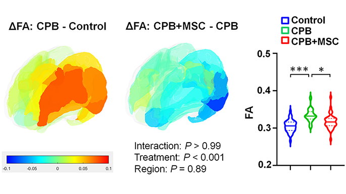
Differences of cortical fractional anisotropy between cardiopulmonary bypass and control (left), cardiopulmonary bypass + mesenchymal stromal cells and cardiopulmonary bypass (center), and 3 groups (right).
A pre-clinical study in the journal JACC: Basic to Translational Science shows that infusing bone marrow-derived mesenchymal stromal cells (BM-MSCs) during cardiac surgery provides both cellular-level neuroprotection for the developing brain and improvements in behavior alterations after (or resulting from) surgery.
What this means
According to lead author Nobuyuki Ishibashi, M.D., Oxidative and inflammatory stresses that are thought to be related to cardiopulmonary bypass cause prolonged microglia activation and cortical dysmaturation in the neonatal and infant brain. These issues are a known contributor to neurodevelopmental impairments in children with congenital heart disease.
This study found that, in a pre-clinical model, the innovative use of cardiopulmonary bypass to deliver these mesenchymal stromal cells minimizes microglial activation and neuronal apoptosis (cell death), with subsequent improvement of cortical dysmaturation and behavioral alteration after neonatal cardiac surgery.
Additionally, the authors note that further transcriptomic analyses provided a possible mechanism for the success: Exosome-derived miRNAs such as miR-21-5p, which may be key drivers of the suppressed apoptosis and STAT3-mediated microglial activation observed following BM-MSC infusion.
Why it matters
Significant neurological delay is emerging as one of the most important current challenges for children with congenital heart disease, yet few treatment options are currently available.
Applications of BM-MSC treatment will provide a new therapeutic paradigm for potential MSC-based therapies as a form of neuroprotection in children with congenital heart disease.
Children’s National Hospital leads the way
The Ishibashi lab is the first research team to demonstrate the safety, efficacy and utility of using cardiopulmonary bypass to deliver BM-MSCs with the goal of improving neurological impairments in children undergoing surgery for congenital heart disease. In addition to this pre-clinical research, a phase 1 clinical trial, MeDCaP, is underway at Children’s National.
Recent additional funding from the NIH will allow the team to identify molecular signatures of BM-MSC treatment and mine specific BM-MSC exosomes for unique cardiopulmonary bypass pathology to further increase understanding of precisely how and why this cell-based treatment shows success.
https://innovationdistrict.childrensnational.org/wp-content/uploads/2023/09/Ishibashi-Cell-Therapy-Study-CNRI-Feature.png385685Innovation Districthttps://innovationdistrict.childrensnational.org/wp-content/uploads/2023/12/innovationdistrict_logo-1-1030x165.pngInnovation District2023-09-06 11:21:532023-09-18 12:05:06Cell therapy mitigates neurological impacts of cardiac surgery in pre-clinical model
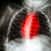
Significant neurological delay is emerging as one of the most important current challenges for children with congenital heart disease, yet few treatment options are currently available.
The research lab of Nobuyuki Ishibashi, M.D., at Children’s National Hospital, recently received $3.3 million in additional funding for research into cell therapy for neuroprotection in children with congenital heart disease. The new support comes from the National Heart, Lung and Blood Institute (NHLBI) of the National Institutes of Health.
The research goal
The overarching goal of the award is to establish detailed molecular signatures from critical cell populations for tissue repair and regeneration at single cell resolution after bone marrow-derived mesenchymal stromal cell (BM-MSC) delivery. The team has shown cellular, structural and behavioral improvements in pre-clinical models after delivery of BM-MSCs through cardiopulmonary bypass for children with congenital heart disease. However, the mechanisms underlying the therapeutic action of BM-MSCs still remain largely unknown. This R01 renewal will address the key knowledge gap.
Why it matters
Significant neurological delay is emerging as one of the most important current challenges for children with congenital heart disease, yet few treatment options are currently available.
The Ishibashi lab has demonstrated the efficacy and utility of using cardiopulmonary bypass to deliver BM-MSCs to improve neurological impairments in children undergoing surgery for congenital heart disease. Most notably, this included development of a phase 1 clinical trial, MeDCaP, at Children’s National.
The big picture
Together with the ongoing clinical trial established from the previous award, identifying molecular signatures of BM-MSC treatment and mining specific BM-MSC exosomes for unique cardiopulmonary bypass pathology will significantly improve understanding of this cell-based treatment. This work will also provide a new therapeutic paradigm for potential cell-free MSC-based therapies for neuroprotection in children with congenital heart disease.
https://innovationdistrict.childrensnational.org/wp-content/uploads/2023/07/x-ray-CHD-FeatureCNRI.png385685Innovation Districthttps://innovationdistrict.childrensnational.org/wp-content/uploads/2023/12/innovationdistrict_logo-1-1030x165.pngInnovation District2023-07-11 15:50:112023-07-14 09:35:26Cell therapy research for neuroprotection in congenital heart disease receives another $3.3 million from NIH
Children’s National Hospital announced a $96 million investment from an anonymous donor family to transform rare childhood brain tumor research and care. The donation, which strengthens our globally recognized leadership in the field, is one of the largest in the hospital’s history.
Children’s National will harness the investment to recruit more top talent and advance the most promising research. This will produce safer, more effective treatments. It also will elevate standards of care to help children with rare brain tumors thrive for a lifetime.
The big picture
Brain tumors are the most common solid tumors affecting children. They are especially challenging in kids because their brains are still developing. The disease and current treatments can put them at risk for lifelong complications.
The anonymous family’s investment provides new hope for patients who face rare and often challenging brain tumor diagnoses — in the Washington, D.C., community and around the world.
“This incredible partnership will lift up one of the nation’s top pediatric brain tumor programs into the stratosphere,” said Kurt Newman, M.D., president and CEO of Children’s National. “It will immediately propel our best-in-class research and care, allowing us to bring new therapies to children with brain tumors. This fundamentally changes the healthcare journey and long-term outcomes for children and their families.”
Why it’s important
This transformational investment will have a far-reaching impact on our ability to save and improve the lives of children with brain tumors. Funds will fuel collaborative breakthroughs across a range of scientific and psychosocial approaches.
The partnership will supercharge highly individualized and promising treatments for children with brain tumors. We will radically transform the research landscape with a focus on:
Low intensity focused ultrasound (LIFU) – Advancing laboratory research and a clinical program designed to treat childhood brain tumors with LIFU therapy
Cellular immunotherapy – Testing new gene-engineered immune cell products and accelerating their integration into standards of care
Rare Brain Tumor Program – Propelling new clinical trials through the hospital’s national and global leadership in pediatric brain tumor consortia. Already, Children’s National is leading a new collaborative with hospitals in North America, South America and Europe to better understand and find novel treatments for these rare diseases
Neurosurgery innovation – Exploring multiple ways to perform safer, more effective neurosurgery and developing new methods to enhance drug/agent delivery
Precision medicine – Recruiting leading scientists to advance biology-informed therapies that can be targeted for children across a spectrum of brain tumors
Good Manufacturing Practices (GMP) facility – Expanding our GMP, one of the first standalone facilities at a children’s hospital in the country, to translate new discoveries into clinical trials more rapidly
Additional priorities including expansion of clinical research infrastructure and growth of bioinformatics,brain tumor repository and molecular diagnostics initiatives
The partnership also transforms how we approach care. It will power our pursuit of psychosocial, behavioral health and neuroscientific initiatives to help kids live well and cope with the unique circumstances of their diagnosis. We will focus on:
Lifetime health and wellness – Building a world-class research and clinical care program to shape a new paradigm for supporting a child’s physical and emotional health during and long after cancer treatment
Child Mental Health & Behavioral Brain Tumor Lab – Establishing a robust neuro-oncology mental health program that delivers timely interventions and specialized psychiatric care for patient well-being
Additional priorities including a new Neuroscience Nursing ExcellenceProgram and growth of psychosocial support activities that bring comfort and encouragement to children during their treatment journey
Children’s National is proud to lead the way to a better future for pediatric rare brain tumor patients and expand our internationally recognized capabilities for neuro-oncology care.
https://innovationdistrict.childrensnational.org/wp-content/uploads/2023/06/child-in-hospital-bed-Feature.png300400Innovation Districthttps://innovationdistrict.childrensnational.org/wp-content/uploads/2023/12/innovationdistrict_logo-1-1030x165.pngInnovation District2023-06-22 12:57:492023-06-22 12:59:13$96 million philanthropic investment will transform rare pediatric brain tumor research and care
“I am a pediatric intensivist, and I am very interested in some of the pathologies and conditions that I come across in the ICU. We hatched this question that revolved around the idea: what can we do for TBI (traumatic brain injury) patients to enhance their cellular regeneration? … We looked at NG2-glia in particular, otherwise known as oligodendrocyte precursor cells. They are about 2-8% of the brain…. Do these cells respond to sleep and circadian rhythm? Is it a factor? Does it help? Does it hurt?”
Find out more about what Terry Dean, M.D., Ph.D., says he has learned about these and other questions through his recent research with interim Chief Academic Officer Vittorio Gallo, Ph.D. They join the Society for Neuroscience in a webinar on the circadian rhythms of these important brain cells and how their regeneration may be used someday to promote healing after brain injuries.
https://innovationdistrict.childrensnational.org/wp-content/uploads/2023/04/Gallo-and-Dean-webinar-screengrab-Feature-CNRI.png385685Innovation Districthttps://innovationdistrict.childrensnational.org/wp-content/uploads/2023/12/innovationdistrict_logo-1-1030x165.pngInnovation District2023-04-20 09:59:512023-09-18 12:03:04In the News: Regenerative brain cells and the circadian clock


 The Divisions of Neurology and Neurosurgery at Children’s National Hospital are proud to host the 35th Annual Pediatric Neurology Update course.
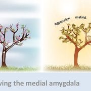


 Children’s National Hospital in Washington, D.C., was ranked as a top hospital in the nation by the

 The Children’s National Research Institute released its
 Several experts from Children’s National Hospital will be sharing their knowledge at the upcoming

 Children’s National Hospital named
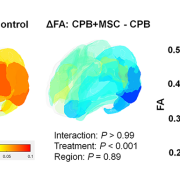
 Children’s National Hospital announced a $96 million investment from an anonymous donor family to transform rare childhood brain tumor research and care. The donation, which strengthens our globally recognized leadership in the field, is one of the largest in the hospital’s history.