
Driving pediatric breakthroughs through 2023
 The Children’s National Research Institute released its 2022-2023 Academic Annual Report. In the report, a summary of the past academic year highlights the accomplishments of each of the institute’s research centers, provides research funding figures and exalts some of the institute’s biggest milestones.
The stories in the report are a testament to the hard work and dedication of everyone at the Children’s National Research Institute.
We celebrated five decades of leadership and mentorship of Naomi Luban, M.D., and her incredible accomplishments in the W@TCH program, which have been instrumental in shaping the future of pediatric research.
We also celebrated innovation, highlighting our recent FDA award to lead a pediatric device consortium, which recognizes our commitment to developing innovative medical devices that improve the lives of children.
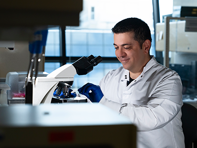
Breakthroughs at the Research & Innovation Campus continued as our researchers worked tirelessly to develop new treatments and therapies that will transform the lives of children and families around the world.
Taking a look at the breakthroughs happening in our now six research centers, we spotlighted the following stories:
- Reflecting on decades of progress in the blood, marrow and cell therapy programs at Children’s National. Our researchers have made significant strides in this field, and we are proud to be at the forefront of these life-saving treatments.
- In genetic medicine, we continue to be a beacon of hope for families facing rare and complex conditions. Our researchers are making incredible breakthroughs that are changing the landscape of pediatric medicine.
- We are also proud to share the $90 million award received from an anonymous donor to support pediatric brain tumor research. The predominant focus of this award is to develop new treatments that will improve outcomes for children with this devastating disease.
- This year, we opened a new Center that enhances our research capabilities in the field of Prenatal, Neonatal & Maternal Health Research. We are excited about the possibilities this new center will bring and look forward to the discoveries that will emerge from it.
- In addition, we are driving future pandemic readiness with the NIH funded Pediatric Pandemic Network. Our researchers are using cutting-edge technology and innovative approaches to prepare for the next pandemic and protect children.
- We are also exploring the potential of artificial intelligence (AI) in pediatric breakthroughs. Our researchers are using machine learning and other AI techniques to develop new treatments and therapies that will transform the lives of children.

 Advanced MRI visualization techniques to follow blood flow in the hearts of cardiac patients. Gene therapy for pediatric patients with Duchenne muscular dystrophy. 3D-printed casts for treating clubfoot. These were among the most popular articles we published on Innovation District in 2023. Read on for our full list.


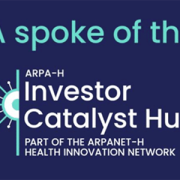
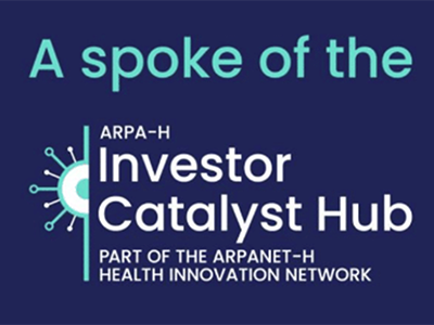 The hospital will advocate for the unique needs of children as part of nationwide network working to accelerate transformative health solutions.


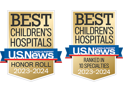 Children’s National Hospital in Washington, D.C., was ranked #5 in the nation on the U.S. News & World Report 2023-24 Best Children’s Hospitals annual rankings. This marks the seventh straight year Children’s National has made the Honor Roll list. The Honor Roll is a distinction awarded to only 10 children’s hospitals nationwide.


 A clinical trial testing a new drug to increase growth in children with short stature. The first ever high-intensity focused ultrasound procedure on a pediatric patient with neurofibromatosis. A low dose gene therapy vector that restores the ability of injured muscle fibers to repair. These were among the most popular articles we published on Innovation District in 2022. Read on for our full top 10 list.