Residents: Frontline defenders against antibiotic resistance?

A recent survey assessed whether residents knew which antibiotics were most appropriate for treating five common pediatric infections, including acute otitis media (ear infection).
Antibiotic resistance continues to grow around the world, with sometimes disastrous results. Some strains of bacteria no longer respond to any currently available antibiotic, making death by infections that were once easily treatable a renewed reality.
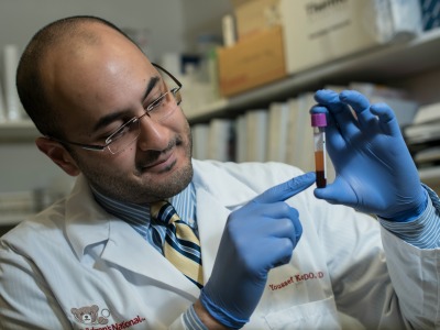
Avoiding this fate is possible, research suggests, if antibiotic prescribers do five essential things correctly: Give the right patient the right medication at the right dose through the right route at the right time. Medical residents – doctors who have finished medical school but are still receiving training at clinics and hospitals by working under more experienced physicians – are key to this strategy since they often are part of the frontline care team that selects and initiates antibiotic therapies. However, it has been unclear whether their prescribing patterns match these five “rights,” says Geovanny F. Perez, M.D., a pulmonologist at Children’s National Health System.
“Residents often decide which antibiotics to start a patient on, so they could become the first line of defense against antibiotic resistance,” Dr. Perez says. “They also could be an important target for education efforts if their prescribing patterns aren’t aligned with current guidelines.”
To determine whether residents are prescribing in ways that best avoid antibiotic resistance, Dr. Perez and colleagues sent an email survey to all 189 residents at two large children’s hospitals: Children’s National, a tertiary care center that serves patients throughout the greater Metropolitan Washington area at its main campus and network of primary care clinics; and Nicklaus Children’s Hospital, the largest freestanding pediatric hospital in South Florida.
The survey was divided into two parts. The first aimed to assess the knowledge of these residents about which antibiotics are most appropriate to treat five common pediatric infections: Acute otitis media (ear infection), group A streptococcal pharyngitis (strep throat), sinusitis (sinus infection), pneumonia and urinary tract infections.
The second part of the survey was meant to ascertain how residents acquired their antibiotic knowledge and prescribing behaviors. It asked about their awareness of antibiograms – a profile of which medications are effective against different local bacterial strains that is updated periodically at most hospitals – whether residents ever prescribed antibiotics for viral infections and the major influences on their prescribing decisions.
About one-half of the residents returned their surveys. Their answers suggested that most of them followed prescribing guidelines for the recommended drugs to treat otitis media, streptococcal pharyngitis and urinary tract infections. However, there were significant variations from guidelines for treating sinusitis and pneumonia, with many residents choosing antibiotics that were against current recommendations.
Additionally, only 3 percent of respondents indicated that they frequently used antibiograms, an important tool in selecting the most effective antibiotics. About one-half indicated that they sometimes used antibiograms, and one-quarter said that they never used an antibiogram. An additional 17 percent disclosed that they did not know what an antibiogram was. Even among those that knew about this important resource, about one-half said that they didn’t know where to access antibiograms specific to their hospitals.
Three-quarters of respondents indicated that they had prescribed antibiotics to patients who they considered to have a viral infection, rather than a bacterial one – a scenario in which antibiotics have no effect. In a follow-up question assessing the reasons for these decisions, 63 percent answered that they were following instructions from an attending physician or senior resident. More experienced physicians also played a more general role in shaping residents’ antibiotic knowledge: About 54 percent of residents said that their general pediatric inpatient attending physician – who oversees their training efforts – was their most influential source of knowledge in this area.
The findings, published in the September 2017 issue of Hospital Pediatrics, provide eye-opening insights into how residents prescribe antibiotics and their motivations for these choices, says Dr. Perez – particularly how the training they receive from mentors steers decisions many residents must make multiple times a day. He adds that antibiotic stewardship programs, which provide instruction to health care providers about current prescribing guidelines and practices, should focus on both residents and their resident charges for maximum impact.
“Ideally, we should be matching the guidelines 100 percent or at least close to it,” Dr. Perez says. “We think this goal is definitely attainable with the right training for both residents and their mentors alike.”