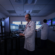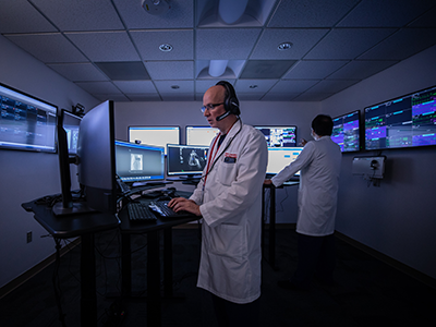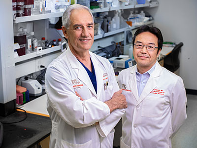
Lung transplant expert Michael Tsifansky, M.D., F.A.A.P., joins Children’s
Earlier this year Michael Tsifansky, M.D., F.A.A.P., joined Children’s National Hospital as an attending physician in the Cardiac Intensive Care Unit and in the Division of Pulmonology and Sleep Medicine. He brings to Children’s National a unique mix of expertise in critical care and pulmonary medicine. That passion for these two subspecialties has also made him one of the country’s leading experts in lung transplant procedures and the recovery from them.
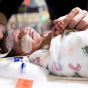
Dr. Tsifansky shared more information about caring for patients with complex lung diseases, especially those with end-stage lung disease. He outlines the patient population for pediatric lung transplants and the arduous process patients endure while waiting for a transplant, undergoing this major procedure, and then recovering from it.
What types of patients undergo lung transplant surgeries?
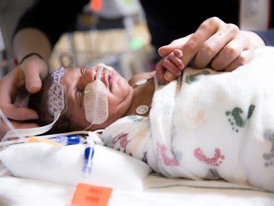
Lung transplantation in children is indicated when the following criteria are met:
- End-stage lung disease
- No reasonable alternative to the established diagnosis
- No medical or surgical alternative to the current course of treatment
- No other organ failure
- Stable social environment
Could you describe the surgery process?
Pediatric lung transplantation may be performed on cardiopulmonary bypass, on extracorporeal membrane oxygenation (ECMO) or off extracorporeal cardiopulmonary support (ECS). The donor’s lungs are kept chilled prior to transplantation and should be transplanted within six to eight hours after removal from the donor. The donor’s main-stem bronchi and pulmonary arteries are connected to those of the recipient, and the donor’s pulmonary venous drainage is connected to the recipient’s left atrium using the donor’s left atrial roof tissue. This procedure typically takes six to eight hours.
Could you describe the recovery process?
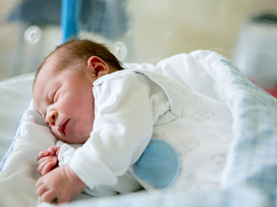
Typically, pediatric lung transplant recipients are extubated and encouraged to sit up four to six hours after the transplant procedure and walk soon afterward. It is important that they be out of bed and moving as soon as possible, and our colleague from Rehabilitation Services (physical and occupational therapists and rehabilitation physicians) will be working with the children toward these goals. After transplantation, pediatric patients will be given discharge instructions with individualized guidelines for a healthy lifestyle. Patients should return to near-normal life approximately three to six months after transplantation.
How long does the recovery process take?
The patient will remain hospitalized for 11-14 days following surgery for acute rehab, titration of antirejection meds and initial healing.
You’ve mentioned that it’s important for transplant patients to get moving as part of recovery. When can a patient begin walking again?
Lung recipients will be assisted into a chair soon after the transplant. Within the first 24-36 hours, the patient is encouraged to take short walks, increasing the distance each day. A physical therapist will work with the patient during their hospitalization to meet their goals. We also encourage patients to exercise on the treadmill regularly while hospitalized. By the time the patient is ready to go home, he or she will be able to easily move around by themselves and do most of their care without assistance. They feel so much better than before transplant and have so much energy that we almost always have to gently limit their activity for a short while to allow their chest incision to heal properly.
What do you see as the next step in pulmonary care for end stage lung disease at Children’s National Hospital?
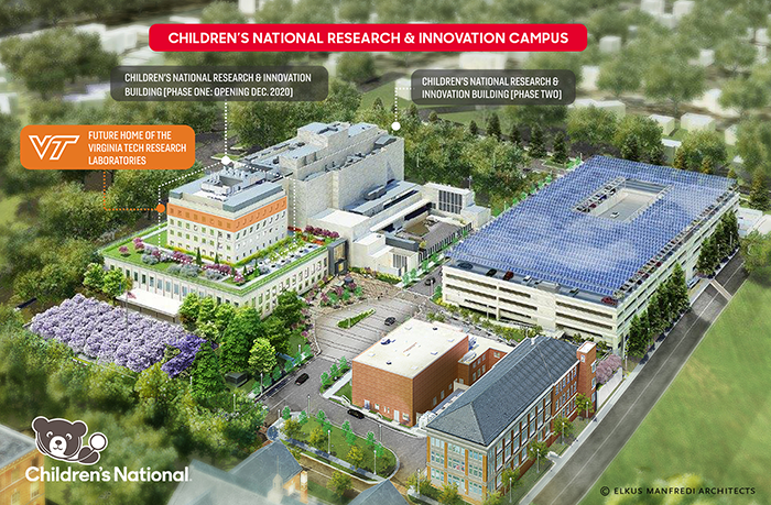
The development of a pediatric-specific lung transplant and respiratory failure program is the natural extension of the hospital’s cystic fibrosis program, heart transplant program and programs in pulmonary hypertension, bronchopulmonary dysplasia and extracorporeal membrane oxygenation for respiratory failure.
At present, there is no local option for a pediatric-specific program that can perform the transplant and provide the necessary comprehensive wrap-around services for patients in infancy up to age 18. As a top children’s hospital, Children’s National is uniquely positioned to provide the highest level of pediatric-specific care to this patient population and allow patients and their families to spend more time at home while undergoing this and other lifesaving treatments.
Dr. Tsifansky hopes to launch a comprehensive pediatric lung transplant and respiratory failure program at Children’s National in the very near future. Stay tuned for future developments from this area.